

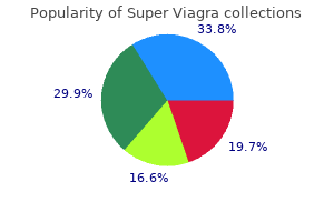
Super Viagra dosages: 160 mg
Super Viagra packs: 10 pills, 20 pills, 30 pills, 60 pills, 90 pills, 120 pills, 180 pills

160 mg super viagra sale
As such erectile dysfunction natural foods 160 mg super viagra, it raises ethical erectile dysfunction pills viagra 160 mg super viagra order with mastercard, philosophical, and social issues that no different procedure does. There has been an exponential increase within the variety of cosmetic procedures performed over the past 2 decades (a 162% increase since 1997 within the United States), with over 1. In trendy societies, the place mobility and huge networks of short-time acquaintances are the norm, "first impression" becomes crucial. As the European Union Bioethics Commission report established,7 there are four essential parts to be thought-about: the worth and which means of magnificence, the that means and vary of the precept of autonomy, the proper objectives of medicine, and the problem of publicly funded well being care. There are exterior and social components at play for patients who determine to undergo an aesthetic process, including social norms and the dominant best of magnificence. These underline the significance of the promotion of numerous magnificence beliefs, by governments and the media. Proper Goals of Medicine Medicine is supposed to be about therapy and disease, whereas aesthetic surgical procedure is about nondisease and enhancement. However, the drawing of clear lines between medicine and aesthetic surgical procedure has been proven to be philosophically unimaginable. Serious suffering that deserves treatment is inside the area of aesthetic surgery as a lot as in traditional medicine. Issue of Publicly Funded Health Care Note Core ethical issues in aesthetic surgical procedure: 1. What emerges in this method as one single criterion underlying these exceptions is patient suffering, often however not solely brought on by social norms. Patient Selection and the Rhinoplasty Consultation the broader social and ethical context of rhinoplasty raises appreciable points. Those with a correctable deformity and cheap expectations who can be treated by plastic surgery 2. Those with no deformity and unreasonable behavior who would be inappropriate candidates for surgical procedure and as an alternative should be referred for psychiatric evaluation three. Significantly interfered together with your social life, schoolwork, job, other activities or other elements of your life Only a affected person who fully understands the goals, dangers, and limitations of the operation can provide real informed consent. Areas of concern20 Use of surgical cosmetic interventions Success of beauty surgical procedure Rates of dissatisfaction with cosmetic surgery Other dangers 0. In a examine of 26 sufferers undergoing forty six procedures in the United Kingdom, rhinoplasty was related to marked dissatisfaction and a rise within the diploma of preoccupation and handicap, with the worst consequence in these with repeated operations. The bottom line, as expressed succinctly by Goode,31 might be distilled as follows: hearken to your intestine emotions and to your staff-a affected person who appears unsuitable for rhinoplasty through the first minutes of the consultation more than likely is. This may be complemented with computerimaging evaluation and manipulation, as discussed later. Preand postoperative images of previous patients may be helpful, although the surgeon should resist the temptation of focusing solely on "poster patients"; certainly, the circumstances where she or he achieved a less than ideal outcome, and even cases of patients who have been unhappy and underwent revision surgery, should be shown and discussed. It is counterproductive, and a few patients may be insulted if the discrepancy between their nose and the perfect nose is analytically described. Patients are probably to use the Internet to gather info, both before and after their session. Printed material and handouts with info that the affected person can take up at residence are also important. Indeed, in a current research, the quality of printed handouts and the data gathered from the Internet have been the elements most strongly correlated with total patient satisfaction with the consent process. Anatomy of the Bony Pyramid the bony vault or pyramid is the upper one-third of the nostril and is fashioned by the nasal bones and the ascending (frontonasal) strategy of the maxilla. Visible are the bony vault, consisting of the nasal bones and the frontonasal process of the maxilla, and the cartilaginous pyramid, consisting of upper and decrease lateral (alar) cartilages. Nasal Bones the nasal bones are cephalically hooked up to the frontal bone, laterally to the ascending means of the maxilla, medially to each other, and posteriorly to the septum. Their caudal end overlaps for a few millimeters the upper lateral cartilage, like a roof tile. Caudally and laterally, they form, along with the ascending strategy of the maxilla, the pyriform aperture. Anatomy of the Cartilaginous Pyramid 2 the decrease two-thirds of the nose are formed by the cartilaginous pyramid. This is a unified, winged structure that includes the upper lateral cartilage and the cartilaginous septum, which articulate with one another in a T- or Yshaped configuration. Surgical Anatomy of the External Nose 419 Scroll of cephalic fringe of lateral crus of alar cartilage. The inner valve is created by the convergence of the septum with the upper lateral cartilage on the level of the pinnacle of the inferior turbinate corresponding to the supratip breakpoint or depression. This is the narrowest part of the higher airway, and any degree of narrowing of this angle can lead to nasal obstruction. This area can be significant histologically, because it constitutes the interface between the (external) squamous epithelium and the (internal) nasal mucosa. Tips and Tricks One of the roles of spreader grafts is the widening of the angle fashioned by the articulation of the septum with the upper lateral cartilage. Lateral and caudally to the lateral crura, fibroareolar tissue lies between them and the pyriform aperture, while laterally and cephalically, there are a couple of small accessory cartilages. More cephalically (between the nasal bones and the pyriform aperture), there are a few sesamoid cartilages. The lateral crus is the widest a part of the alar cartilage and is tightly adherent to the overlying nostril pores and skin. Caudally, the upper lateral cartilage articulates with the alar cartilage in the scroll space. Usually the cephalic fringe of the alar cartilage overlaps the caudal edge of the upper lateral cartilage, though a quantity of configurations have been described. The alar cartilage is thus comprised of the medial, middle or intermediate, and lateral crura. They form two arches, with the medial crus converging within the midline and thus forming the columella, and the lateral crus supporting the lateral wall of the nasal vestibule. The medial crura converge in the midline (columellar phase of the medial crura) and diverge more inferiorly, toward the nasal backbone (medial crural footplates). The center crus consists of the domal section, containing the tip-defining point, and the lobular section. Tips and Tricks the domal phase of the intermediate crus of the alar cartilage can take numerous shapes, and its configuration defines to a big extent the shape of the nasal tip (boxy, bifid, etc. The area cephalic to the tip is called the supratip space and the area slightly below it, the infratip. The domal section of the intermediate crus and the angle of the medial crura and their approximation of the domes are all important elements that define the tip shape, rotation, and projection. The projection and rotation of the tip are regulated by the relative size of the medial crura (anterior stand, A) and the two lateral crura (lateral stands, B). Tips and Tricks Endonasal rhinoplasty may lead to loss of tip support by disruption of the scroll area via an intercartilaginous incision, whereas external rhinoplasty disrupts the attachment of medial crura to the septum, the interdomal ligaments, and the soft tissue envelope.

Cheap super viagra 160 mg line
Benign erectile dysfunction with new partner super viagra 160 mg purchase with mastercard, radial sclerosing lesions may have this appearance however biopsy is important to set up histology erectile dysfunction doctors in south jersey super viagra 160 mg order free shipping. Skin retraction and nipple retraction carry vital threat of malignancy and require tissue biopsy. Segmental: Calcifications restricted to a phase or wedge-shaped portion of the breast might come up inside a single ductal system and its branches. Diffuse/scattered: Calcifications that seem to be randomly distributed all through the breast are referred to as diffuse or scattered. Compared to linear or Unusual Findings Focal pores and skin thickening may be associated with benign or malignant ideologies. Skin thickening may be present with benign circumstances such as an infection and venous obstruction. Focal asymmetry describes an space of asymmetry that lacks the appearance of a real mass. Physical examination has poor specificity with solely 4% of symptomatic women found to have malignancy (14). The objective of mammography on this setting is to characterize the palpable finding and assess the steadiness of the breast. Breast ultrasound is used extensively within the setting of a symptomatic affected person along with mammography. Use of ordinary practice guidelines is really helpful for imaging and management of symptomatic girls (24). A suspicious clinical finding ought to bear surgical consultation even when imaging is unfavorable. In this example, the mass may be localized for the surgeon to ensure proper excision. Skin Changes or Inflammatory Breast Findings Women presenting with inflammatory breast findings are referred for diagnostic mammography and infrequently sonography. The distinction between inflammatory most cancers and an infection is frequently not possible by imaging alone as both might show pores and skin modifications, interstitial edema, and abnormal axillary lymph nodes. Unless a suspicious mass or calcifications are found which might direct biopsy, pressing scientific analysis is really helpful. Sonography could detect a fluid assortment consistent with abscess which may be confirmed with aspiration. For those ladies initially treated with antibiotics for suspected mastitis, very short-term scientific reevaluation to guarantee resolution is crucial. Palpable Mass or Thickening Individuals with a palpable breast abnormality corresponding to a discrete mass or focal thickening or nodularity ought to bear diagnostic imaging prior to biopsy since biopsy can alter mammographic and sonographic appearances. Women 30 years and older are beneficial for mammography and sonography; girls youthful than 30 are initially evaluated with sonography, though mammography may be essential in sure circumstances (24). Approximately 5% to 15% of sufferers with a palpable cancer could have a falsenegative mammogram. This quantity is higher for girls with extremely dense breasts and lower for ladies with extraordinarily fatty breasts. An individual clinician could overestimate mammography efficiency due to the low incidence of most cancers within the symptomatic inhabitants. Of a typical group of 250 diagnostic sufferers referred for mammography, there will be 10 cancers. Only 1 of the 250 could have most cancers and a false-negative mammogram (assumes a four per a hundred cancer incidence and a 10% false-negative rate) however this low quantity is due primarily to the low incidence of cancer rather than the superb mammographic efficiency. If the mammogram is adverse, ultrasound is performed of the palpable space since most cancers with false-negative mammography shall be identified as abnormal by sonography. A affected person with a adverse mammogram and unfavorable ultrasound within the setting of a palpable finding is at very low risk of malignancy. The false-negative fee of combined ultrasound and mammograms at experienced breast facilities is 0% to 3% (25�28). The management of sufferers with palpable findings with unfavorable mammogram and ultrasound is dependent upon the scientific evaluation. Axillary Lymph Node Presentation of Breast Cancer Less than 1% of girls with breast most cancers will current with an axillary mass discovered to represent metastatic breast cancer in axillary lymph nodes with normal breast bodily examination. Diagnostic mammography should be carried out of both breasts to assess for occult breast most cancers. Symptomatic Pregnant and Lactating Women Sonography is the preliminary imaging evaluation of pregnant and lactating women, many whom have never been screened due to younger age though mammography could also be also useful in some instances to assess for calcifications (31). The radiation dose of a two-view mammogram to the uterus or fetus is lower than 1 per 10,000 the breast dose (0. Extensive calcifications associated with ductal carcinoma in situ by mammography are typically a contraindication to breast-conservation therapy. Multicentric disease by mammography can additionally be a contraindication to breast-conservation remedy. The impression on Bloody or Serous Discharge Mammographic sensitivity could also be as little as 10% for intraductal cancer presenting as nipple discharge (29). If the mammogram is unfavorable, retroareolar breast ultrasound may be performed to assess for intraductal masses or other findings. Filling defects or stenoses require surgical excision to decide histologic trigger. Magnification mammography is routinely used within the setting of breast cancer manifested as microcalcifications to assess extent. Following lumpectomy with unfavorable pathologic margins, mammography is really helpful in instances with malignant calcifications to guarantee excision. Suspicious residual calcifications should be topic to reexcision prior to radiation remedy. The reported sensitivity of mammography for detection of in-breast recurrence is variable (35). Mammographic look of cancer in some high-risk groups could also be just like the inhabitants normally. However, because these are young women, mammography may be much less sensitive as a outcome of greater frequency of dense breasts and possible aggressive tumor biology. The use of digital mammography in young women with dense breasts could improve sensitivity (3). Fibroglandular tissue attenuates x-rays and produces a white ("dense") area on a mammogram. Entirely fats Scattered fibroglandular densities Heterogeneously dense Extremely dense (7). Gynecomastia presents as retroareolar mammographic density without calcification which may be uneven. Enlarged breast due to extreme adipose tissue ("pseudogynecomastia") seems as fats on mammography and requires no additional analysis. The mammographic findings of male carcinoma are similar to feminine most cancers however microcalcifications are uncommon. Mammography is very delicate for breast cancer detection due to the lack of breast tissue in most men with negative predictive values of 99% to one hundred pc reported (38). Because some unusual forms of gynecomastia could appear mass-like by mammography, biopsy is required in these cases to set up the analysis.
Syndromes
- Foot pain, sores on the feet, or blue toes
- Increased amount of urine produced
- A large area at the base of the tongue, or a tongue that is large compared to the mouth
- Blood in urine
- Coma
- For more information call your car seat manufacturer, car manufacturer, or the State Highway Safety Office.
- Severe headache
- Loss of control over bowels or bladder
Super viagra 160 mg buy generic online
Sclerosing adenosis is normally an incidental discovering erectile dysfunction medication options cheap 160 mg super viagra otc, however might current as a mammographic abnormality (microcalcifications erectile dysfunction treatment wikipedia discount super viagra 160 mg on-line, distorted architecture) or a mass lesion (also generally known as nodular adenosis or adenosis tumor). This lesion is composed of distorted epithelial, myoepithelial, and sclerotic stromal components arising in association with the terminal duct lobular unit. This lobulocentric sample is key to the correct prognosis of sclerosing adenosis and its variants, and is greatest appreciated at low energy microscopic examination. The epithelium in sclerosing adenosis might undergo apocrine metaplasia, and is then referred to as apocrine adenosis. The apocrine metaplastic cells may show cytologic atypia, elevating the differential prognosis of invasive carcinoma if the lesion is examined at high microscopic energy with out accounting for the lobulocentric architecture appreciated at low energy (12). Because of the distorted glandular pattern of sclerosing adenosis, this lesion may be confused with a lowgrade invasive carcinoma, significantly tubular carcinoma. Although sclerosing adenosis consists of distorted, elongated, or obliterated glands and tubules, tubular carcinoma consists of angulated tubules with open lumens. The stroma of sclerosing adenosis is fibrotic or sclerotic compared with the desmoplastic stroma of invasive carcinoma. Importantly, versus tubular carcinoma, sclerosing adenosis incorporates myoepithelial cells, which can be highlighted by immunohistochemistry. In the unique examine of Dupont and Page (3), 70% of the biopsies showed nonproliferative lesions. The solely group of patients within the nonproliferative category with an elevated threat of creating breast cancer was that with gross cysts plus a family history of breast most cancers. It must be noted that, although Dupont and Page initially included fibroadenomas among the nonproliferative lesions, the outcomes of a subsequent examine by these investigators indicated a higher relative risk for breast most cancers among sufferers with fibroadenoma than for patients with nonproliferative lesions (10). As a outcome, fibroadenomas at the second are included among the many proliferative lesions with out atypia (see the section on fibroadenomas). Proliferative Lesions without Atypia Included throughout the group of proliferative lesions with out atypia are traditional ductal hyperplasia (11) (also generally known as moderate or florid hyperplasias of the standard type), intraductal papillomas, sclerosing adenosis, and radial scars (3). Usual ductal hyperplasias are intraductal epithelial proliferations more than four epithelial cells in depth. They are characterised by a bent to bridge and often distend the concerned space. These spaces are sometimes slit-like and organized across the periphery of the proliferation, with their long axes parallel to the basement membrane. The cells are cytologically benign, however the pattern simulates that of an invasive carcinoma. The cells might type tufts, micropapillations, arcades, bridges, stable, and cribriform patterns (11). A second cell inhabitants with options much like these seen in usual ductal hyperplasia can additionally be sometimes present. These cells are monomorphic, evenly spaced, and dyshesive, with spherical or oval, often eccentric nuclei and pale cytoplasm usually with intracytoplasmic vacuoles. In addition to involving lobular units, the cells of atypical lobular hyperplasia can also contain ducts (14). It is necessary to note that with the growing use of mammographic screening, atypical hyperplasias are being diagnosed extra incessantly than prior to now. For instance, when a biopsy is carried out due to a palpable mass, atypical hyperplasia is seen in solely about 2% to 4% of cases (3). In contrast, atypical hyperplasia was recognized in 12% to 17% of biopsies performed due to the presence of mammographic microcalcifications (15). Near the center of this area is a proliferation of relatively uniform epithelial cells with monomorphic, round nuclei just like those seen in low-grade ductal carcinoma in situ. However, these cells comprise only a portion of the proliferation throughout the house. The acini of this lobule comprise a proliferation of small uniform cells, that are dyshesive, and are equivalent to the cells that comprise lobular carcinoma in situ. Family History Columnar Cell Lesions and Flat epithelial Atypia Lesions of the breast characterized by enlarged terminal duct lobular models lined by columnar epithelial cells are being encountered increasingly in breast biopsies performed because of mammographic microcalcifications. Some of those lesions characteristic banal columnar cells in either a single layer (columnar cell change) or showing stratification and tufting, however without complex architectural patterns (columnar cell hyperplasia). In other columnar cell lesions, the lining cells exhibit cytologic atypia, most commonly of the low-grade, monomorphic type. Additional studies are wanted to higher understand the biological nature and the level of subsequent breast most cancers danger associated with these lesions. There is common settlement that the presence of a household historical past of breast most cancers in a first-degree relative (mother, sister, or daughter) is related to a slight enhance within the breast cancer danger in women with proliferative lesions without atypia (3�7). The influence of household history on breast most cancers risk in women with atypical hyperplasia is much less clear, however. Time since Biopsy Information relating to the relationship between time since benign breast biopsy and breast most cancers risk is out there from several research. In the Nashville examine, ladies with prolifera- Factors Modifying Breast Cancer Risk in Women with Biopsy-Proven Benign Breast Disease A number of factors appear to modify the breast most cancers risk related to biopsy-proven benign breast disease, including a household historical past of breast cancer, time since biopsy, menopausal status, and the looks of the background breast tissue. The regular epithelial cells in the acini of this terminal duct are replaced by a population of columnar epithelial cells with round, monomorphic nuclei. Similarly, in the Mayo Clinic research, an extra breast cancer risk was seen amongst girls with biopsy-proven benign breast disease for at least 25 years after the benign breast biopsy (5). Among sufferers with atypical hyperplasia, the relative danger was persistently elevated past 15 years (16). More data are needed to clarify further the relationship between time since biopsy and breast cancer threat for women with benign breast illness, notably for girls with atypical hyperplasia. Another issue of medical importance is the affect of postmenopausal hormone substitute remedy on the danger of breast most cancers in women with biopsy-proven benign breast illness. Clinical follow-up research have shown that ladies who take hormone substitute therapy are at elevated danger for developing breast cancer (19). The use of A study from the Mayo Clinic has suggested that the presence of lobular involution within the background breast tissue of a benign breast biopsy is related to a significant decrease in the risk of breast cancer. Furthermore, in that research the presence of lobular involution modified the risk in women with proliferative lesions without atypia and in these with atypical hyperplasia. Laterality of Risk Breast cancers that develop among women with atypical hyperplasia could happen in either breast. Overall, approximately 60% of cancers that develop in girls with atypical hyperplasia occur in the ipsilateral breast; an extra of ipsilateral cancers is seen significantly within the first 10 years after the benign breast biopsy (2,5). These observations suggest that the concept that atypical hyperplasias characterize only danger indicators is overly simplistic and that, in a minimum of some cases, these lesions may act as direct (albeit nonobligate) precursors to invasive breast cancer (23). They further point out that the chance amongst girls with biopsy-proven benign breast illness is influenced by different factors as properly. To counsel individual sufferers properly, an understanding of the distinction between relative danger and absolute danger is important. The relative threat for breast cancer represents the incidence of breast cancer among girls within a sure subpopulation divided by the incidence of breast cancer in the reference population. The magnitude of the relative danger is very dependent on the breast most cancers incidence in each the examine group and the reference population.
Buy super viagra 160 mg lowest price
Hypertension is commonly thought-about extra likely to erectile dysfunction pump.com generic 160 mg super viagra overnight delivery prolong the length of spontaneous epistaxis erectile dysfunction needle injection super viagra 160 mg purchase mastercard, but neither this nor an overall association between adult epistaxis and hypertension has been recognized. Liver disease must also be thought-about, as it will have an effect on coagulation not solely by reducing the manufacturing of most coagulation components, but also by adversely influencing the clearance of activation merchandise. Hepatic failure and cirrhosis could present with the complete spectrum of issue deficiencies when all procoagulant and inhibitory elements are decreased. High-output coronary heart failure may develop as a consequence of shunting of blood between the arterial and venous circulation. The condition is inherited as an autosomal dominant genetic disorder, resulting in a weakness of blood vessel partitions and vascular malformations. It is characterised by telangiectasias on the skin and mucosal surfaces, which can induce severe and persistent epistaxis and arteriovenous malformations of the inner organs (lungs, gastrointestinal tract, brain, liver, and spine). The phrases anterior and posterior bleeds had been described before the diagnostic use of endoscopes and lack precision. It has been suggested that anterior and posterior should check with the connection to the road of the piriform aperture. The logical corollary is that medical epistaxis should be managed in conjunction with, and even solely by, doctor colleagues, whereas surgical epistaxis is extra amenable to the interventions of otolaryngologists, maybe using adjunctive interventional radiology. Etiology 511 the incidence of epistaxis from the septum and lateral nasal wall varies between reports. Endoscopic evaluation has described 70% of bleeds as arising from the nasal septum. Vascular Anatomy of the Nose the nasal mucosa is a dynamic structure with a rich vascular supply. The arterial provide is derived from both the interior and exterior carotid arteries, with anastomoses between the 2 techniques inside the nasal mucosa. The arteries travel inside the mucoperiosteum and mucoperichondrium, excluding branches that pass along bony canals in the inferior and middle turbinates. The sphenopalatine foramen itself lies at the posterior end of the center turbinate. Anteriorly, blood flows alongside the facial artery to the superior labial artery before coming into the anterior section of the nostril. Although the variety of branches varies in accordance with the reviews, ranging from 2 to 10, bifurcation of the maxillary artery earlier than the sphenopalatine foramen is widespread and happens in 89% of cases. Lee at al21 famous that in 38% of instances, some portion of the artery could run anterior to the posterior maxillary wall. Furthermore, in 50% of cases, a branch of the inferior turbinate artery extends anterosuperiorly to supply the posterior fontanelle. These variations offer an explanation for the etiology of large-volume postsurgical posterior epistaxis following routine restricted maxillary antrostomy. The lateral branch provides the lateral wall of the nose and the inferior turbinate. The medial department passes over the anterior sphenoid wall, 1 cm above the posterior choana, to the posterior septum, the place it divides into three parts. The posterior nasal artery, typically referred to because the septal artery, has been described as passing through its personal foramen posterior to the sphenopalatine foramen. Internal Carotid Artery System the internal carotid arterial blood travels along the ophthalmic artery via the superior orbital fissure to the orbital apex, then anteriorly to give off the anterior and posterior ethmoidal arteries. Within the cranial cavity, each arteries branch, giving rise to terminal branches that supply the dura of the anterior fossa; some branches run inferiorly to supply the superior nasal cavity. The anterior ethmoid artery is the bigger and most fixed of the 2; the posterior ethmoid artery could also be absent in up to 30%. Additionally, the bone over the paraclivial and parasellar carotid could additionally be dehiscent inside a well-pneumatized sphenoid sinus. This anatomical relationship supplies the chance for large epistaxis derived from this phase of the inner carotid artery secondary to trauma or idiopathic aneurysmal/pseudoaneurysmal dilation. This is the Woodruff venous plexus, which contains massive, thin-walled veins with little or no muscle or fibrous tissue within a thin mucosa comparatively devoid of different Venous Drainage System Generally, the veins comply with the course of the arteries inside the nasal mucosa. However, there are emissary branches that result in the cavernous sinus and comparable branches from the pterygoid plexus that also cross to the cavernous sinus. James Little was a professor of surgical procedure in New York in 1879 when he described the blood provide and administration of a bleeding "ulcer" of the anterior nasal septum in four patients. Wilhelm Kiesselbach, a lecturer in otology at the University of Erlangen in Germany, subsequently described three. Clinical Features the features of epistaxis are finest described based on the scientific presentation. Children usually current in the outpatient clinic with a historical past of recurrent nosebleeds. Adults may current to the clinic with recurrent spontaneous nosebleeds, however their medical history is usually more complicated, significantly with advancing years. Pregnancy is associated with an increased incidence in nosebleeds as a end result of elevated nasal blood circulate from hormonal modifications. Indeed, the prevalence of epistaxis in pregnant versus nonpregnant ladies has been discovered to be considerably elevated (20. The patients often have either a single episode or a short sequence of episodes main as a lot as a significant unilateral bleed. It may be tough to differentiate which is the bleeding side in profuse bleeds. Patients presenting as emergencies with ongoing or recent bleeding will usually be agitated and distressed. They may also be hypovolemic, anemic, and have other comorbidities that may be exacerbated by the acute bleeding episode. In the United States, the American Heart Association Basic Life Support protocol guides prioritization of consideration in the management of emergency situations. Once that is deemed secure, consideration may then be turned toward controlling the bleeding whereas simultaneously establishing vascular entry for the availability of quantity replacement and hemodynamic support. Acute nontraumatic epistaxis happens spontaneously in varied situations, and sufferers will only go to the hospital if the bleeding is persistent and heavy, typically resulting in hospital admission for several days. The preliminary administration is due to this fact to apply first-aid measures to management and minimize the blood loss. Lying down ought to be prevented, until the patient feels faint, thus decreasing the risk of aspiration and minimizing the amount of blood ingested, as this is a potent gastric stimulant and will lead to vomiting. Sitting forward decreases the blood flow to the nasopharynx and also permits blood entering the pharynx to be spat out. The affected person ought to be suggested to pinch the soft part of the nostril, below the nasal bones, and hold for 10 to 15 minutes with out releasing, while respiration through the open mouth. Assessment and treatment ought to happen in a suitable room with suction and specialist equipment available. Medical and nursing employees should wear appropriate protecting clothing and aprons, as the danger of contamination by blood is excessive. The nasal cavities ought to then be sprayed with an area vasoconstrictor and topical anesthetic resolution.
Order super viagra 160 mg mastercard
Greater fat intake after analysis was associated with a nonsignificantly worse survival end result erectile dysfunction causes medications 160 mg super viagra purchase. However erectile dysfunction doctors in tulsa generic super viagra 160 mg free shipping, greater protein consumption, primarily from poultry, fish, and dairy sources, was associated to a better prognosis, even after controlling for protein consumption previous to prognosis. Similarly, higher intake of trans fat and saturated fat after prognosis of breast most cancers was associated with greater general mortality, although not breast-cancer-specific mortality (511). This is important because with early prognosis and good remedy, the large majority of girls will survive their breast cancer, however they proceed to be at risk for illnesses of girls normally. In a current pooled analysis, alcohol consumption after prognosis of breast most cancers has overall not been related to survival (512), although a touch vital increase in recurrence was seen in postmenopausal women. Also in a pooled evaluation, higher soy consumption after diagnosis was related to a nonsignificantly lower danger of breast-cancer-specific mortality, and a considerably decrease danger of breast cancer recurrence (513). Also, common use of supplements of vitamin E and vitamin C was related to decrease danger of breast most cancers recurrence (514). Several randomized trials have been performed among girls with early-stage breast most cancers to decide the consequences of dietary change on recurrence or mortality. In one trial, 2,437 ladies with breast cancer had been randomized to a low fat food plan or their ordinary diet and adopted for a median of 5 years (515). Dietary fats intake was lowered to 33 grams per day in the intervention group compared to fifty one grams per day within the management group, and weight was also six kilos lower in the intervention group. In another trial among three,088 ladies, one group was assigned to a food plan excessive in fruits, vegetables, and fiber and low in fat (516). The improve in fruit and vegetable consumption was large, and documented by a 50% increase in blood carotenoid degree, however the reported distinction in fat consumption was small (-15%), so this examine primarily examined the benefit of increasing fruit and vegetable intake. Enthusiasm for the hypothesis that dietary fats consumption was responsible for the high rates of breast most cancers charges in Western countries was primarily based largely on the weakest type of epidemiologic evidence-ecologic correlation research. Excess power intake in relation to physical activity during adulthood, which accelerates progress and the onset of menstruation during childhood, leads to weight acquire in center life and thus can contribute considerably to breast cancer danger. These effects of vitality steadiness clearly account for an essential part of worldwide differences in breast most cancers rates. Alcohol intake, even at very low levels, is a well-established danger issue for breast most cancers, and research demonstrating that even average alcohol consumption will increase endogenous estrogen ranges present a potential mechanism, thus supporting a causal interpretation. Diet throughout childhood has been comparatively unstudied, but current proof suggests that larger intake of soy products and decrease consumption of pink meat throughout this era might reduce threat of breast cancer. Other latest findings counsel that characterization of breast cancers by hormone receptor standing, and probably other options, could also be necessary in research of food plan. Although our understanding of food regimen and breast most cancers is incomplete, evidence may be considered conclusive that breast most cancers threat could be decreased by avoiding weight acquire throughout grownup years and by limiting alcohol consumption. Although much less conclusive, some evidence suggests that breast cancer danger could be modestly reduced by limiting consumption of pink meat throughout early adult life, by changing saturated fat with monounsaturated fat, and by consuming extra fruits, vegetables, and whole grains (which characterizes the Mediterranean dietary pattern). A variety of potential mechanisms have been proposed including changes in menstrual cycle characteristics, lowering intercourse hormones and insulin-like development components, and/ or bettering immune function (519,520). The mechanisms by which physical activity reduces publicity to hormones range by interval of life. Young ladies taking part in strenuous athletic training similar to operating and ballet dancing have delayed menarche (522�524), which is known to cut back danger of breast cancer, and even moderate-intensity bodily activity might delay menstruation (398). This impact of exercise at young ages may be reflected in lower physique weight and physique fat, both of which are determinants of delayed menstruation (399,522). A later menarche is associated with a later onset of normal ovulatory cycles and decrease serum estrogen concentrations throughout adolescence (525). Further, a substantial degree of ovarian dysfunction may happen even amongst physically active girls who appear to have regular menstrual cycles (528). Among older ladies, levels of past and present physical exercise affect fats stores (522,523,528�531), which after the menopause are main websites of conversion of androstenedione to estrogen (532,533). A number of epidemiologic studies have reported an inverse affiliation between physical exercise and postmenopausal breast cancer, although the evidence is less constant for premenopausal breast cancer (519,520,534� 537). However, there are a variety of features regarding this association that remain unclear. Methodologic variations in physical activity assessment are likely to have contributed to inconsistencies in examine outcomes. Studies have differed in the ages at which bodily exercise was assessed, methods for measuring intensity, frequency, and duration of bodily exercise, definition and categorization of physical activity levels (including consideration of solely leisure, or leisure and occupational, activity), and age at breast cancer diagnosis. However, outcomes have diversified even amongst research which have tried to assess physical exercise at related times in life utilizing comparable tools. One of the strongest reductions in breast most cancers danger related to increased physical activity was reported in a population-based case-control research of girls younger than 40 years (538). This was the primary study explicitly devoted to the relationship between physical activity and breast cancer, and it was also the primary to use an in depth physical exercise evaluation instrument to quantify the common number of hours per week of leisure physical exercise over the reproductive life span, beginning at menarche. These researchers concluded from their varied analyses that lifelong bodily exercise is the important exposure of interest with regard to breast cancer threat. Since publication of this research, many other research (537,539�554) have assessed the connection between lifetime physical activity and breast cancer risk. In considered one of these studies (547), outcomes assist those reported above, with reduced danger of breast cancer in premenopausal girls with greater lifetime bodily activity. In distinction, one other examine found no association between exercise in earlier intervals of life and postmenopausal breast cancer (548). Types of activity are extensively varied across people, as nicely as across research. Broad categories of leisure, household, and/or occupational activity have been assessed in plenty of research. The findings of inverse associations with family and occupational bodily activity, however not with recreational exercise, suggest that residual confounding by sociodemographic and reproductive components are a minimum of partly liable for the noticed inverse relationships. Among types of leisure actions, some studies have noticed stronger associations for extra average or vigorous actions, in contrast with much less intense actions (555). However, even brisk strolling seems to be beneficial, as was reported in one research (556). A case-control research conducted amongst premenopausal and postmenopausal girls in city Shanghai (544) found vital inverse dose�response relationships between years of (recreational) exercise participation and breast most cancers threat, in addition to between lifetime occupational activity and breast most cancers threat. It has been hypothesized that prime ranges of physical activity during adolescence are significantly necessary with respect to influencing breast most cancers risk. However, different research which have examined the association between physical activity during adolescence and breast cancer threat have found little evidence for a protecting impact. Indeed, some research have observed stronger associations with more modern, or later in life, bodily activity. In distinction to the detailed measurement of lifetime bodily exercise employed by a few of the studies talked about earlier, a relatively simple measure of physical activity was used in a potential cohort research of Norwegian girls aged 20 to 54 years at baseline (558).
Buy discount super viagra 160 mg online
This adenosis is characterised by a haphazard proliferation of small glands composed of a single layer of epithelial cells erectile dysfunction at age 29 proven 160 mg super viagra. The glands are comparatively rounded and many include eosinophilic secretions of their lumens erectile dysfunction usmle buy generic super viagra 160 mg on-line. Those with related invasive carcinomas must be managed as for the stage of the invasive carcinoma. This lesion is characterized by a central fibroelastic core containing entrapped benign glands. Radiating from this core are ducts that show a variety of adjustments, including cysts and epithelial hyperplasia. They have been described in the literature by a variety of different names, together with sclerosing papillary proliferation, nonencapsulated sclerosing lesion, indurative mastopathy, and radial sclerosing lesion. The time period complicated sclerosing lesion is sometimes used for related lesions bigger than 1 cm in measurement or for these lesions with a quantity of fibroelastotic areas in shut contiguity. First, they might, on mammographic, gross, and microscopic examination, simulate breast carcinomas. Second, the connection between the presence of radial scars and subsequent breast most cancers has lengthy been a matter of controversy (see discussion below). Radial scars are most frequently incidental microscopic findings in breast biopsies performed for other indications (60,61). The reported incidence of radial scars varies from 4% to 28% with more modern studies closer to the 5% to 7% vary (61,62). Several research have discovered radial scars to be bilateral and multicentric, with these frequencies reported to be as high as 43% and 67%, respectively (60). They are sometimes a number of, with as many as 31 lesions having been noticed in a single breast. On gross examination, radial scars are irregular, graywhite, and indurated with central retraction-an appearance identical to that of scirrhous carcinoma. On microscopic examination, radial scars are characterized by a central zone of fibroelastosis from which ducts and lobules radiate, exhibiting various benign alterations, similar to microcysts, apocrine metaplasia, and proliferative modifications, such as florid hyperplasia and papillomas. Within the central area of fibroelastotic stroma, smaller entrapped ducts are present, which are often distorted or angular in appearance. These ducts are lined by a number of layers of epithelium and an outer myoepithelial cell layer. The presence of these myoepithelial cells may be confirmed immunohistochemically with markers such as clean muscle myosin heavy chain, p63, and calponin. The relationship between radial scars and breast most cancers has fascinated investigators for many years. The remark that the entrapped epithelial elements within the central zone of fibroelastosis in radial scars might mimic tubular carcinoma led several authors to postulate that radial scars represent an early section in the growth of some breast cancers (63). The presence of invasive, in situ carcinoma, or each in some radial scars has been cited as additional support for the idea of their malignant potential (64). To define additional the connection between radial scars and breast cancer, Sloane and Mayers (64) reviewed 126 radial scars and complicated sclerosing lesions. They discovered that carcinoma and atypical hyperplasia had been extra frequent in radial scars larger than 6 to 7 mm than in smaller radial scars and in radial scars in ladies older than 50 years than in youthful ladies. Studies of the frequency of radial scars in ladies with breast cancer compared with these with out cancer have, nevertheless, yielded conflicting results relating to their potential premalignant nature (65). Until recently, the malignant potential of radial scars postulated in these observational reviews had not been validated by scientific follow-up studies. The few obtainable follow-up research that existed have been characterised by small patient numbers and lack of suitable controls. The outcomes of 1 case-control research recommend that girls with a biopsyproven radial scar are at increased danger for subsequent breast most cancers. In that study, the presence of a radial scar was related to a twofold enhance in breast cancer threat, independent of the histologic class of benign breast illness (61). Moreover, the presence of a radial scar additional increased the breast most cancers danger in girls with other types of proliferative breast illness, significantly these with proliferative lesions without atypia. Therefore, radial scars are probably greatest considered markers of generalized elevated breast most cancers danger. Given that in situ and invasive carcinomas seem to be more frequent in larger than smaller radial scars (64), the chance that a minimum of some radial scars symbolize direct cancer precursors must also be thought-about. Most authorities agree that the finding of radial scar on a core needle biopsy is a sign for excision (67). The pathogenesis of radial scars is uncertain, as are the reasons for their affiliation with an increased threat of breast cancer. It is engaging to postulate that a disturbance within the normal reciprocal stromal�epithelial interplay exists in radial scars. This may be a mirrored image of a more generalized perturbation of the interaction between stromal and epithelial cells in the breast, a phenomenon postulated to be necessary in breast most cancers pathogenesis. There can also be an association with prior trauma or surgical procedure, notably the presence of breast implants. Patients sometimes current with a palpable mass which is usually related to pores and skin retraction or fixation to the underlying pectoral muscle. Microscopically, fibromatoses encompass interlacing bundles of spindle-shaped cells surrounded by collagen. The cells show minimal to no cytologic atypia, and mitoses are only infrequently encountered. The proliferation tends to surround and entrap preexisting ducts and lobules without destroying them. Fibromatosis may exhibit keloid-like areas where collagen is increased, and the periphery of the lesion could additionally be extra mobile, with lymphocytic aggregates also current. On electron microscopic and immunohistochemical examination, most of the tumor cells have the options of fibroblasts and myofibroblasts. These tumors happen extra commonly in African American than white ladies, and usually seem between puberty and menopause, implicating some hormonal issue of their development. Granular cell tumors of the breast most commonly occur in the higher, inside quadrant in distinction to carcinomas, which happen most regularly within the higher, outer quadrant. Patients present with a palpable mass that could be associated with pores and skin retraction or fixation to chest wall skeletal muscle tissue. The similarity of granular cell tumors to carcinoma can additionally be evident on mammographic examination, on which they resemble scirrhous carcinoma. Gross examination of the lesion reveals a gray-white to tan firm tumor which could be gritty when minimize with a knife; these options additional help the impression of carcinoma. Microscopically, these lesions are identical to granular cell tumors in other sites, consisting of a poorly circumscribed proliferation of cells during which the most attribute feature is distinguished granularity of the cytoplasm. On electron microscopic examination, these granules correspond to secondary lysosomes.
Cardo (Artichoke). Super Viagra.
- Are there safety concerns?
- Dosing considerations for Artichoke.
- What other names is Artichoke known by?
- Irritable bowel syndrome, water retention, snakebites, kidney problems, anemia, arthritis, liver problems, preventing gallstones, high blood pressure, and other conditions.
- High cholesterol.
- Preventing alcohol-induced hangover.
- How does Artichoke work?
- Upset stomach symptoms such as nausea, vomiting, flatulence (gas), and stomach pain.
- What is Artichoke?
Source: http://www.rxlist.com/script/main/art.asp?articlekey=96817

Generic super viagra 160 mg online
It additionally helps to establish medial and lateral rotation of the zygoma erectile dysfunction joke order super viagra 160 mg free shipping, including the place of fragments of the zygomatic arch erectile dysfunction treatment vancouver super viagra 160 mg amex. The coronal scan finest demonstrates horizontal buildings, such as the ground and roof of the orbit and vertical sinus walls11�13 (Table 29. Approaches (Including Endoscopic Management) Management Timing of Repair In general, most surgeons try to repair facial fractures as quickly as potential. There are exceptions, particularly when the affected person has sustained life-threatening accidents that have to be stabilized earlier than it would be thought-about protected to proceed with surgical intervention. Because facial fractures typically heal adequately when repaired after a delay, is has been assumed that To repair fractures of the craniofacial skeleton, the bones must be visualized and the fractures lowered. In the previous, because of concerns about issues related to fracture exposure, fractures have been decreased indirectly using suspension wiring to stabilize the occlusal fragments to the more stable bones superiorly (upper face and cranium). Because of advances within the management of congenital craniofacial anomalies that required broad publicity for repair and reconstruction, incisions were developed that are now routinely used for the administration of fractures of the craniofacial skeleton. Occasionally, a restricted fracture may be approached via a laceration, however normally, lacerations might want to be extended, and this will usually lead to unsatisfactory exposures and scars. Exposure of the upper face, including the frontal sinuses, supraorbital rims, nasal root (including the medial orbit), zygomaticofrontal region, and zygomatic arches, is achieved by way of a coronal incision. When elevating this flap, it is essential to protect the pericranial layer, as this can be needed for restore of a skull base defect and preservation of the separation between the contaminated nasal/sinus area and the sterile cranial cavity. However, if publicity is needed down to the level of the zygomatic arch, lateral elevation might be needed. When elevating over the temporal area, care have to be taken to avoid damage to the temporal (frontalis) branch of the facial nerve. When this exposure is used, it is necessary to make sure to close/resuspend the temporalis fascia on the end of the case to avoid midfacial droop. The frontozygomatic area could be instantly exposed by way of the upper lid blepharoplasty incision. The infraorbital rim and orbital flooring could also be approached through a transconjunctival or decrease lid (transcutaneous) subciliary incision. However, unintentional violation of the orbital septum will increase the risk of decrease lid malposition postoperatively. The various is to make the incision posterior to the orbital septum (closer to the fornix). Although this incision immediately violates the orbital fat, when repairing fractures, the orbital fats might be exposed due to the fracture anyway. The pores and skin and muscle could also be incised collectively, or a stepladder method could additionally be used, elevating the pores and skin a quantity of millimeters before going via the orbicularis oculi muscle. Note that within the older affected person with lower lid laxity, a lower lid tightening can be incorporated. Elevation within the subperiosteal plane provides exposure of the entrance face of the maxilla up to and together with the malar eminence, and it extends medially along the piriform aperture up to the nasal bone. When further exposure is needed, a midface degloving approach may be included. This requires circumferential incision of the nares, and nasal stenosis is subsequently one of the risks. However, it does provide extra superior exposure of the medial infraorbital rims and the nasal bones. Endoscopic Approaches For the mid- and upper face, endoscopic approaches may be used for administration of fractures of the anterior wall of the frontal sinuses and for orbital fractures. Some have used the endoscope to help manage fractures of the zygomatic arch, however this system has so far not been included into the practices of most surgeons. The anterior frontal sinus bone could be explored using the identical method as is usually used for endoscopic forehead lifting. Two paramedian incisions are made above the hairline (so that the incisions are camouflaged by the hair), and elevation is performed subperiosteally all the method down to and under the fractures. In some nondisplaced, linear posterior wall fractures, trephination has revealed the presence of brain herniating into the sinus, a discovering that resulted in a change within the remedy plan. Keep in thoughts that even a nondisplaced fracture was probably associated with transient displacement in the course of the insult. For orbital floor fractures, an endoscope could additionally be positioned via the conjunctival or subciliary incision for endoscopic help. Alternatively, a sublabial incision could additionally be used to access the anterior wall of the maxillary sinus, which is opened. The endoscope is then placed into the sinus and used to visualize the fracture and the herniated orbital contents. The medial orbital wall may be approached via the pores and skin or mucosa, or it might be reached transnasally. The transnasal method uses commonplace sinus surgery methods to reach the lateral wall of the ethmoid, which is the medial wall of the orbit. Care have to be taken to avoid inadvertent penetration into the orbit due to the herniation of the orbital contents into the sinus. Reduction of Fractures, Fixation/Stabilization of Fractures Maxillary Fractures For all fractures that involve tooth-bearing bone, reestablishment of the occlusion is the first priority. As a rule, arch bars are positioned on the teeth (when present) as the LeFort I Non-displaced/immobile Displaced/mobile �Soft food plan �Reduce occlusion �Stabilize buttresses (rigid fixation). Once the occlusion has been correctly established, consideration is turned to fixing the tooth-containing bones to the intact bones superior to them. Generally speaking, a whole Le Fort I fracture traverses the four midfacial buttresses, the 2 medial buttresses along the piriform apertures, and the two lateral buttresses alongside the solid zygomaticomaxillary bone. If the continuity of these 4 areas is reestablished with inflexible fixation, the Le Fort I fracture is repaired. There is a large variety of plate sizes and shapes obtainable to the surgeon, though most will fix these stable bones with 1. It is essential to make certain that two firmly positioned screws are positioned on both side of every buttress. When a gap 1 cm is current throughout a single buttress, bone Management 555 Maxillary Alveolar Fractures Maxillary alveolar fractures are sometimes difficult to stabilize, as fixation with screws dangers damage to tooth roots. An arch bar is definitely useful, though care must be used to keep away from inadvertent extraction of enamel. When the midface has been impacted, it often rotates superiorly, so that fixing the occlusion could be misleading, as the mandible could also be pulled forward, in flip pulling the condyles out of impartial position. In such circumstances, exposure of the nasal root is often necessary, and disimpaction could require attaching a wire or screw to this space and pulling it ahead. Some surgeons choose to use Rowe midfacial disimpacters, which grasp the palate via the nose and mouth. Once disimpaction has been achieved, the occlusion must be set while guaranteeing that the mandible is neutrally positioned. The majority of the exposure for fixation is completed by way of the sublabial incision. Care is taken to keep away from trauma to the infraorbital nerves (frequently already injured) through the elevation and restore.
160 mg super viagra buy mastercard
It is critical that suppliers counsel sufferers appropriately about these results and retain the flexibility to recontact them if the variant turns into reclassified erectile dysfunction pills for diabetes 160 mg super viagra discount overnight delivery. If an affected individual at excessive danger is the primary to be examined within the family erectile dysfunction treatment home veda order 160 mg super viagra mastercard, a adverse end result may arise owing to a selection of possibilities, similar to 1. A mutation could be present within the gene/s analyzed, but was not detectable by the method/s used. A uncommon mutation in another gene or mutations in multiple genes could be implicated, for which testing could or may not be out there. With respect to the latter possibility, it could be very important bear in mind that phenocopies can happen inside families as breast cancer is a standard illness. However, ovarian most cancers is less more doubtless to be a phenocopy provided that it happens a lot much less incessantly than breast most cancers and is a major predictor of discovering a deleterious mutation. Not surprisingly, the incidence of those founder mutations is considerably greater when chosen Jewish populations are studied, corresponding to sufferers with breast or ovarian most cancers (72). Myriad Genetics provides an Ashkenazi Jewish panel with the three frequent mutations which is considerably cheaper than comprehensive analysis. Founder mutations have also been described in different European and non-European populations, similar to those with Icelandic, Norwegian, Dutch, or French Canadian ancestry. It is necessary for clinicians to determine whether focused testing is appropriate. Testing for a Familial Mutation Finally, as soon as a mutation in a most cancers susceptibility gene is recognized, relations could also be provided testing for less than the only mutation. As talked about, one exception to this is for Ashkenazi Jewish people, who ought to usually be tested for all three founder mutations no matter which one is segregating within the family. If instances of cancer are present, and especially if these are suggestive of an inherited predisposition, the patient should still have an elevated risk of cancer and his or her medical administration plan may must take this under consideration. The prevalence of recurrent or "founder" mutations is pronounced in people of To illustrate concepts in result interpretation, consider Pedigree 2. If this statement can be replicated in quite a few households, the accumulation of such information at the facet of statistical approaches, would add additional credence to this assumption. This example underscores the significance of providing genetic testing to an affected particular person first. In this situation, rather than a check outcome offering reassurance, the affected person must be counseled that she continues to be thought-about to be at high risk for breast cancer. There are two kinds of definitive take a look at outcomes: (a) a optimistic result refers to the identification of a deleterious mutation related to elevated most cancers risks; and (b) a true unfavorable result means that a mutation previously identified in a blood relative has been ruled out. These differences sometimes stay stable and even dissipate in the course of the 12 months following testing (77,78). Two small research that targeted on individuals unaffected with cancer found no variations on psychosocial outcomes between carriers and noncarriers at three years or five years posttesting (78). These research additionally spotlight the potential modifying role of risk decreasing surgical procedure on psychosocial outcomes. Although these studies are reassuring, need for warning exists in interpreting these outcomes owing to the broad variability in emotional responses to testing and the choose nature of research samples to date. Further, extra analysis is required to determine whether or not the largely optimistic outcomes associated with genetic testing in managed analysis applications may be replicated in community settings by which extensive genetic counseling could not always be supplied or the place genetic counseling is provided through telephone or the web (84). Finally, the participants in most of those studies have been overwhelmingly white, well-educated, and of excessive socioeconomic standing. The short-term influence on ladies who receive positive check results is less consistent. This mixture of decreased misery among these receiving unfavorable test results and secure or elevated distress among those that obtain Ethical Issues in Genetic Counseling and Testing for Hereditary Breast Cancer Genetic counseling and testing for hereditary most cancers threat typically raises many advanced points due to the uncertain however usually predictive nature of knowledge obtained; potential risks and limitations of testing; and since genetic take a look at results, particularly positive results, have implications, not only for the individuals examined, however for their members of the family as well. In this part, the following main themes will be highlighted: (a) the importance of knowledgeable consent; (b) predictive testing in youngsters; (c) duty to warn; and (d) obligation to recontact. The means of genetic counseling affords sufferers with a possibility to make knowledgeable selections and to actively consent to genetic testing. Documentation of this discussion should be made, and, if required, patients should signal a written consent kind prior to genetic testing. An concern that continues to garner a significant quantity of consideration is the issue of testing children for susceptibility to grownup onset cancers. Most skilled societies agree that, normally, genetic testing for minors should happen when medical benefits accrue in childhood (87). However, particular person circumstances, including the maturity of the minor and his or her capacity to present assent/consent, and the household issues should all be explored during the means of genetic counseling and with the involvement of different suppliers such as the pediatrician and a psychologist. Another matter associated to family communication considerations what obligations people should inform their relatives about genetic risk and the ethical obligations of clinicians to ensure that relations of the tested patient are informed about this threat. On one hand, affected person autonomy and respect for privateness are critically necessary, however there are circumstances when it may be argued that providing benefit. From a legal standpoint, the well-known Tarasoff case set the precedent for a breach of confidentiality between health care provider and affected person when imminent hurt is foreseeable and preventable (89). In this case from 1976, a patient mentioned together with his psychotherapist his intention to kill a woman, which he in the end did. Indeed, the logistics of figuring out and instantly contacting relatives usually show to be prohibitive. Although several organizations worldwide have developed tips that outline the exceptional circumstances during which it may be permissible to override affected person confidentiality to disclose genetic test outcomes, steering from organizations such as the American Medical Association and the American Society of Clinical Oncologists could be very practical (91,92). These guidelines stress the significance of preand posttest counseling as a possibility for suppliers to explain dangers to relations and their expectations about household disclosure, and to supply help to sufferers to accomplish this aim. In addition, consent forms can embody language concerning the role that the supplier and patient will play in figuring out and notifying at-risk relatives, including circumstances, if any, under which patient confidentiality could additionally be breached. It is useful to reiterate implications to relatives in a summary letter to the affected person, as properly as facilitating the process of disclosure by giving sufferers assets to assist accomplish this objective. Finally, given the various developments in most cancers genetics, the problem of whether or when to recontact sufferers has been raised. Or, extra just lately, suppliers are grappling with whether or not excessive danger patients who obtained uninformative results must be recontacted about the availability of multi-gene panel testing. Changes in management recommendations may also immediate questions about whether or not and which sufferers to recontact. In addition, abstract letters to patients can specify that sufferers check in with the clinic at defined time intervals or that they want to check dependable resources for important updates. In summary, genetic counseling and testing for hereditary most cancers danger might yield many potential benefits to individuals and their households. In some instances, however, affected person values and preferences and the potential for adverse outcomes need to be balanced carefully when considering ethically challenging issues. This part summarizes current information concerning the advantages and limitations of those interventions. It is necessary to note that many of the suggestions for screening or danger reduction on this group of women at high risk are primarily based on nonrandomized knowledge or professional opinion (50). An further unresolved concern is the concern that radiation publicity, both in the type of prior chest x-ray or mammograms, may improve the danger of breast most cancers in mutation carriers (102�104).
Cheap super viagra 160 mg on-line
The integrity of the lamina must be verified on the preoperative imaging erectile dysfunction pumps review generic 160 mg super viagra amex, as nicely as intraoperatively erectile dysfunction at age 28 160 mg super viagra discount, by regularly pressing on the eye. Powered instrumentation ought to be used with care alongside the lamina papyracea and with the opening of the blade away from the lamina papyracea, notably within the presence of a dehiscence in the bony margins. If used properly, the HajekKofler and Kerrison punches are very secure choices for the removal of septations in delicate areas, such as alongside the lamina papyracea. The threat with noncutting instruments is of penetrating by way of the lamina; due to this fact, the tip of these devices ought to never be oriented instantly toward the lamina papyracea during dissection, but somewhat held in an upright orientation parallel to the lamina. Clearance of the Skull Base the first step in this process is to establish the position of the cranium base by identifying the anterior wall of the sphenoid leading to the cranium base or within the sphenoid sinus by performing a wide sphenoidotomy, as described above. Bony septations can then be fractured with a curet, which ought to be held in an upright orientation and used to apply drive in a posterior-to-anterior course. The risk of damaging the fovea ethmoidalis increases when devices are pushed in a posterior direction. The resulting bony fragments are removed utilizing Blakesley forceps, and the remaining extreme mucosa is fastidiously d�brided away. During the ethmoid dissection, the anterior ethmoidal artery is normally positioned behind the second lamella of bone posterior from the frontal sinus, which regularly corresponds to the superior attachment of the anterior face of the bulla ethmoidalis; it ought to be visible after thorough clearance of the skull base. The Ethmoid and Sphenoid Sinuses Clearance of the Lamina Papyracea It is important to achieve full removing of bony septations in the area of the ethmoid and sphenoid sinuses. The anterior wall of the agger nasi cell is eliminated with a Hajek-Kofler punch flush with the frontal process of the maxilla. The remaining frontal recess dissection includes full elimination of all cells that encroach on the frontal ostium, thereby maximizing the natural ostium of the frontal sinus with out enlarging it. At the tip of the frontal recess dissection, the surgeon ought to be ready to clearly visualize the skull base, including the anterior ethmoidal artery, the frontal ostium, and the roof of the frontal sinus. There must be no residual cell buildings on the lamina papyracea or on the medial facet of the frontal beak. Preoperatively, the maximum diameter may be determined on an axial scan at the stage of the olfactory bulb; in most sufferers, this is 22 18 mm. A septal window that removes the high anterior septal cartilage and bone is created. If necessary, utilizing the fluorescein-stained saline flushed through minitrephines as a information, the frontal process of the maxilla is drilled. The lateral restrict of the dissection may be decided by exposing small areas of the undersurface of pores and skin. Once the frontal sinus is entered bilaterally, the frontal beak is eliminated till the anterior wall of the frontal sinus runs smoothly into the nasal cavity without any ridge. The maximum posterior restrict is achieved by identifying the primary olfactory nerve fiber that types the anterior boundary of the olfactory fossa. Drilling with the aid of picture guidance allows for the gradual elimination of bone underneath the olfactory bulb, thereby clearly defining the T-shaped anterior projection of the cribriform plate. Failure to obtain the maximum dimension of the frontal sinus ostium is mostly because of insufficient reduction of the bone over the olfactory bulb and is predictable when an inverted U shape is evident instead of a more fascinating oval shape. Tips and Tricks Long-term success of the frontal drill-out procedure is extra doubtless when the dimension of the frontal neo-ostium is maximized, resulting in an oval shape somewhat than an inverted U form. References 373 Frontal sinus saline douches are begun inside 2 hours of completion of surgical procedure to wash out any blood clots from the frontal ostium. Immediately prior to removal of the cannulas, 5 mL of steroid and antibiotic cream (not ointment) are injected into every frontal sinus. Nasal saline irrigation is begun on the day after surgery, and all sufferers obtain 5 to 10 days of broad-spectrum antibiotics. Continued medical remedy is crucial for a successful end result within the administration of fungal rhinosinusitis. Adequately visualizing and grading the severity of disease inside the maxillary sinus c. Ongoing medical management is essential and consists of oral corticosteroids, antibiotics, and copious saline irrigations within the immediate postoperative period; continued saline irrigations and topical corticosteroids are used as upkeep therapy thereafter. Key Points � Fungal rhinosinusitis can be categorized as acute invasive, granulomatous invasive, persistent invasive, fungal colonization, fungus ball, and eosinophilic fungal rhinosinusitis, together with allergic fungal rhinosinusitis; these have substantial variability of their prevalences, pathophysiologies, clinical symptoms, and prognoses. Curr Opin Otolaryngol Head Neck Surg 2009;17(1):43�49 374 21 Fungal Rhinosinusitis 2. Eosinophilic mucus chronic rhinosinusitis: medical subgroups or a homogeneous pathogenic entity Management of allergic fungal sinusitis with postoperative oral and nasal steroids: a controlled examine. Impact of surgical remedy on paranasal fungal infections in bone marrow transplant sufferers. Sinus surgery combined with antifungal therapy is efficient in the therapy of invasive Aspergillus sinusitis in neutropenic patients with most cancers. A prospective, randomised, placebo-controlled trial of postoperative oral steroid in allergic fungal sinusitis. Treatment of continual rhinosinusitis with high-dose oral terbinafine: a double-blind, placebo-controlled study. Topical antifungal treatment of continual rhinosinusitis with nasal polyps: a randomized, double-blind medical trial. Amphotericin B irrigation for the remedy of continual rhinosinusitis without nasal polyps: a randomized, placebo-controlled, double-blind research. The incidence of issues utilizing new landmarks and a modified strategy of canine fossa puncture. The incidence of problems after canine fossa puncture carried out throughout endoscopic sinus surgery. Isolation of fungi by standard laboratory strategies in sufferers with chronic rhinosinusitis. Detection of fungal organisms in eosinophilic mucin using a fluorescein-labeled chitin-specific binding protein. H�ufigkeit und Nachweis von Pilzen und eosinophilen Granulozyten bei chronischer Rhinosinusitis. Comparison between polymerase chain reaction and fungal culture for the detection of fungi in sufferers with continual sinusitis and regular controls. Amphotericin B and lysine acetylsalicylate in the combined treatment of nasal polyposis related to mycotic an infection. Fungal cultures of various components of the upper and decrease airways in chronic rhinosinusitis. Characterization of fungi in continual rhinosinusitis using polymerase chain response and sequencing. Eur Arch Otorhinolaryngol 2008;265(6):651�655 376 22 Approaches to the Frontal Sinus Christos Georgalas and Wytske Fokkens Summary. Christian Albert Theodor Billroth Introduction the persisting curiosity in frontal sinus surgical procedure testifies to our incapability, after greater than 2 centuries, to provide you with a easy, protected, and constantly effective method. The anatomy of the frontal sinus provides the necessary thing to this conundrum: a big bony cavity lined with mucus-producing epithelium that drains in the nose by way of a slim, convoluted recess lined by mucosa with a excessive propensity for scarring. Not not like attempts to reopen and preserve the patency of different slender mucosal-covered pathways (the subglottic trachea involves mind), every method resembles a battle against the pure tendency of circumferential mucosa to heal by secondary intention and granulation. Although it will be elegant to current the history of surgical approaches to the frontal sinus as a easy evolution from extra traumatic (external) procedures to less Summary Both the surgical indications and the surgical strategies acceptable for the frontal sinus have lengthy been a matter of controversy.
Purchase 160 mg super viagra with mastercard
A affected person with a historical past of proper frontal mucocele drained at an outdoor institution offered with proptosis and eye pain erectile dysfunction support groups generic super viagra 160 mg without prescription. All air cells encroaching on the frontal sinus outflow tract top 10 causes erectile dysfunction cheap super viagra 160 mg amex, corresponding to agger nasi and suprabullar cells, are utterly eliminated to increase the probabilities of long-term frontal patency. Evacuating a frontal sinus mucocele using picture guidance-based suction without performing a complete frontal recess dissection invites recurrence. A full frontal sinus dissection may be completed with a 0- or 30-degree 4-mm nasal endoscope; we additionally routinely use a 70-degree endoscope for visualization during dissection. If minimal osteoneogenesis is current, the frontal recess may be dissected with frontal sinus devices. Care should be paid to preserving the mucosa surrounding the outflow tract to assist enhance long-term patency. In instances with abundant osteoneogenesis, a 70-degree diamond bur could also be useful to help 612 32 Acquired Noninfectious, Nonneoplastic Disorders of the Nose and Paranasal Sinuses V Rhinology: the Multidisciplinary Interface. Stenting after frontal sinusotomy is controversial and primarily based on surgeon choice. If the decision to place a stent is made, it should be a delicate, snug material, corresponding to Silastic or different inert materials, and eliminated within 2 weeks to stop bacterial biofilm formation on the stent itself, which may create a nidus for irritation. A wide maxillary antrostomy is then created by resection of the posterior fontanelle flush with the posterior wall of the sinus. Patients with prior Caldwell-Luc procedures sometimes had their maxillary mucosa stripped, typically resulting in a contracted, hypoplastic maxillary sinus with multiple recurrent, well-loculated mucoceles. Patients with a history of facial trauma can even develop laterally positioned mucoceles throughout the maxillary sinus. These patients typically require a modified endoscopic medial maxillectomy, typically along side a Caldwell-Luc procedure for efficient drainage and marsupialization of the mucoceles. Ethmoid mucoceles are approached with a whole ethmoidectomy and removal of the anterior, inferior, and medial features of the mucoceles. The numerous bony partitions throughout the ethmoid sinus pose an increased danger of growing mucoceles within the postsurgical cavity as scarring and secondary obstruction develop. In sufferers with isolated ethmoid mucoceles along the cranium base, especially within the setting of prior paranasal sinus surgical procedure, the prognosis of an isolated encephalocele should be entertained. Sphenoid mucoceles may be approached via both a transnasal/transseptal or conventional transethmoid method. Commonly, in postsinus and/or pituitary surgical procedure patients, the anatomy is significantly altered. In such instances, the superior turbinate is essentially the most reliable landmark if current. The inferior onethird is eliminated to establish the natural ostium, and a wide sphenoidotomy is carried out. Postoperative Care Postoperatively, nasal saline irrigation and topical nasal steroids are critical in helping to clear the operative site of debris and maintain patent ostia. All walls of the ethmoid mucocele except the lateral (medial orbital wall) have been removed. This giant mucocele with significant skull base erosion recurred following sphenoidotomy at an out of doors institution. A transnasal/transseptal approach was carried out to completely open the face of the sphenoid sinus. In the setting of mucopyocele or positive intranasal cultures, sufferers are placed on a culturedirected antibiotic routine. Patients are monitored and d�brided postoperatively until healing and reestablishment of regular mucociliary clearance pathways are full, which takes a minimal of 6 weeks and sometimes can take months. Outcomes In 1989, Kennedy et al14 reported a sequence of 18 sufferers handled with endoscopic marsupialization with a 0% recurrence at an 18-month common follow-up interval. This success was replicated in a quantity of subsequent studies, and in 2000 Har-El and DiMaio12 reported the largest series to Silent Sinus Syndrome 615 date of 103 patients with 108 paranasal sinus mucoceles with a 0. This process can ultimately result in prolapse of the orbital contents, enophthalmos, and ensuing ocular symptomatology. Subsequently, in 1981, Wilkins and Kulwin16 reported a series of 5 sufferers, each of whom presented with spontaneous enophthalmos and ptosis unassociated with antecedent orbital trauma. The authors noticed that the sufferers each had unexpected defects within the orbital flooring that have been attributed to their chronic, ipsilateral maxillary sinusitis. In 1994, the term silent sinus syndrome was introduced by Soparkar et al17 within the ophthalmology literature. This group coined the term silent sinus syndrome to describe this constellation of ocular and sinonasal findings. None of their 14 sufferers had indicators or symptoms suggestive of intrinsic inflammatory rhinosinusitis. The authors hypothesized that this phenomenon could have been caused by bone transforming in the maxillary sinus secondary to ostial obstruction in the setting of "hormonal and native influences" in an try and explain the findings of a contracted maxillary sinus without symptoms in younger adults. The common duration of the progressive, attribute orbitopathies till presentation is three months (range 10 days�2 years). This enclosed cavity in sure settings is believed to develop air resorption, thus creating a suction effect of negative stress inside the maxillary antrum. Scharf et al26 measured inspiratory and expiratory pressures inside the maxillary sinus in anesthetized rabbits. They demonstrated that patent unperturbed ostia had an isobaric stress studying in animals, however transient blockade of the maxillary sinus ostia first produces elevated pressures throughout the sinus, adopted by a protracted period of measurable unfavorable pressure (20 mm Hg). Symptoms such as infraorbital facial pressure, pain, nasal congestion, and 616 32 Acquired Noninfectious, Nonneoplastic Disorders of the Nose and Paranasal Sinuses V Rhinology: the Multidisciplinary Interface purulent rhinorrhea are notably absent. Patients will often complain of double vision, but they generally have regular ocular motility and vision. Alternatively, the middle meatus may be obscured on the endoscopic exam due to lateral displacement of the middle turbinate toward the uncinate course of. Classic imaging findings seen here include atelectasis of the best maxillary sinus partitions compared with the left aspect, lowered proper maxillary antral volume and opacification, and increased right orbital volume with clear descent of the orbital ground. Exceedingly uncommon problems within the differential are orbital fats atrophy, Recklinghausen disease (absence of the sphenoid wing), linear scleroderma, Parry-Romberg syndrome (progressive hemifacial atrophy), and pseudoenophthalmos. In their evaluation of the literature, Brandt and Wright19 recognized that as sufferers have increased bone changes, they appear to have decreased symptoms, although the exact purpose for this remains unclear. This allows for full resolution of any attainable infectious brokers discovered within the operated antrum prior to placement of a international material implant, and likewise permits for the potential for pure resolution of orbital findings and subjective complaints. The main concern throughout treatment is inadvertent entry into the orbit as a result of underlying anatomical modifications from the disease course of. Traditional uncinectomy techniques, corresponding to using a sickle knife to incise the uncinate on the anterior attachment to the lacrimal bone, is discouraged, as this can lead to penetration through the lamina papyracea.