

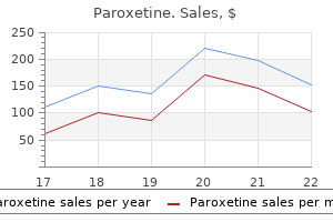
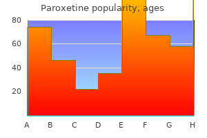
Paroxetine dosages: 20 mg, 10 mg
Paroxetine packs: 30 pills, 60 pills, 90 pills, 120 pills, 180 pills, 270 pills, 360 pills

Buy 10 mg paroxetine
Procedures are required to make certain that energy peaking symptoms 7 generic paroxetine 20 mg with mastercard, uniformity medicine urology cheap 20 mg paroxetine amex, linearity, decision, and sensitivity fall inside normal norms. When analyzing the emissions from some extent supply of radioactivity, the photomultiplier tubes directly overlying the point supply in both the X or the Y planes detect probably the most scintillation occasions (greatest number of "counts"). Position logic circuitry is used to place this specific photon emission in a given set of X, Y coordinates. These advances include multidetector imaging systems that enable for a smaller size imaging system, permitting imaging rooms to use much less ground house. Solid-state, semiconductor-based detectors, similar to cadmium zinc telluride, have the potential to enhance energy and spatial resolution, decrease scan occasions, scale back radiation exposure, and permit for simultaneous dual-isotope imaging in small footprint imaging systems. Image Generation When a scintillation occasion has been localized and falls inside the required power window for detection, the generation of an image can happen. First, the analog signal obtained from the power peak must be converted to a digital sign using an analog-to-digital converter. This digital signal can be reworked from a spatial area to a frequency domain by way of the Fourier transformation. When the occasion localized digital data is within the frequency domain, it might be used for the construction of an image. This methodology requires the raw projection knowledge to be handed via a so-called ramp filter to omit sure frequencies (filter "cutoff") and enhance different frequencies ("power") to optimize picture high quality. Filters operate to take away inherent reconstruction artifacts (particularly the "star" artifact inherent in again projection), optimize the signal-to-noise ratio in picture reconstruction, and supply picture enhancement. This method allows for speedy image reconstruction, but loss of image info occurs due to filtering. Iterative methods use mathematical equations to model the actual imaging physics and geometry of the acquisition and to reconstruct the picture after discriminating the image into pixels. An understanding of those principles helps the clinician choose the appropriate and most secure imaging research for the affected person and acknowledge potential sources of imaging error and artifacts. Radioactive decay can produce alpha particles, beta particles, positrons, and gamma photons. Photons work together with matter through the photoelectric impact, Compton scatter, and pair manufacturing. Attenuation happens when photons interact with matter, proportional to the attenuation coefficient for the interacting matter. I I Gamma cameras are composed of a collimator, a scintillation crystal, a light-weight pipe, photomultiplier tubes, a pulseheight analyzer, position circuitry, an analog-to-digital converter, and a show device. The relationship between the diploma of coronary stenosis and the maximal hyperemic response was first reported greater than 30 years in the past. Nonreversible myocardial perfusion defects usually relate to necrosis or infarction. Current imaging protocols enable the correct evaluation of relative regional perfusion and myocardial perform at relaxation and stress based on regional blood move heterogeneity. Redistribution is believed to symbolize areas of ischemic but viable myocardium, whereas fastened, nonredistributing defects are thought to represent nonviable, fibrotic scar. When 201Tl alone is used, quite lots of different acquisition protocols of stress imaging have been employed, together with redistribution and reinjection imaging. Overall sensitivity of a number of stress-redistribution-reinjection research averaged 85% with a decrease specificity (averaging 47%), suggesting that this protocol tends to overestimate the potential for contractile function restoration. After an intravenous injection, the initial myocyte uptake is principally determined by regional myocardial perfusion, whereas the integrity of the cell membrane is predominantly essential for delayed imaging of tracer retention (potassium ion complete distribution). A unfavorable mitochondrial gradient charge is essential for its accumulation and retention within the myocyte. This lack of significant redistribution signifies that separate rest and stress injections are commonplace with 99mTclabeled compounds. Different acquisition protocols can be used with these brokers, together with 2-day stress/rest, same-day rest/stress, same-day stress/rest, and dual-isotope protocols. Two-Day Protocol From a technical perspective, to optimize imaging high quality, the 2-day stress/rest is probably certainly one of the most most popular acquisition protocols. The primary advantage is the utilization of two high doses of Tc 99m labeled compounds, which permits high-quality photographs to be obtained because of the elevated excessive count fee. The stress research should be carried out first as a result of the rest study can be omitted if the stress study is normal. Obviously, the major drawback is the delay in reporting of the final evaluation. If the research is performed for the diagnosis of myocardial ischemia, the stress portion should be accomplished first as a end result of that may avoid the reduction of distinction that a previously resting injection would have on a stress-induced defect. If detection of viable myocardium or evaluation of the reversibility of a perfusion defect is the indication, efficiency of the resting examine first could additionally be preferable. As with all Tc 99m labeled compounds, imaging ought to begin between 60 and ninety minutes after injection to allow hepatobiliary clearance and to minimize subdiaphragmatic exercise if vasodilators had been administered. To improve the washout of gastrointestinal activity from liver and gallbladder, fluids or a fatty meal can be suggested. Dilsizian and colleagues10 described the utility of quantitative Tc 99m sestamibi imaging when the severity of decrease in Tc 99m sestamibi uptake within irreversible defects was thought of or when an extra redistribution picture was acquired after the remaining injection for detection of dysfunctional but viable myocardium. A significant inverse linear relationship has been described between Tc 99m sestamibi uptake and myocardial fibrosis in biopsy specimens. These tracers might prove to be of more value within the near future, contemplating the key position that oxidative metabolism plays in preservation of myocardial function. Dual-Isotope Protocols Dual-isotope imaging protocols utilizing Tc 99m labeled compounds and 201Tl are based on the power of the Anger digital camera to acquire information from the 2 totally different energy windows representing each radiotracer. Separate acquisition instances can reduce the necessity of downscatter correction that can diminish 201Tl distinction images, resulting in an overestimation of defect reversibility; this might be achieved by buying 201Tl knowledge units earlier than the administration of Tc 99m due to the very limited (2. One of the most important advantages is the potential of measuring contractile operate and the left ventricular ejection fraction. The principal distinction between stress methods pertains to the mechanisms used to disclose regional myocardial blood circulate abnormalities as an indication of coronary stenosis. It is important to select the most applicable check by the indication on a patient by patient foundation. When the aim is to consider train tolerance, the duration of the exercise, signs developed, and hemodynamic changes are the first factors to consider. Exercise testing is performed on the treadmill in accordance with the Bruce protocols and allows the assessment of different hemodynamic variables, corresponding to exercise capability, blood stress, and coronary heart fee responses. It is crucial that the intravenous injection of the radiotracer be performed at maximal stress and that train continue for no less than an extra 60 seconds to guarantee optimal myocardial concentration. The conventional goal of the check as a suitable stage of cardiac workload has been the achievement of no less than 85% of the utmost predicted coronary heart fee (220 - age). A maximal stress take a look at might satisfy diagnostic purposes if it goes beyond the hemodynamic threshold of triggering the ischemic symptoms.
Paroxetine 20 mg generic on-line
It can be related to a "cleft" anterior mitral valve leaflet medicine 10 day 2 times a day chart paroxetine 10 mg buy discount line, which leads to mitral regurgitation symptoms youre pregnant best 10 mg paroxetine, or defects within the atrioventricular or membranous interventricular septum. The patent foramen ovale usually outcomes from abnormal resorption of the septum primum through the formation of the foramen secundum. An abnormally giant foramen ovale can happen as a outcome of faulty improvement of the septum secundum. The defect lies instantly adjacent to the atrioventricular valves, either of which may be deformed and incompetent. These defects usually lie high within the atrial septum close to the entry of the superior vena cava, and are typically related to anomalous proper higher pulmonary venous return. An uncommon inferior sort is related to partial anomalous return of the right decrease pulmonary vein. Anomalous drainage can be into the right atrium, the superior vena cava, or the inferior vena cava. Coronary sinus opacification, which precedes proper atrial opacification, confirms the prognosis. Larger defects are associated with substantial shunting, which can lead to volume overload of the best atrium, proper ventricle, and pulmonary arteries. If left untreated, the left-to-right shunting might lead to pulmonary hypertension, proper ventricular failure, decreased right ventricular compliance, and probably right-to-left shunting (Eisenmenger syndrome). Cardiac auscultation reveals a traditional S1, mounted splitting of S2, and a systolic outflow murmur on account of the increased flow into the main pulmonary artery. The development of pulmonary hypertension leads to narrowing of the splitting of S2 and accentuation of the pulmonary closure element. The pulmonic systolic murmur decreases in intensity, and a diastolic pulmonic regurgitation murmur may appear. The commonest manifestations of this condition are improvement of fatigue, dyspnea on exertion, and train intolerance generally in the course of the third and fourth a long time. The improvement of palpitations related to supraventricular arrhythmia is the most common symptom in adults. Occasionally, sufferers might current with paradoxical embolization or recurrent respiratory an infection. Over time, pulmonary strain will increase because of increased pulmonary resistance, and Imaging Technique and Findings Radiography A plain chest radiograph. Until pulmonary resistance increases, resulting in pulmonary hypertension, the parenchymal vessels prolong farther towards the pleura than expected, and seem to taper normally. The left-to-right shunt quantity hundreds the right coronary heart, resulting in right atrial and ventricular dilation and clockwise (leftward) cardiac rotation. B, Lateral radiograph reveals filling of the retrosternal area by the dilated major pulmonary artery and right heart, and no evidence of left coronary heart enlargement. The central (extrahilar) pulmonary arteries stay enlarged, however the parenchymal pulmonary artery segments turn into vasoconstricted, producing the everyday appearance of acute change of pulmonary artery caliber seen in pulmonary hypertension. Careful evaluation of the peripheral pulmonary artery segments reveals extension toward the pleura past that present in a normal examination, however in smaller caliber vessels. As lengthy because the shunt is left-to-right, right coronary heart enlargement and a standard left heart are seen. In the rare circumstance of shunt reversal, progressive right coronary heart decompression and left coronary heart enlargement could additionally be seen. The left subclavian (1 arrow) and left bracheocephalic (2 arrow) veins and superior vena cava (3 arrow) have been opacified. Evaluation of the situation, measurement, and direction of shunt could be performed by use of colour move Doppler and echocardiographic contrast brokers. Important info corresponding to estimated pulmonary artery pressure and the stress of further cardiac chambers may additionally be obtained. Accurate evaluation of pulmonary-to-systemic flow (Qp/ Qs) ratio may be obtained and corresponds to the shunt fraction. The acute change in caliber of the pulmonary arteries from hilar to parenchymal branches (arrowheads) indicates pulmonary artery hypertension. During the percutaneous closure, intracardiac echocardiography, which is now applied to many other percutaneous methods, could also be used for the guidance of the restore procedure. Several hundred heartbeats are sampled, quantified, and displayed in an endless-loop cine format for qualitative visible analysis and quantitative interpretation and analysis. After a single labeling process, serial studies can be obtained for periods of four to 6 hours. The left anterior oblique view is used for qualitative evaluation of world left ventricular ejection fractions and stroke volumes using a radioactivity count-based strategy throughout the cardiac cycle. This method includes sampling through the initial transit of the bolus by way of the central circulation. Note the dilated proper decrease lobe pulmonary vein (pv), and left (1 arrow) and proper (2 arrow) pulmonary arteries. The medial (membranous) aspect of the interventricular septum (1 arrow) and the medial inferior (primum) facet of the interatrial septum (2 arrow) are deficient. Invasive catheterization permits estimation of the magnitude of the shunt (Qp/Qs ratio) and measurement of the pulmonary artery pressure. In this case, partial anomalous pulmonary venous return is normally current and it affects the right upper pulmonary vein. B, Slice knowledge nuclear photographs also present significant right ventricular dilation (arrows). In addition to the excessive surgical mortality and morbidity threat, closure of the defect in these patients worsens end result. Elective closure in sufferers with small defects is controversial because patients with small defects typically have a good prognosis, and the risk of cardiopulmonary bypass is probably not warranted. The benefit of catheter closure of small secundum defects particularly in these patients remains to be determined. Definitive medical evaluation of atrial septal defect by magnetic resonance imaging. Magnetic resonance and computed tomographic evaluation of congenital coronary heart disease. Cross-sectional echocardiographic evaluation of atrioventricular septal defect: primary morphology and preoperative risk elements. The anatomical kinds of atrial septal defect: their incidence and clinical analysis. Clinical and echocardiographic assessment of a right-to-left shunt throughout an atrial septal defect secondary to tricuspid regurgitation. Transesophageal threedimensional echo evaluation of sinus venosus atrial septal defect.
Diseases
- Renal agenesis meningomyelocele mullerian defect
- Leukomelanoderma mental retardation hypotrichosis
- Conjunctivitis ligneous
- Osteosarcoma limb anomalies erythroid macrocytosis
- Cardiomyopathy spherocytosis
- Eronen Somer Gustafsson syndrome
- Hypopigmentation oculocerebral syndrome Cross type
- Richieri Costa Guion Almeida syndrome
- Adenocarcinoma of esophagus
Buy 10 mg paroxetine fast delivery
The dissection to expose the internal medicine knowledge 10 mg paroxetine buy with visa, exterior jnc 8 medications paroxetine 10 mg order fast delivery, and common carotid arteries should keep away from harm to the superior laryngeal, hypoglossal, glossopharyngeal, marginal mandibular, and vagus nerves. Manipulation of the carotid body must be minimized to prevent bradycardia or hypotension. Systemic heparin is given, and vascular clamps are applied on the proximal uninvolved frequent carotid artery and the distal nondiseased internal carotid artery and exterior carotid artery. Many surgeons recommend placement of a brief lived shunt from the widespread carotid artery to the distal inner carotid artery to keep prograde circulate to the brain through the endarterectomy. Others use shunting selectively, primarily based on variables such as internal carotid artery backpressure measurements, operative electroencephalography, sensory evoked potentials, or statement for neurologic occasions throughout regional anesthesia. The artery is then incised longitudinally, and the plaque is separated from the frequent carotid artery and the interior carotid artery. Great care is taken to depart behind a smooth luminal floor with no debris or intimal flaps. The arteriotomy is normally closed with an artificial or vein patch to forestall restenosis and to cut back perioperative stroke danger. Asymptomatic Carotid Stenosis the Veterans Affairs Cooperative Study Group16 enrolled men with asymptomatic carotid stenosis with diameter discount of 50% or more. In sufferers with 60% or extra diameterreducing stenosis, the projected 5-year ipsilateral stroke price for surgically treated patients was 5. The procedure can be carried out by way of a 2- to 3-inch-long vertical or transverse cervical incision (A). Vessel loops are used to control the widespread, inner, and external carotid arteries after cautious dissection (B). A shunt can be used to keep cerebral perfusion, after which the plaque is dissected free by way of a vertical arteriotomy (C and D). The main consequence measured was the composite of stroke or demise within 30 days of the procedure. Whereas the outcomes supplied necessary information, this research had some limitations that precluded a definitive answer. The research had aimed to randomize 1900 patients however needed to be terminated because of insufficient enrollment. Of the patients enrolled, demographics and lesion traits had been similar within the two treatment teams. The safety and efficacy of any new therapy have to be clearly established in a controlled, appropriately powered, adequately monitored clinical trial, with appropriately credentialed operators, beneath Institutional Review Board and U. The frequent carotid artery is cannulated most commonly through a transfemoral method, and the lesion is visualized (A). The sheath is superior to inside a couple of centimeters proximal to the lesion, and an embolic protection gadget (in this case a filter device) is deployed distal to the lesion (B). Protected balloon angioplasty is performed to enable safe passage of the stent (C), after which the stent is deployed (D, on this case, a nitinol tapered stent). Such procedures should be performed solely by correctly credentialed physicians in a monitored trial setting. These attainable indications include carotid stenosis within the presence of prior cervical irradiation, anatomically inaccessible lesions (generally above the C2 vertebra), and severe medical comorbid conditions. Procedure Routinely, two preprocedural antiplatelet brokers (aspirin and clopidogrel) are used. Transbrachial or direct carotid exploration and cannulation may be undertaken in chosen patients with severe aortoiliac disease. A 6F lengthy sheath is positioned throughout the widespread carotid artery proximal to the lesion. Intermittent hand-injection angiography is performed throughout the complete procedure to affirm appropriate balloon and stent placements; bone landmarks are used for the same objective. Reporting: Information for the Referring Physician A clinically detected carotid bruit or neurologic symptoms attributable to the carotid artery territory (stroke, transient ischemic attack, amaurosis fugax) should prompt a noninvasive evaluation of the carotid arteries and referral for evaluation of possible carotid artery stenosis. Because sufferers must survive several years to derive a benefit from revascularization, the risks of providing the process to high-risk people must be rigorously thought-about. Randomized clinical trials: how will results affect clinical practice in the management of symptomatic and asymptomatic extracranial carotid occlusive disease Ischemic stroke subtype incidence amongst whites, blacks, and Hispanics: the Northern Manhattan Study. Quantitative correlation of plaque localization with move velocity profiles and wall shear stress. Carotid endarterectomy in asymptomatic patients-is distinction angiography necessary Color-flow duplex scanning of carotid arteries: new velocity standards based on receiver operator characteristic analysis for threshold stenoses used in the symptomatic and asymptomatic carotid trials. Significance of plaque ulceration in symptomatic patients with high-grade carotid stenosis. Progress report of prognosis following surgery or nonsurgical remedy for transient cerebral ischemic assaults and cervical carotid artery lesions. Management of atherosclerotic carotid artery illness: medical follow pointers of the Society for Vascular Surgery. Kirsch, and Ulrike Hamper Stroke stays the third main cause of death and is a major explanation for morbidity within the United States. Whereas the heart is the number one supply, 20% to 30% of strokes are believed to be secondary to embolus from plaque or thrombus on the carotid bifurcation. Risk components for illness at the carotid bifurcation include atherosclerosis, hypertension, diabetes mellitus, hyperlipidemia, hypercholesterolemia, obesity, and smoking. Patients with danger factors for carotid plaque, carotid bruits, and signs of stroke or transient ischemic assaults are sometimes referred for analysis of the carotid arteries, which could be performed with ultrasonography, computed tomographic angiography, magnetic resonance angiography, or standard angiography. Of these potential screening modalities, carotid ultrasound examination is essentially the most available, least invasive, and least expensive. Hence, this technique has vital interobserver and intraobserver variability as properly as poor reproducibility. However, if the carotid arteries are too deep to visualize with the linear array transducer in a affected person with a short, thick neck, a decrease frequency curved array transducer may be necessary for enough penetration. Discrepancies between the different velocity criteria as properly as with the gray-scale or colour Doppler estimation of proportion stenosis and the waveform pattern must be defined. The affected person is examined in the supine position with the neck prolonged and slightly turned to the contralateral side. Plaque echotexture ought to be characterised as hypoechoic, heterogeneous, or echogenic. The use of spatial compounding and harmonic imaging will also improve gray-scale resolution. The color acquire must be optimized by slowly rising the achieve till color speckles are famous within the surrounding gentle tissues. The achieve is then decreased until the colour pixels are seen only within the vessel lumen. If the achieve is set too excessive, the colour pixels will overwrite or "bleed" over plaque, obscuring visualization of the true extent of plaque burden, particularly during systole, and stenoses may be overlooked. If the acquire is too low, sensitivity to blood circulate will be decreased, and false-positive diagnoses of occlusion or stenosis shall be made. The color velocity scale must be adjusted such that colour fills the lumen reaching to the vessel wall. Therefore, the color velocity scale may should be changed slightly as one interrogates the totally different vessels within the neck.

10 mg paroxetine purchase with visa
In these circumstances medications 73 10 mg paroxetine order otc, quicker acquisition in the neck medicine vs medication paroxetine 10 mg discount with visa, where the vessels are bigger, and slower acquisition in the head, the place the circle of Willis arteries are smaller, is possible. Peak carotid artery opacification depends on numerous elements, such as patient weight, cardiac output, and recirculation of blood pool. A large-bore intravenous catheter of at least 20 gauge in the right antecubital fossa is greatest. The quantity and rate of injection range broadly, depending on the variety of detectors out there (Table 91-2). Recently, highconcentration contrast material of 370 to four hundred mg I/mL has been proven to present earlier and better peak enhancement with decrease injection rates of roughly four mL/ sec. The saline flush also minimizes the artifact of extremely concentrated contrast within the subclavian vein, which might cause streak artifacts close to the aortic arch and takeoff of the great vessels. The window stage settings are important to the correct interpretation of carotid stenosis. C, Oblique axial images at the degree of most stenosis are mechanically generated primarily based on the center line demonstrated in B. Attention must be paid to not miss a number of lesions at the target artery because tandem lesions are a standard function in atherosclerosis. The automated monitoring could not work constantly near contrast-filled veins or bone. The user is usually provided with a tool to rapidly appropriate this goal pathway back into the carotid lumen. D, At the level of most stenosis, the indirect axial image reveals both delicate plaque and calcifications in the carotid plaque. E, Automated share diameter and area stenosis are calculated primarily based on minimal decoding doctor interaction. Larry Tanenbaum, Department of Radiology, Mount Sinai School of Medicine, New York. The standing of the lumen as easy, irregular, or ulcerated also wants to be reported. Any tandem stenoses elsewhere within the carotid artery, in addition to the status of the circle of Willis, are necessary to describe. Finally, any vital adjoining delicate tissue or bony abnormalities must be reported. Magnetic resonance imaging of vulnerable atherosclerotic plaques: current imaging strategies and molecular imaging probes. Systematic evaluate of computed tomographic angiography for assessment of carotid artery disease. What are current preprocedure imaging necessities for carotid artery stenting and carotid endarterectomy Analysis of pooled data from the randomised controlled trials of endarterectomy for symptomatic carotid stenosis. Carotid endarterterectomy in asymptomatic patients-is contrast angiography essential Measuring carotid stenosis on contrast-enhanced magnetic resonance angiography: diagnostic efficiency and reproducibility of three completely different strategies. Can magnetic resonance angiography and duplex ultrasonography exchange contrast arteriography Effects of doubling and tripling the spatial resolution in normal three-dimensional contrastenhanced magnetic resonance angiography of carotid artery disease. Noninvasive detection of steno-occlusive illness of the supra-aortic arteries with threedimensional contrast-enhanced magnetic resonance angiography: a potential, intra-individual comparative analysis with digital subtraction angiography. A distinction dose discount study for three-dimensional excessive spatial resolution contrast-enhanced magnetic resonance angiography of supraaortic arteries at three. The underlying reason for a thoracic aortic aneurysm can typically be predicted by its location and morphologic features and by the age of the affected person. An instance is that of the ascending aortic aneurysm, which by its location is related to the additional issues of whether to replace the aortic valve, to reimplant the coronary arteries, and to restore the arch vessels. In addition, assessment of global cardiovascular operate is paramount in directing applicable therapy strategy. Moreover, some problems, such as spinal cord ischemia, are germane to surgical restore of thoracic aortic aneurysms, and imaging can present a highway map that will permit potential modification of surgical method to scale back the probabilities of such issues. Of note, aneurysms because of systemic arterial disease have much less male preponderance and a more superior age at presentation than abdominal aneurysms do, resulting in greater comorbid disease. The function of such a classification affects the seek for associated vascular lesions, the surgical method, and the potential problems. These scenarios require an "aneurysm mentality" as a result of saccular aortic dilations are at specific danger for rupture and are thus additionally categorised as aneurysms. False aneurysms (also generally recognized as pseu- Atherosclerosis Atherosclerosis impacts the aortic wall in many ways, similar to via erosion of the interior elastic lamina and subsequent exposure of the medial layer of the aortic wall to the pulse stress or through ischemia of the media from decreased blood supply because of illness within the vasa vasorum. Another mechanism of aneurysm formation is thru development of atherosclerotic plaque ulceration to a penetrating atherosclerotic ulcer with breakage of the intimal layer. Atherosclerotic aneurysms are related to hypertension, coronary artery illness, and stomach aortic aneurysms. Cystic Medial Necrosis As the name implies, cystic medial necrosis affects the medial layer of the arterial wall; degeneration of the smooth muscle creates "cystic areas," leading to a fusiform aneurysm. The pathophysiologic process entails the aortic root, resulting in dilation of the annulus of the aortic valve. Associated aortic regurgitation could require concomitant alternative of the aortic valve. Marfan syndrome is the most typical of the connective tissue processes, with an incidence of 1:10,000. The elastin-depleted aorta is stiffer and more prone to dilation because it incurs higher pulse pressure than the usually distensible aorta does. Plaque ulceration eventually breaks by way of the intimal layer, causing a penetrating atherosclerotic ulcer, which can lead to an aneurysm with a focal bulge or saccular configuration (B). The syndrome is characterised by fragile arterial tissue that not only is vulnerable to aneurysm, dissection, and rupture but can even make surgical restore difficult. Unlike Marfan and LoeysDietz syndromes, which have a predilection for the aortic root, Ehlers-Danlos syndrome extra often impacts the visceral arteries. Trauma Blunt thoracic trauma with a sudden deceleration mechanism injures the aorta at its points of relative fixation (due to shear), which includes the aortic isthmus (in the region of the ligamentum arteriosum), the hiatus, and the aortic root, in descending frequency. Injury might cause transection or intimal disruption and, if unrecognized, may be manifested as a saccular aneurysm that will involve disruption of the intima and media (pseudoaneurysm or false aneurysm). The arteriopathy tends to be more systemic than in Marfan syndrome, and the postoperative surveillance should issue each the repaired artery and the remote arteries including the intracranial circulation. The sinotubular junction could be effaced, leading to a "spring onion" appearance to the aortic root and ascending aorta. Oblique coronal multiplanar reformatted picture before (A) and 2 years after (B) uncomplicated restore of dilated aortic root. Note the everyday appearance of a repaired aorta with kinking at the anastomosis (arrow). Note the distinction in appearance of an ascending aortic aneurysm that spares the root (B). Inflammation and Infection Infection may primarily involve the artery and secondarily result in aneurysm, or an aneurysm might become infected. The commonest website of an infection is the ascending aorta close to the sinus of Valsalva.
Purchase 20 mg paroxetine otc
Ultrasonography Abnormalities on echocardiography symptoms 7 dpo bfp buy discount paroxetine 20 mg, including increased or decreased wall thickness symptoms 4 weeks pregnant 20 mg paroxetine buy otc, ventricular dilation, useful impairment, mitral regurgitation, impaired diastolic relaxation, and pericardial effusion, have been reported in 14% to 40% of sufferers with sarcoidosis. Echocardiography is usually the primary examination performed when the prognosis is suspected, and may show regional wall movement abnormalities and thickening of the interventricular septum, with brilliant echoes suggesting infiltration. Alternatively, the ventricles may seem thinned with world dysfunction and aneurysm formation. Diastolic dysfunction may be seen during the initial interstitial inflammatory stage when systolic perform remains to be regular. Patients can also present with congestive coronary heart failure, cor pulmonale, supraventricular and ventricular arrhythmias, conduction disturbances, ventricular aneurysms, pericardial effusions, mitral valve abnormalities, and sudden cardiac demise. Acute myocardial inflammation ensuing from sarcoid infiltration could also be seen as areas of focal thickening with elevated sign depth on T2-weighted black blood photographs. Late adjustments embody wall thinning and delayed hyperenhancement thought to reflect persistent scarring. These changes could additionally be tough to distinguish from chronic infarction, although they tend to be in a noncoronary distribution and should spare the subendocardium. Echocardiography is beneficial to assess operate and focal wall motion abnormalities in typical locations for sarcoidosis, but is comparatively nonspecific. Imaging Techniques and Findings Radiography Plain radiographs present no information regarding cardiac sarcoidosis. The extent of delayed hyperenhancement correlated with illness duration, ventricular function, mitral regurgitation, and presence of ventricular tachycardia. Nuclear Medicine Thallium 201 scintigraphy myocardial perfusion research usually show segmental areas of decreased uptake in the ventricular myocardium that disappear or lower in size during stress or after intravenous dipyridamole administration. Gallium 67 scintigraphy has additionally been used to present cardiac and extracardiac illness, for follow-up of lively disease, and as a information for potential websites for biopsy. More just lately, Tc 99m sestamibi has been used as a perfusion agent, with a reverse distribution much like that described in thallium. According to the Japanese tips, eight of 21 sufferers were diagnosed with cardiac sarcoidosis. Angiography Coronary angiography is usually regular in sufferers with cardiac sarcoidosis. Cardiac amyloidosis generally has a poor prognosis, with a mean survival of 13 months for patients presenting with main amyloidosis. Oral chemotherapy, including melphalan and prednisone, has shown restricted advantages to sufferers with cardiac involvement. Stem cell transplantation has shown promising outcomes for treatment of major amyloidosis; however, the mortality associated with transplantation is five instances larger in amyloidosis compared with other hematologic malignancies. Medical remedy for endomyocardial eosinophilic disease contains anticoagulation, diuretics, and digitalis. Corticosteroids, hydroxyurea, cytotoxic drugs, and imatinib all have been employed, with variable outcomes. Histologically, these patients have interstitial fibrosis with elevated amounts of collagen, glycoprotein, triglycerides, and cholesterol within the myocardial interstitium. Differential analysis between constriction and restriction in these patients is especially troublesome as a result of radiation may induce pericardial fibrosis and constriction. Endocardiectomy has been performed in sufferers with eosinophilic endomyocardial disease, with comparatively excessive operative mortality. On imaging, visualization of a thickened pericardium, often with focal distortion of the ventricular contour and atrial enlargement, permits assured diagnosis of constrictive pericarditis. Etiologies embrace amyloidosis, eosinophilic endomyocardial disease, siderotic cardiomyopathy, sarcoidosis, radiation, storage diseases, diabetes, and idiopathic. Amyloid heart disease: new frontiers and insights in pathophysiology, analysis, and administration. Frequency and distribution of senile cardiovascular amyloid: a clinicopathologic correlation. Prognostic significance of Doppler measures of diastolic function in cardiac amyloidosis: a Doppler echocardiography research. Echocardiographic findings in systemic amyloidosis: spectrum of cardiac involvement and relation to survival. Prognostic significance of ultrasound myocardial tissue characterization in patients with cardiac amyloidosis. Detection of left ventricular systolic dysfunction in cardiac amyloidosis with pressure price echocardiography. Assessment of restrictive cardiomyopathy of amyloid or idiopathic etiology by magnetic resonance imaging. Cardiac adjustments in systemic amyloidosis: visualization by magnetic resonance imaging. Pitfalls in analysis and medical, echocardiographic, and hemodynamic findings in endomyocardial fibrosis: a 25-year expertise. Endomyocardial fibrosis and intracardiac thrombus occurring in idiopathic hypereosinophilic syndrome. Idiopathic hypereosinophilic syndrome: magnetic resonance imaging findings in endomyocardial fibrosis. Endomyocardial fibrosis in Churg-Strauss syndrome assessed by cardiac magnetic resonance imaging. The position of Doppler left ventricular filling indexes and Doppler tissue echocardiography in the assessment of cardiac involvement in hereditary hemochromatosis. Cardiac iron determines cardiac T2*, T2, and T1 in the gerbil model of iron cardiomyopathy. Cardiovascular T2-star magnetic resonance for the early diagnosis of myocardial iron overload. Evaluation of the accuracy of gadolinium-enhanced cardiovascular magnetic resonance within the prognosis of cardiac sarcoidosis. Other distributions of hypertrophy include apical, midventricular, and concentric. In patients with vital septal hypertrophy, heart failure can also be attributed to left ventricular outflow tract obstruction. Septal thickening of 13 to 15 mm or extra and a septal-to-posterior wall ratio of 1. Short axis Short axis Long axis (four-chamber view) Orthogonal to coronary sinus Velocity-encoded cine of multiphase imaging relatively freed from movement, as nicely as three-dimensional reformations. Specifically, echocardiography is commonly of limited worth within the assessment of the anterior and lateral left ventricular wall and of the ventricular apex. B, Horizontal long-axis images are divided into three segments-septum, apex, and free wall. First-pass images both earlier than and after administration of coronary vasodilator agent are analyzed to decide perfusion reserve. First-pass gadolinium imaging has correlated decreased perfusion reserve to each the location and extent of ventricular hypertrophy. This axial black blood image shows hypertrophy of the apical region of the left ventricle, with a maximal thickness of three.
Syndromes
- Recognize of risks and limits of the science of medical care and that health care providers are human and can make mistakes.
- Unpasteurized dairy foods such as milk or cheese
- Haemophilus influenzae vaccine (Hib)
- National Institute of Neurological Disorders and Stroke - www.ninds.nih.gov/disorders/myasthenia_gravis/detail_myasthenia_gravis.htm
- Light-headedness, especially with or after activity or exercise
- During that time the mother can express the milk or pump her breasts (to stay comfortable and maintain the flow of milk) while feeding the baby formula.

Paroxetine 10 mg order overnight delivery
They usually have single proper ventricles that in the end fail in the face of systemic afterload treatment bee sting 10 mg paroxetine proven. They could have valvular disease that will increase the stress or quantity load of the only ventricle medications you can give your cat 20 mg paroxetine order with visa. The second goal is the characterization of physiology, including valvular disease, myocardial operate, and move evaluation. Both the area of coarctation of the aorta and the collaterals are identified (arrows). In the current period, they typically bear several surgical procedures in the course of the first several years of life to present a secure cardiopulmonary physiology. The first of those procedures is usually performed in the neonatal interval and is directed at recruiting the one ventricle because the systemic pumping chamber and providing managed pulmonary blood circulate. The subsequent procedures involve sequential conversion to a physiology of separated systemic and pulmonary circulations with passive pulmonary blood flow (elimination of intracardiac "mixing"). The final circulation is named the Fontan circulation after the French surgeon who developed and first performed it in humans. Images ought to extend from the diaphragm to the arch vessels to start to assess cardiac and vascular anatomy. Black blood imaging could additionally be useful if anatomy is in query or if there are metallic units in place inflicting artifact on cine imaging. First, two-dimensional planes are prescribed throughout the aorta as well as the vessels supplying blood move to the pulmonary vascular bed. Twodimensional circulate assessment throughout the atrioventricular valves as properly as the superior and inferior venae cavae is usually useful for assessment of atrioventricular valve regurgitation and for corroboration of different circulate and volumetric data. A seven-dimensional circulate method may prove helpful on this anatomy and physiology as it may possibly obtain move in all vessels in a single acquisition. First, as a end result of the passive nature of pulmonary blood flow, they develop high right-sided filling pressures, dilated Fontan pathways, and atrial arrhythmias. As a results of high filling pressures, they often form systemic-to-pulmonary venous collaterals, which lead to cyanosis. Serial examinations must be performed, the frequency of which must be dictated by scientific status. Clinical functions of cardiovascular magnetic resonance in congenital coronary heart disease. Thoracic cardiovascular anomalies in kids: evaluation with a fast gradient-recalledecho sequence with cardiac-triggered segmented acquisition. Comparison of gated single-photon emission computed tomography with magnetic resonance imaging for analysis of left ventricular perform in ischemic cardiomyopathy. The vary of regular values of cardiovascular structures in infants, kids, and adolescents measured by magnetic resonance imaging. Delayed-enhancement cardiovascular magnetic resonance identifies fibrous tissue in children after surgical procedure for congenital heart disease. Magnetic resonance imaging analysis of myocardial perfusion and viability in congenital and bought pediatric heart illness. Free-breathing, three-dimensional coronary artery magnetic resonance angiography: comparability of sequences. Diagnostic performance of coronary magnetic resonance angiography as in contrast in opposition to standard x-ray angiography: a meta-analysis. Effect of continual sustainedrelease dipyridamole on myocardial blood flow and left ventricular function in patients with ischemic cardiomyopathy. Prognostic value of cardiac magnetic resonance stress tests: adenosine stress perfusion and dobutamine stress wall movement imaging. Prognosis of negative adenosine stress magnetic resonance in patients presenting to an 30. White Imaging of coronary atherosclerosis has trusted coronary angiography as the gold commonplace since Sones and Shirey developed the technique at the Cleveland Clinic in the Nineteen Sixties. Although the in vivo demonstration of susceptible plaques, permitting for prevention of plaque progression, is but elusive, it may be only a matter of time earlier than coronary angiography is changed by noninvasive techniques as the primary software for coronary atherosclerotic imaging. The prevalence is affected by age, gender, genetic predisposition, and bought threat components. Approximately 10% of males 50 to 70 years old who die of noncardiac causes have obstructive coronary lesions at autopsy. Typically, the interaction between endothelial injury, inflammation, lipid influx, angiogenesis, thrombosis, and cell dying ends in the pathologic hallmark of atherosclerosis-the atheroma, or core of cellular particles left by remnants of easy muscle cells, macrophages, and pink blood cells. As imaging of the vessels becomes extra refined, the identification of atherosclerotic plaque composition will become as essential as diploma of stenosis, which is at the limit of the aptitude of current modalities similar to angiography. There is a great morphologic heterogeneity of atherosclerotic plaque, which reflects the stage of progression and the number of pathways concerned after intimal injury. Inflammatory, thrombotic, proliferative, and apoptotic pathways typically lead to lipid accumulation, which is currently the major goal of newer imaging methods. B, Higher magnification of the thin cap, with quite a few foam cells and parts of cholesterol crystals at the top of the image. In most males, and men and women older than 50 years, atheromatous plaques dominate in most coronary lesions. In younger sufferers, especially girls, obstructive coronary disease might happen within the absence of serious cell dying and necrotic core formation. The identification of the 2 forms of plaque is crucial if imaging strategies evolve that particularly goal the identification of lipid-rich necrotic core. Much current imaging technology is focused on the identification of necrotic materials as a result of that is the precursor lesion of the most common substrate for coronary atherothrombosis. Thin Cap Atheroma and the Vulnerable Plaque the expansion of the atheroma, or necrotic core of the lipid-rich atherosclerotic plaque, results in thinning of the overlying fibrous cap. An arbitrary fibrous cap thickness of 65 �m has been designated because the criterion for the "weak" plaque or skinny cap fibroatheroma. The goal of imaging to detect skinny cap fibroatheroma and distinguish it from fibroatheroma with thicker caps remains elusive, however might ultimately allow the prophylactic therapy of lesions susceptible to rupture with the purpose of preventing atherothrombosis. A more modern modification of this staging schema3 outlines growth of atheroma. The normal human intima contains a small cushion of clean muscle cells and matrix, which is present from delivery and accentuated at department points; this lesion has been termed adaptive or diffuse intimal thickening. A, Significant stenosis with areas wealthy in extracellular lipid (clear areas), which consists largely of esterified ldl cholesterol and phospholipid, but no necrotic core formation. C, Thrombus overlying a easy muscle cell�rich plaque, with cleared areas indicative of lipid swimming pools (pathologic intimal thickening), however little or no necrotic core formation. D, Higher magnification of the plaque erosion, showing the platelet-rich thrombus in the lumen. Nevertheless, these patients develop significant luminal narrowing, and luminal thrombus could occur by mechanisms other than rupture of the skinny fibrous cap. Because plaque erosion thrombi may be small and nonocclusive, they lead to layering of smooth muscle cell�rich plaque that expands and not using a requisite necrotic core, typically with little outward expansion (positive remodeling). The consequence of subocclusive plaque rupture is commonly plaque enlargement and outward growth (remodeling) of the arterial wall.
Order paroxetine 10 mg otc
Is early primary restore for correction of tetralogy of Fallot comparable to medications hypertension 10 mg paroxetine generic overnight delivery surgical procedure after 6 months of age The impact of pulmonary valve substitute after tetralogy of Fallot restore: a matched comparison medicine quizlet paroxetine 10 mg order without prescription. Remodeling of the proper ventricle after early pulmonary valve replacement in children with repaired tetralogy of Fallot: assessment by cardiovascular magnetic resonance. Preoperative thresholds for pulmonary valve substitute in sufferers with corrected tetralogy of Fallot using cardiovascular magnetic resonance. Indications and timing of pulmonary valve alternative after tetralogy of Fallot repair. Chronic pulmonary valve insufficiency after repaired tetralogy of Fallot: diagnostics, reoperations and reconstruction prospects. Right ventricular dysfunction and pulmonary valve substitute after correction of tetralogy of Fallot. Aortic root dilatation in tetralogy of Fallot long-term after repair-histology of the aorta in tetralogy of Fallot: proof of intrinsic aortopathy. Quantitative morphometric analysis of progressive infundibular obstruction in tetralogy of Fallot. Demonstration of coronary arteries and major cardiac vascular constructions in congenital coronary heart illness by cardiac multidetector angiography. Accurate quantification of pulmonary artery diameter in sufferers with cyanotic congenital heart disease utilizing multidetector-row computed tomography. Right ventricular diastolic operate in children with pulmonary regurgitation after repair of tetralogy of Fallot: volumetric analysis by magnetic resonance velocity mapping. For anatomic obstructive lesions, the ductus arteriosus is usually patent, providing retrograde flow into the pulmonary circulation. Real-time two-dimensional, multiprojectional, transthoracic echocardiography with standard gray-scale and colour Doppler strategies readily evaluates the cardiac chambers, interatrial and interventricular septa, valves, systemic and pulmonary venous connections, central pulmonary arteries, and thoracic aorta (including arch sidedness). In select purposes, such as with valvular disease, three-dimensional echocardiography can supply larger structural detail. Concurrent with the structural evaluation, circulate, function, and hemodynamics are all assessed. In the diagnostic algorithm at many centers, catheterization with angiography follows echocardiography. Clinical workup and initial management will rely upon prenatal diagnosis, obstetric course, comorbid symptoms, physical examination, and preliminary diagnostic imaging. Single- and two-projection chest radiographs afford elementary cyanotic cardiac illness evaluation, together with heart size and contour, degree of pulmonary vascularity, and aortic arch sidedness. In parallel, the airway, lung parenchyma, pleura, and thoracic skeleton are analyzed for various or comorbid noncardiovascular causes. Flow obstruction could happen at the tricuspid valve, infundibulum, pulmonary valve, or a mix thereof, whereas the shunting might occur at the atrial or ventricular septal ranges. Radiation dose can be lowered and contrast medium obviated if the process is restricted to a right heart catheterization with out angiography. An anteroposterior chest radiograph reveals an enlarged right atrium and ventricle associated with delicate decreased pulmonary vascularity, consistent with a right-sided obstructive lesion. Echocardiography was subsequently carried out, demonstrating important pulmonary stenosis. Both can generate cardiac chamber quantity and qualitative and quantitative functional information. Image postprocessing can readily be facilitated for information units from each modalities. Assessment of body techniques outdoors of the thorax will add appreciable examination time. The aim is to survey and define the thoracic and upper abdominal cardiovascular and noncardiovascular morphology. Cine sequences should then be obtained for qualitative and quantitative cardiac chamber and valve useful evaluation. Next, velocity maps are obtained for hemodynamic analysis, focusing on at a minimal the ventriculoarterial valves and supravalvular segments. Regions of interest may also be placed on the branch pulmonary arteries, atrioventricular valves, central systemic veins, and pulmonary veins. Advantages embody short examination instances, upkeep of excessive diagnostic image quality in the presence of devices and metallic materials, and nonthoracic cardiovascular and multisystem organ analysis with the identical bolus of contrast medium. A, An anteroposterior chest radiograph reveals average cardiomegaly with a dominantly enlarged right atrium (arrowheads), an absent major pulmonary artery section (asterisk), and decreased peripheral pulmonary vascularity. Voltage and amperage must be lowered, balancing the expected diagnostic and image qualities. For most pediatric purposes, pertinent morphology may be evaluated in a single series without electrocardiographic gating. Usually, the septal and posterior leaflets are involved, with insertion on the margin of the inlet and trabecular right ventricular zones, both immediately or by way of anomalous chordae tendinae and papillary muscles. The diploma of septal and posterior leaflet displacement and irregular morphology has broad variability. In distinction to the septal and posterior leaflets, the anterior leaflet usually has dysplastic morphology, however maintains normal attachments. It is enlarged, with a sail look and fibrous strands, which may be muscularized. Rarely, the anterior leaflet could have abnormal displacement alongside the best ventricular anterior free wall, leading to tricuspid stenosis. In 10% of cases, the anterior leaflet could have downward displacement, obstructing the ventricular inlet, leading to an obstructing, imperforate tricuspid valve. Both portions are sometimes dilated, with a loss of myocytes, resulting in decreased contractility. In addition to these components, tricuspid insufficiency and potential anterior leaflet stenosis or obstruction decrease ahead flow, contributing to the diminished proper ventricle filling volumes. The conduction pathway abnormalities result in dysrhythmias; the most common are atrial tachycardias and supraventricular and ventricular arrhythmias. Clinical Manifestations Ebstein anomaly exhibits nearly equal distribution between women and men. Infants born with Ebstein anomaly are mostly full time period, with a weight between 2 and 4 kg. The neonate and toddler may present with feeding intolerance, whereas children and older sufferers might use squatting to alleviate symptoms. Physical examination could also be outstanding for a systolic and fewer commonly a diastolic murmur, systolic thrill, digital clubbing, and precordial deformity. In a four-chamber projection, apical leaflet displacement into the right ventricle is identified by recognizing that it extends past the atrioventricular degree of the mitral valve insertion. Atrialized and native portions of the best ventricle are outlined based mostly on the insertion degree.
10 mg paroxetine amex
A left-sided ductus arteriosus is usually seen with this entity symptoms dengue fever generic paroxetine 20 mg with mastercard, passing between the descending aorta and left pulmonary artery medicine advertisements paroxetine 10 mg generic with amex. Right aortic arch with mirror-image branching is almost at all times related to congenital heart anomalies, mostly tetralogy of Fallot, reported in 25% of cases. The left subclavian arises from a retroesophageal diverticulum, which is the remnant of the regressed dorsal segment. Because this situation forms a complete vascular ring, these patients usually present with nonpositional stridor because of airway compression. Instead of originating from the arch, an isolated left subclavian artery arises from the left pulmonary artery through the ductus arteriosus. The embryologic origin of this anomaly is hypothesized to involve regression of the fourth arch as well as a portion of the sixth arch, with migration of the seventh intersegmental artery to the level of the sixth arch, forming the communication between the ductus and the pulmonary artery (which arises from the left sixth arch) and the left subclavian artery (arising from the seventh intersegmental artery). Although isolated subclavian artery has been reported on the proper, it occurs far more commonly on the left. Right aortic arch with isolated left subclavian artery is nearly always associated with congenital coronary heart disease, most commonly with transposition and tetralogy of Fallot. Left-sided aortic arch with aberrant right subclavian artery is the most common aortic arch anomaly, with an occurrence of 1: 200. B, Reconstructed three-dimensional picture reveals a sagittal indirect view of the right-sided aortic arch with aberrant left subclavian arising from a retroesophageal diverticulum. A, Contrast-enhanced axial image via the chest shows a vascular structure (arrow) posterior and to the right of the trachea (T) and esophagus (E). Note the three vessels in the suspected location of the great vessels on this image. This affected person has anomalous origin of the left vertebral artery (asterisk) from the arch, accounting for this look. In this explicit patient, it passes posterior to the esophagus, which is the commonest course. C, Coronal indirect reformatted image by way of the arch exhibits the aberrant right subclavian arising from Kommerell diverticulum and ascending superiorly along the best mediastinum. This aberrant artery mostly passes posterior to the esophagus, but it could cross between the trachea and esophagus in 18% and anterior to the trachea in 4%. The aortic arch is usually situated between the second costosternal junction on the right and the T4 vertebral body. A cervical arch typically passes posterior to the esophagus and can be associated with each left and proper arches. The aortic arch branches may additionally be anomalous in origin with this entity as properly. On plain movie radiography, a cervical arch is manifested as a superior mediastinal mass with tracheal deviation. B, Maximum depth projection fast spoiled gradientrecalled picture of the aortic arch shows the three aortic branches arising from the arch, which extends cranially in relation to the good vessel origins. Clinically, sufferers can current with a pulsatile neck mass but usually are asymptomatic. The right frequent carotid arises from the best brachiocephalic artery, most commonly at the level of the sternoclavicular joint. In approximately 12% of the inhabitants, the origin is above the sternoclavicular joint. The frequent carotid arteries are invested in a carotid sheath, which incorporates the internal jugular vein and the vagus nerve as nicely, with the artery medial to the vein and anteromedial to the nerve. The frequent carotids course obliquely cephalad, passing posterior to the sternoclavicular joints and deep to the sternocleidomastoid muscle, to the extent of the thyroid cartilage, approximately C4, the place they bifurcate into the interior and exterior carotid arteries. The bifurcation is the placement of the carotid body (also called the carotid glomus or glomus caroticum), which is a cluster of chemoreceptors that can give rise to glomus tumors. In approximately 80% to 85% of sufferers, the inner carotid artery is posterior or posterolateral to the exterior carotid. It courses from the bifurcation on the degree of the thyroid cartilage to the posterior aspect of the mandibular neck, the place it branches into the superficial temporal and inner maxillary arteries. The exterior carotid has several branches, which usually come up in the following order: superior thyroid, ascending pharyngeal, lingual, facial, occipital, and posterior auricular arteries, followed by the terminal branches, the maxillary artery and superficial temporal artery. The inner carotid remains inside the carotid sheath and retains the identical relationship to the jugular vein and vagus nerve as the common carotid artery. The older classification delineated 4 segments: the cervical, petrous, cavernous, and cerebral segments. The newer classification system described by Bouthillier17 divides the internal carotid into the following seven segments: cervical (C1), petrous (C2), lacerum (C3), cavernous (C4), clinoid (C5), ophthalmic (C6), and communicating (C7). Three-dimensional reconstructed picture of the external carotid exhibits its branches in the following order: superior thyroid, lingual, facial, ascending pharyngeal, occipital, posterior auricular (which arises from the occipital in this patient), superficial temporal, and maxillary arteries. The carotid bifurcation and the proximal portion of the cervical phase lie within an external anatomic landmark generally known as the carotid triangle. The triangle is defined anatomically by the sternocleidomastoid muscle posteriorly, the omohyoid muscle anteriorly, and the stylohyoid and digastric muscles superiorly. The petrous section of the interior carotid artery travels in the carotid canal of the petrous bone, taking a short vertical course earlier than coursing medially and horizontally by way of the canal. The lacerum segment is a brief section of the interior carotid artery that begins at simply above the foramen lacerum (typically crammed with fibrocartilage in living patients) and continues to the petrolingual ligament. On exiting the carotid canal, the cavernous phase begins on the petrolingual ligament, ascends to the posterior clinoid process, then passes alongside the body of the sphenoid bone anteriorly, and at last programs vertically on the medial facet of the anterior clinoid process, where it perforates the dura and becomes the clinoid phase at the proximal dural ring. The clinoid section includes the portion of inner carotid artery between the proximal dural ring and the distal dural ring, where it then becomes the ophthalmic phase, which is intradural. Lateral anatomic diagram depicting the seven inside carotid artery segments: C1, cervical phase (the bulb is indicated by stippling, the ascending phase by horizontal lines); C2, petrous phase; C3, lacerum segment; C4, cavernous section; C5, clinoidal phase; C6, ophthalmic segment; C7, speaking phase. The ultimate phase of the interior carotid artery, referred to as the communicating section, passes between the optic and oculomotor nerves, giving rise to the posterior communicating artery and the anterior choroidal artery earlier than bifurcating into the terminal branches of the inner carotid artery, the anterior cerebral artery and the middle cerebral artery. Variant Anatomy the level of the carotid bifurcation may be variable, most commonly above C4 when it happens. Huber17a reported the bifurcation at C4-5 in 48% of 658 bifurcations and at C3-4 in 34%. The most stable anatomic landmark for the bifurcation is the superior thyroid cartilage. Illustration of the carotid triangle delineates the anterior border by the omohyoid muscle, the posterior border by the sternocleidomastoid muscle, and the superior border by the digastric muscle. The lingual and superior thyroid artery branches from the external carotid are also depicted along with the interior and external carotid arteries. Less frequent variations include a single trunk giving rise to both carotids and a standard origin with the left subclavian, resulting in symmetric bilateral brachiocephalic trunks, occurring in 1. These anastomoses mostly happen with the interior carotid but have been reported with the widespread carotid and exterior carotid arteries as well. A primitive trigeminal artery is the most typical of those, with an incidence between zero. With Saltzman sort I, the midbasilar and posterior speaking arteries are usually hypoplastic.
Paroxetine 10 mg overnight delivery
The whole attenuation can then be measured directly by inserting a source of exercise on the edge of the field of view and buying counts with the affected person within the scanner and evaluating to counts acquired with nothing within the scanner treatment models paroxetine 20 mg discount on line. This technique required several minutes of information assortment to obtain good statistics and carefully represents the typical place of the dynamically altering the thorax contents noticed during the emission examine medicine to stop vomiting 20 mg paroxetine with amex. The number of detected events is tremendously elevated if the scanner is operated in three-dimensional mode. Unfortunately, the variety of confounding events (randoms and scatter) is increased as well as the variety of good events (true). At some point, the detriment of dealing with the dangerous events outweighs the benefit of amassing more good occasions. At some level, it was realized that if the detected occasions are restricted to those occurring only within a plane, the variety of detected occasions will be many instances less than it could possibly be. On the opposite hand, it takes rather more computer memory and processing energy to reconstruct photographs when nonplanar occasions are additionally included. The computing threshold was passed around 1999, and in fashionable scanners, the acceptance of events is opened up in order that any pair of detectors can record an occasion. On the other hand, many extra random, scatter, and prompt gamma events are also recorded. However, if the scanner is operating in three-dimensional mode, scatter into neighboring planes will be recorded. Because of this, the fraction of recorded occasions which are scattered in a threedimensional scanner can method 50%. In many imaging conditions, when the time window could be lowered to roughly 6 ns or much less, the benefit of collecting more occasions outweighs the disadvantages. Segmentation takes benefit of there being just a few main tissue types within the field of view, similar to bone, tissue, and lung. The advantage of this method is that it reduces variation and noise in the picture. So, an appropriate calibration curve needs to be a mix of two or more linear curves that cover the range of attenuation generally found in the physique. Attenuation Mismatch Artifacts can come up from improper registration between the transmission and emission scans. Intra-scan movement is obvious by mismatched boundaries and uptake in areas not normally related to perfusion or elevated metabolism. Note the decreased blood pool to myocardium contrast and obvious lack of decision. The respiratory cycle is generally monitored by use of a bellows, chest band, or infrared tracking system, of which the sinusoidal part is then divided right into a predetermined number of bins, commonly 10. Monitoring of the cardiac cycle is performed with an electrocardiograph; the section is similarly divided into a preset number of bins between successive R�R waves, generally eight bins. The drawback of this method is that the collected immediate occasions are distributed into many separate images (~80), and reconstruction of a single picture results in poor quality due to the low variety of counts. Therefore, a number of gates are often added together to improve picture high quality on the sacrifice of motion-free image information. The motivation is that the free-breathing state offers a more accurate illustration of attenuating structures present within the emission examination. This approach increases the axial sampling, which suppresses motion artifacts and leads to blurring when linear interpolation algorithms are used in the reconstruction. In this case, the cardiac and respiratory phases are unfold along the axial course. Scattered events add to the background, however as a end result of small-angle scattering is more possible, scatter adds extra events to the strains of response that move through the central areas of the body. For example, a picture that incorporates scatter would have greater obvious exercise on the middle. On the opposite hand, if the scatter correction is overzealous, there might be a lower in apparent activity near the middle. Scatter correction is a tough drawback because to calculate the amount of scatter, you must know the situation of each the radioactivity and the scattering material. Photons can scatter off any electron in the physique, however contemplating each risk is unwieldy. The calculation turns into a lot simpler if all the scattering is taken into account to occur from a single scattering supply. Scatter events that originate exterior of the sector of view tend to contribute a uniform or very slowly various background all through the field of view. The following algorithm is commonly used to account for scatter from unknown sources. The scatter calculation is carried out as described before for the recognized scatterers. The latter condition may be logistically troublesome in facilities performing cardiac perfusion examinations by using adenosine to stress the patient. The means of coregistration has not yet been automated; therefore, it stays a supply of variability launched by individual consumer preferences in subjectively assessing alignment high quality. It is recommended that the emission research be reconstructed with out corrections to scale back the probability of mistaking a low-count region for an anatomic edge. Variability in the coregistration can additional be reduced by implementing a top quality management procedure. This assumes that after random occasions correction, the one events exterior of the patient are from scatter. The important level right here is that practical scatter correction is a two-step process: step one estimates the shape of the scatter distribution, and the second scales it to match the information outside the affected person. However, in the 82Rb case, there are also apparent random occasions exterior the body. Therefore, if the scatter distribution is scaled to match the background, it will be made artificially excessive. Often, these photopenic areas overlap the guts and might be learn as a defect, leading to a false-positive examine outcome. As pc power continues to increase, more and more subtle algorithms can be utilized to estimate and to right for scatter. On first consideration, most individuals consider scattered photons to be contaminants that must be discarded from the data set. So, the line of response from a scattered event is prone to cross near the actual source of exercise. Currently, state-of-the-art reconstructions model the scatter and subtract it from the primary information set. One is a direct calculation of the picture from the data, and the other is an iterative approach that calculates successive approximations that result in the final image. On the other hand, a number of assumptions must be made concerning the knowledge for the calculation to be performed.

Discount 10 mg paroxetine visa
B treatment 8mm kidney stone paroxetine 20 mg buy lowest price, On the colour Doppler image symptoms narcolepsy 20 mg paroxetine discount mastercard, however, the color overwrites and obscures the plaque. The Doppler angle must be calculated in reference to a vector parallel to the course of blood move in the residual lumen (yellow line) quite than parallel to the vessel wall. Additional gray-scale and color or energy Doppler photographs in addition to spectral Doppler tracings should be obtained as needed wherever extensive plaque burden, vessel narrowing, or color aliasing is seen. Normal Findings the normal carotid arteries have a thin, common echogenic wall with out focal areas of calcification, intraluminal plaque, or thrombus. Color should fill the vessel lumen homogeneously with a slight central improve in color depth in maintaining with regular parabolic or laminar circulate. Where the carotid bulb widens, a helical blood circulate sample or reversal of peripheral circulate is a normal finding, particularly in youthful sufferers, and is believed to be as a end result of boundary layer separation. Whereas all segments of the extracranial carotid arteries normally show a sharp systolic upstroke and skinny spectral envelope, the amount of diastolic circulate varies in every vessel, reflecting the oxygen consumption and peripheral vascular resistance of the vascular mattress provided. The angle cursor must be placed parallel to the path of blood circulate in the color jet or vessel lumen rather than parallel to the vessel wall. The course of blood circulate will, actually, parallel to the vessel wall generally, but the jet of blood may travel tangentially or obliquely in relationship to the vessel wall if plaque is irregular or uneven. In such instances, the angle correction cursor should be placed parallel to the jet of blood as seen on colour Doppler imaging. Finally, the spectral Doppler acquire must be optimized; the acquire ought to be elevated till background speckles appear on the spectral tracing after which readjusted downward till the background is homogeneously black. Apparent spectral broadening with "fill in" of the spectral envelope can be spuriously created if the spectral Doppler gain is about too excessive. Whereas the amount of diastolic move might differ from patient to patient, it ought to be symmetric right to left in a given particular person. Hence, the authors advocate that if the Doppler examination is to be used as a screening test, lower thresholds with greater sensitivity should be used. However, if the Doppler examination is intended as a diagnostic take a look at with out anticipating confirmation by some angiographic imaging modality, then specificity must be emphasised and better thresholds are really helpful. Note shade mosaic just distal to the narrowing of the vessel lumen indicative of increased velocity of flow in the post-stenotic jet. By visible inspection, the share diameter reduction is estimated at almost 90%. Incorrect Gain If the colour gain is set too excessive, the colour will bleed over the vessel wall, obscuring plaque and stenosis. Measurements ought to be made at an identical point in the course of the arrhythmia in every vessel section if the arrhythmia is regular. However, a markedly irregular heartbeat does introduce a measure of unreliability to the Doppler criteria. Ventricular conduction defects, medications (including common cardiac medicine similar to afterload reducers like propranolol), and hypothyroidism might end in brachycardia. Prior radiation therapy, carotid dissection, arteritis, or fibromuscular dysplasia must be considered when a long-segment stenosis is noted, though diffuse atherosclerosis may also be the trigger. In addition, the echotexture and surface contour of the plaque should be assessed. Prospective studies have proven that hypoechoic, irregular plaque is associated with an elevated danger of neurologic events and elevated fee of plaque development. However, on shade Doppler imaging, hypoechoic plaque is readily noticed as a signal void. Similarly, surface irregularities are often finest seen when outlined by shade Doppler flow. Indentations or divots in plaque identified on color Doppler imaging can, in fact, be endothelialized and due to this fact not be true ulcers. The identical limitation is true for angiography, and plaque ulceration stays primarily a histologic analysis. If the acquire is ready too low, plaque will seem falsely hypoechoic, raising unnecessary concern. The share diameter reduction is usually best depicted on longitudinal images. However, if the plaque is irregular or eccentric, if there are quite a few foci of plaque, or if the vessel lumen is tortuous, transverse images can be extraordinarily useful, ensuring that the longitudinal image is really midline and never too lateral or off axis. If the longitudinal picture is obtained over an eccentric focus of plaque or too near the vessel wall, the degree of stenosis will be overestimated. If the vessel lumen is obscured by shadowing from calcified, echogenic plaque, imaging from totally different planes. Causes of long-segment stenosis include radiation remedy, carotid dissection, and arteritis (including fibromuscular dysplasia). B, However, the hypoechoic plaque (arrow) is clearly outlined by colour move on the colour Doppler image. Longitudinal gray-scale (A) and colour Doppler (B) pictures of the left carotid bulb reveal circulate (arrow) undermining a focus of echogenic, shadowing plaque. Gray-scale (A) and color Doppler (B) pictures demonstrating a divot or pit (arrow) within a large space of echogenic plaque with an irregular surface. This 79-year-old woman has extreme aortic stenosis with a imply aortic valve gradient of 58 mm Hg and valve space of 0. In addition, changes in the Doppler waveform can also provide clues to uncommon iatrogenic illness within the neck, corresponding to carotid dissections, pseudoaneurysms, and arteriovenous fistulas. Albeit somewhat arbitrarily, abnormalities of the Doppler waveform may be subdivided for the purpose of research and description into adjustments that primarily affect systole, diastole, or the entire cardiac cycle. Parvus Tardus Waveform A parvus (diminished) tardus (delayed) waveform occurs distal to a severe proximal stenosis. The parvus tardus waveform phenomenon turns into extra pronounced the extra distal to the stenosis that the vessel is sampled. In addition, the pattern of distribution of the parvus tardus waveform inside the vessels within the neck helps pinpoint the placement of the proximal stenosis. The bisferiens waveform may not be visualized in all vessels and could additionally be visualized only intermittently in some patients. Note oscillating peak systolic velocities on this 69-year-old man with historical past of atrial fibrillation, global hypokinesis, and ejection fraction of 30%. Pulsus Bisferiens Waveform Rarely, two systolic peaks of similar velocity with an interposed midsystolic retraction or deceleration may be noticed. Whereas this has been described within the literature as mostly associated with aortic regurgitation (particularly if it coexists with aortic stenosis) and hypertrophic cardiomyopathy,24 it might extra probably be a results of adjustments in compliance of the vessel wall. In our expertise, a bifid systolic peak could also be seen in healthy, athletic younger people or in aged sufferers with out known underlying coronary heart illness. Although the quantity of diastolic move could differ from particular person to particular person, the diastolic move sample should be symmetric right to left in the identical individual. However, intrinsic myocardial illness, hypocalcemia, and impairment of venous return have been postulated as attainable causes. On event, a unilateral knocking waveform can be seen in the setting of distal vasospasm, arteritis, or carotid dissection.