

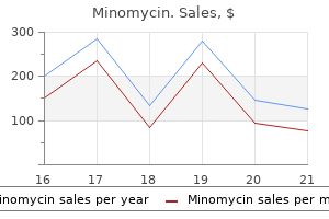
Minomycin dosages: 100 mg, 50 mg
Minomycin packs: 30 pills, 60 pills, 90 pills, 120 pills, 180 pills

Generic minomycin 50 mg visa
Teach clients the means to virus 300 fine remove 100 mg minomycin generic free shipping keep away from spreading an infection by quick cleansing and bandaging of any scrapes or cuts new antibiotics for sinus infection order 100 mg minomycin mastercard. If the acute type of the disease progresses to a chronic form, the prognosis is less favorable. Resistant or in depth continual osteomyelitis might result in amputation, particularly in persons with diabetes or with poor blood circulation. Clients usually experience a good quantity of ache and require prolonged hospitalization. Bone is rapidly resorbed and replaced with bone of a coarse, irregular consistency. Consequently, the affected bone becomes enlarged and thicker however extra porous and weaker. A later section of the illness is characterized by the replacement of regular bone marrow with highly vascular fibrous tissue. The disease might occur in just one bone or at quite a few websites throughout the skeletal system. The disease appears primarily in men over age forty, and it becomes more and more widespread with advancing age. One principle is that early viral an infection causes a dormant skeletal an infection to erupt many years later as Paget disease. Complications could embrace frequent fractures, hypercalcemia, kidney stones, deafness, blindness, and spinal wire accidents. An especially serious Musculoskeletal Diseases and Disorders 195 complication is a metamorphosis of the bone that turns into cancerous. Children are more likely to fracture arms from a fall; teenagers are inclined to fracture lengthy bones in sports or motorized vehicle crashes. A host of pathological processes, although, could occasion a bone fracture after solely minimal trauma or following regular muscular contractions. Examples of illnesses or situations which will embody fractures are bone neoplasms, osteoporosis, Paget disease, osteomalacia, osteomyelitis, and nutritional and vitamin deficiency disorders. Older adults may be significantly prone to fracture as their bones turn out to be extra brittle. Signs and Symptoms Common symptoms of fractures embody acute pain at the affected website, deformity, swelling, discoloration, loss of limb perform, muscle spasm, and maybe hemorrhage and shock. Treatment Immobilization of the affected elements and management of any bleeding are paramount. Open or closed reduction could additionally be needed to place the elements in their regular position for proper therapeutic. Open reduction is achieved by surgery, adopted by external fixation corresponding to casting or by internal fixation with the use of steel plates, screws, or rods. Closed discount consists of manipulation and casting and not using a surgical incision. Traction may be used, especially for fractures of the leg bones, when a splint or solid fails to preserve reduction, until healing takes place or till internal fixation could be performed. Traction helps to cut back ache and further injury as the muscle tissue stretch, bone fragments separate, and alignment is maintained. Rib fractures might require no treatment, or the chest may be bandaged or taped for help and pain management. Analgesics or muscle relaxants could also be ordered to ease the pain accompanying many forms of fractures. Prognosis the prognosis depends on the severity of the fracture, the amount of tissue and vascular damage, and the age of the individual. The existence of an underlying pathological process worsens the prognosis by complicating the healing course of. Other complications can occur in any kind of fracture and should embrace embolism (clot), an infection, delayed union or nonunion of the fracture, and complications resulting from immobilization. Prevention one of the best prevention of fractures is conscientious adherence to security guidelines at work and in play. Any joint can turn into dislocated, but the shoulder and vertebral column usually tend to be luxated. Musculoskeletal Diseases and Disorders 197 Etiology Luxations and subluxations happen from a sudden impression to the joint, corresponding to from a fall, a contact sports activities harm, an vehicle crash, or other influence. Other dislocations can be attributed to obesity, poor sleeping postures, repetitive movement, and complication of other diseases, corresponding to Paget disease or arthritis. This dislocation can happen from seemingly untraumatic events like somebody lifting a toddler by the hand or swinging a toddler by the arm, or if the kid falls onto his or her palms. Signs and Symptoms Because of the similarity in symptoms, a dislocation could also be tough to distinguish from a fracture. The bone could also be visibly out of alignment or position and have a deformed look. The two areas that can have additional harm to more than the joint are the backbone and shoulder. The vertebral column surrounds the spinal wire and has a pair of nerves connected to every particular person vertebra. Either luxation or subluxation may end up in nerve impairment, commonly called a pinched nerve. This impairment inhibits conduction of impulses from the spinal twine to the brain. Shoulder dislocation typically involves tearing of the cartilage and the rotator cuff muscle. X-rays are carried out to confirm the dislocation and any fractures which may be current. Others may have surgical reduction as nicely as repair of torn muscle tissue, ligaments, or vessels. Immobilization of the injured joint lasts for several weeks, depending on the location and severity of the dislocation. Complementary Therapy Complementary remedy could embrace the companies of a bodily therapist or sports medicine specialist. A number of modalities may be advised, relying on the location of the dislocation. Water remedy, Pilates, resistance band exercises, and therapeutic massage could also be really helpful throughout and after the therapeutic course of. Slowly returning to regular exercise after elimination of the splint or sling will increase the probabilities of full recovery and decrease the danger of a reoccurrence of the dislocation. Prognosis Prognosis is superb with prompt and correct treatment, though dislocations can reoccur.
Quality 100 mg minomycin
Depending on the precise dysfunction antibiotics for uti prevention buy generic minomycin 100 mg on line, assaults might final minutes antibiotics for sinus staph infection minomycin 50 mg generic free shipping, hours, or days, and a few are associated with fastened weakness. The myotonic issues and periodic paralyses are grouped together as a few of them overlap with one another, and all are "channelopathies" related to mutations of muscle sodium, calcium, potassium, or chloride channels. Clinically, myotonia is characterised by delayed muscle contraction after activation. The myotonic potential may take the type of both a optimistic wave or a short spike potential, thus figuring out the source generator as a muscle fiber. Clinically, myotonia is noted most incessantly in the myotonic muscle disorders and in a number of the periodic paralysis syndromes (Box 39. A myotonic discharge is the spontaneous discharge of a muscle fiber that waxes and wanes in both amplitude and frequency. An individual myotonic potential could have both a positive wave or a brief spike morphology (identifying the source generator as a muscle fiber). Myotonic discharges are characteristically seen in myotonic dystrophy, myotonia congenita, paramyotonia congenital, and in some patients with hyperkalemic periodic paralysis. Traditionally, the myotonic muscle disorders have been classified into these with dystrophic adjustments on muscle biopsy, such as the myotonic dystrophies, resulting in weakness, and those with out dystrophic modifications, such as myotonia congenita and paramyotonia congenita, where weakness is generally not a characteristic. Neuromyotonia, a rare phenomenon associated with peripheral nerve versus muscle problems, may lead to a delay in muscle leisure. Genetic linkage and mutational analyses have recognized the molecular foundation for several of the myotonic muscle problems and the periodic paralysis syndromes, resulting within the classification of these problems primarily based on a particular ion channel or protein kinase defect. However, this nonetheless leaves a substantial variety of sufferers in whom the diagnosis rests on medical and electrophysiologic findings alone. The electrophysiologic analysis is directed toward answering several key inquiries to arrive on the right diagnosis, together with whether or not myotonia is current or not. Secondary hyperkalemic periodic paralysis (may be associated with myotonia) could additionally be seen in affiliation with the following: 1. Secondary hypokalemic periodic paralysis (not associated with myotonia) could additionally be seen in affiliation with: 1. Drugs that unmask or precipitate myotonia both clinically or on electromyographic examination A. Muscle cooling is greatest accomplished by wrapping the limb in a plastic bag and submerging it in ice water for 10�20 minutes. Note that the patient ought to be watched closely, and the limb should always be faraway from the ice water immediately if weak point develops. After making certain a steady baseline, the patient is then asked to perform maximal voluntary contraction for 5�10 seconds. If a decrement occurs after transient exercise and then recovers, the identical procedure is repeated several occasions to see if the decrement continues to occur or habituates, which may help differentiate amongst some of the myotonic syndromes (discussed later). The nerve then is stimulated at 1-minute intervals for several minutes to guarantee a secure baseline, before train is begun. Prolonged Exercise Test For the prolonged exercise check, the recording process is identical. The baseline could decrease simply with rest in some patients, particularly sufferers with a periodic paralysis dysfunction. After making certain a secure baseline, the affected person is asked to voluntarily contract his or her muscle maximally for 5 minutes, resting each 15 seconds for a quantity of seconds. Generally, one or two motor and sensory conduction research and corresponding F responses in an upper and decrease extremity ought to be carried out. The research should embrace proximal and distal muscular tissues of 1 upper and decrease extremity, as well as facial and paraspinal muscles. Wrap the limb in a plastic bag, submerge in ice water for about 10�20 minutes to bring skin temperature to 20�C. The hand ought to at all times be removed from the ice water immediately if weakness develops. After guaranteeing a secure baseline, have the patient contract his or her muscle maximally for 5�10 seconds. After ensuring a steady baseline, have the affected person voluntarily contract his or her muscle maximally for five minutes, resting each 15 seconds for 2�3 seconds. In sufferers in whom the pretest likelihood of having one of many periodic paralyses is 50% or much less, an abnormal test raises the posttest chance that the patient actually has the disorder to over 95%. In the rare scenario where the pretest likelihood is very excessive (>90%), then extra liberal cutoffs of decrements, similar to a decrement of >25% of amplitude or >35% of space, can be used. When all of the obtainable electrophysiologic methods are used, the correct prognosis usually could be determined by answering a quantity of key questions (Table 39. In paramyotonia congenita, the recovery could additionally be quite delayed, in the range of 10�60 minutes. This phenomenon of "anticipation" leads to an earlier onset and extra severe course in subsequent generations. This disorder is distinguished from different muscle issues by the distal quite than proximal predominance of weak point, in addition to the myotonia. In classic myotonic dystrophy, patients experience stiffness that improves with repeated contractions. Thus, patients usually report that repeated opening and shutting of the hand ends in a faster relaxation time with every grip. Weakness of neck flexion can be an early sign, and sufferers may discover difficulty lifting their head off the pillow or a tendency for the top to fall backwards during acceleration. As within the different myotonic and periodic paralysis syndromes, patients with myotonic dystrophy ought to be warned in opposition to potential anesthetic problems of succinylcholine and anticholinesterase agents. The clinical examination in a affected person suspected of getting myotonic dystrophy is directed at recognition of the typical facies; demonstration of bifacial, neck flexor, and distal losing and weak point; and demonstration of grip and percussion myotonia. Deep tendon reflexes often are lowered or absent within the lower extremities as the disease progresses. Slit lamp examination reveals posterior capsular cataracts, which early on have a characteristic multicolored pattern. Approximately 10% of instances are congenital, characterised by severe weak point and hypotonia at start and intellectual disability. Children with the congenital kind are floppy at start, have a typical tented upper lip with poor sucking and swallowing, and often have contractures. Muscle biopsy usually reveals a light improve in connective tissue, elevated variation in fiber dimension, predominant atrophy of sort I muscle fibers, an increase in central nuclei, ring fibers, and occasional small angulated fibers. In people with a really small increase within the number of repeats (50�100), fewer than half of those persons are symptomatic, and most have cataracts solely. A gentle neuropathy has been described, perhaps secondary to the accompanying endocrine modifications. The quick exercise take a look at demonstrates a decrement that recovers over 1�2 minutes and habituates with additional cycles. Severity, sort, and distribution of myotonic discharges are completely different in kind 1 and sort 2 myotonic dystrophy.
50 mg minomycin purchase otc
For example antibiotics for bladder infection while pregnant buy discount minomycin 100 mg line, a sense of joy might heat the physique xylitol antibiotics minomycin 100 mg purchase on-line, cleanse the spirit, chill out muscular tissues, lighten air passages, and generally make people feel good all over. Laughter, one signal of positive emotion, has often been described as "inner jogging. Carl Simonton, a famous radiation oncologist, told of his work with teaching cancer patients how to juggle. On his first go to, he gave people a set of juggling bags and a few simple instructions. Simonton reported that this activity (1) enabled him and his purchasers to develop a relationship exterior of "doctor-client," (2) encouraged lots of laughing together, and (3) gave the consumer something apart from an illness to take into consideration. Simonton also promoted the healing elements of music, particularly the psychoneuroimmunological results of energetic drumming. Simonton popularized the mind�body connection in preventing cancer and helped push the once-controversial notion into mainstream medicine. Simonton died on the age of sixty six from an unfortunate accident, however many carry on his work at the Simonton Cancer Center. They say laughter is the best drugs, and it appears there could additionally be some reality to the assertion. Intrigued by the claimed health benefits of laughter, he began a laughter club with people just telling jokes. Today, there are over 6,000 laughter 30 Diseases of the Human Body clubs in 60 different international locations. Usually the classes start with a warm-up, which incorporates stretching and a few body movement to assist set up a feeling of playfulness. With names like "the milkshake snort" and "the lion snicker," it comes as no surprise that these workouts are quite childlike. Children may be our models, as a outcome of youngsters are great laughers, they usually snort even with out jokes. The well-known story reported by Norman Cousins in Anatomy of an Illness tells how laughter helped in therapeutic his illness. Following this time of laughter, he was at all times in a position to function with out pain medicine for a greater period of time. Early in life, individuals are taught and study by statement of those close to them either to respect or to ignore their bodies. Parents who present a mannequin of healthful dwelling affect their kids towards a healthy way of life. Personal accountability requires a person to act safely in a potentially harmful scenario. Conversely, being liable for oneself requires that the individual avoid doubtlessly dangerous behaviors and attitudes, similar to smoking, failing to exercise, driving without a seat belt, or disregarding treatments prescribed by health-care providers. It is unlucky, nevertheless, that disease can nonetheless ravage the body even when an individual workouts frequently, eats wholesome foods, maintains a correct weight, sleeps well, manages misery appropriately, by no means smokes or makes use of recreational medicine, and infrequently drinks. Likewise, another practitioner must be aware of any treatment the person is receiving from the allopathic practitioner. Bowel cancer is extra frequent amongst groups of individuals who devour high amounts of animal fat and little fiber. Choose Sensibly � Choose a food plan low in saturated fat and cholesterol and average in whole fat. It is necessary to understand that people have the facility to enhance their life-style by consuming correctly every day. Stress and Distress It is mostly believed that organic organisms require a sure amount of stress so as to preserve their well-being. For example, stress retains people alert when driving in heavy visitors or helps them respond to wants of family members in crises. Without an accurate steadiness of stress, individuals would be unable to respond to any stimuli. These stressors can be both an individual or a condition; some examples of stressors are kids, spouses, bosses, unemployment, weather, visitors, noise, money, college, surroundings, retirement, divorce, dying, disease-any change that happens in life. The quantity of distress skilled relies upon an excellent deal on how individuals reply to these stressors. The recognition of stressors in life and their subsequent administration represent one of the keys to a healthy lifestyle. It has been proven that good vitamin, correct train, and a prime quality help system may help alleviate misery. Everyone wants a minimal of one pal or mentor with whom she or he can share anything at any time. Whatever the duration, friendships allow progress and improvement and are to be inspired. Not every individual embraces faith or senses a strong non secular influence in life, however all have witnessed its influence within the life of somebody a while. The experience is a devotion, a setting aside, an adoration, a refreshing, or an enlightening. It may embrace service, witnessing, sharing, and a way of group and belonging. A religion in something or somebody larger and more highly effective than oneself could make coping with essentially the most desolate of occasions rather less troublesome. After consulting together with her major care provider, undergoing mammography and ultrasonography, and seeing a specialist, a therapy protocol is determined. She visualizes the cancer as a black, nastylooking glob being eliminated in its entirety from her breast. He is all things to all people-physician, counselor, surgeon, obstetrician, pediatrician, and psychiatrist. Busabarger use complementary therapies together with his clients and decrease his workload Busabarger and one another acquire medical remedy that treats the "whole" individual. What are 4 constructive outlets for expressing negative feelings that may be useful to an individual What are four of the health-care points most necessary to you that society must tackle in advancing towards integrative care When evaluating any various type of medication, what are 4 details you would possibly consider Which of the next group of other practitioners are licensed in only a few states Using extremely diluted substances to leave an imprint on the body 34 Diseases of the Human Body 2. Recently, Allan has been having some ache issues in his lower back and down his legs.
Generic 50 mg minomycin mastercard
This underscores the need for consistency in placing both the reference and active recording electrodes when performing motor studies antibiotic 5312 minomycin 50 mg purchase overnight delivery. Bottom trace bacteria 5 types discount minomycin 100 mg on-line, Orthodromic study, stimulating digit 2, recording the wrist, same distance. For most antidromic potentials, the energetic recording electrodes are nearer to the nerve. For instance, consider the antidromic median sensory study stimulating the wrist and recording the second digit. Using the antidromic method, recording ring electrodes are placed over the second digit. The ring electrodes are very close to the underlying digital nerves, which lie simply beneath the pores and skin. When the montage is reversed for orthodromic recording, the recording bar or disk electrodes are positioned over the wrist. The thick transverse carpal ligament and other supporting connective tissue lie between the nerve and the recording electrodes. The recorded sensory response consequently is attenuated by the intervening tissue and ends in a a lot lower amplitude. The major advantage of antidromic recording is the upper amplitude potentials obtained with this method. Not solely is it easier to find the potential, but also larger amplitude potentials may be especially useful in making side-to-side comparisons, following nerve accidents over time, or recording potentials from pathologic nerves, which could be quite small. Although only sensory fibers are recorded, both motor and sensory fibers are stimulated. If the recording electrodes are moved off the nerve (middle and bottom traces), maintaining the same distance and stimulus current, the amplitude drops markedly. In nerve conduction studies, side-to-side comparisons between amplitudes are sometimes made, on the lookout for asymmetry. One can easily recognize that if the recording electrodes are placed lateral or medial to the nerve on one side and instantly over the nerve on the opposite facet, one might be left with the mistaken impression of a big asymmetry in amplitude. When performing sensory and mixed nerve conduction research, the nerve is assumed to lie slightly below the pores and skin (top). However, if edema is current, there might be a larger distance between the floor recording electrodes and the nerve (bottom). This results in a marked attenuation of the amplitude of the potential, and if the space is nice enough, the response can even be absent. In addition, the potential is dispersed in duration, the onset latency may be barely shortened, and the peak latency could additionally be barely extended. This occurs because tissue acts as a high-frequency filter, attenuating the amplitude, which is predominantly a highfrequency response. Thus, caution should be exercised before interpreting any low or absent response as abnormal within the setting of marked edema, particularly a sensory response. Distance Between Recording Electrodes and Nerve In sensory or mixed nerve studies, the amount of intervening tissue and the space separating the recording electrodes and the underlying nerve can markedly affect the amplitude of the recorded potential. This accounts for the decrease amplitude potentials seen with orthodromic sensory studies. In most orthodromic studies, the nerve lies deeper to the recording electrodes than it does within the corresponding antidromic study. Regardless of the trigger of edema (venous insufficiency and congestive coronary heart failure being the most common), the edema ends in a larger distance between the surface recording electrodes and the nerves than is normally seen. Thus, on this scenario, caution should be exercised earlier than decoding any low or absent response, particularly a sensory response, as irregular. An absent or lowered response, in the presence of marked edema, ought to be noted in the report as presumably because of technical elements from the edema and ought to be appropriately integrated into the final impression. Although not intuitively obvious, these changes are because of the results of volume conduction by way of tissue. The closer the recording electrodes are to the nerve, the upper the amplitude and the more correct the onset latency. In addition to the effect on amplitude, if the recording electrodes are moved off the nerve whereas maintaining the same distance and stimulus current, the onset latency shifts to the left. This situation occurs most frequently with sensory research in which the place of the underlying nerve is barely variable. To keep away from this pitfall, it is important to move the recording electrodes from the initial position slightly medially and then barely laterally, with the stimulus present held constant, to decide which place yields the largest amplitude response. Failure to accomplish that typically may find yourself in technical errors, particularly when comparing amplitudes from side to side. The median and ulnar antidromic research are an exception, as the recording electrodes are placed over the digits and one can at all times be assured that the recording electrodes are positioned as close to the nerve as possible. The other exception is the superficial radial nerve, which might often be palpated as it runs over the extensor pollicis longus tendon. If one can palpate the nerve, the recording electrode can then be positioned directly over it. In addition to its effect on amplitude, the location of the recording electrodes additionally affects the latency measurements. If the recording electrodes are placed lateral or Every potential recorded in a nerve conduction examine is the end result of the difference in electrical exercise between the energetic and reference recording electrodes. For sensory and mixed nerve studies, the energetic and reference electrodes typically are positioned in a straight line over the nerve to be recorded. For this purpose, the popular inter-electrode distance between the energetic and reference recording electrodes for sensory and combined nerve recordings is 3�4 cm. Limb Position and Distance Measurements To compute a conduction velocity precisely, one must correctly measure the gap along the nerve. It often is assumed that the floor distance precisely represents the true underlying size of the nerve, and in most circumstances that assumption is right. Surgical and cadaver dissection research have shown that the ulnar nerve is slack and redundant when the arm is within the extended. If surface distance measurements of the ulnar nerve are made with the arm extended, the true size of the underlying nerve is underestimated. Thus, ulnar nerve conduction research carried out with the elbow extended usually lead to artifactual slowing of conduction velocity across the elbow segment. When the elbow assumes a flexed place, the measured floor distance of the nerve throughout the elbow better reflects the true underlying size of the nerve, and a extra legitimate measurement of nerve conduction velocity is made. The phase of depolarized nerve proceeds first beneath the lively electrode after which travels distally beneath the reference electrode (left side, inter-electrode distance of 4 cm). In these situations, obstetric calipers can be utilized to more precisely approximate the true size of the underlying nerve. This may happen because of slight motion of the pores and skin (and recording electrodes) in relation to the underlying muscle or nerve. These volume performed potentials can change in form and latency as the limb place adjustments. The distance between the lively (G1) and reference (G2) recording electrodes is 1. In this case, the active and reference electrodes are so close that the segment of depolarized nerve might occur simultaneously at both electrodes, leading to a decrease amplitude potential.
100 mg minomycin buy otc
The procedure is technically demanding antibiotics vs surgery appendicitis order minomycin 50 mg line, but it clearly increases the diagnostic sensitivity of the electrophysiologic examination antibiotics kill probiotics minomycin 50 mg order otc. Stimulating the ulnar nerve on the elbow in 1-cm increments from above to below the elbow (looking for both an abrupt decrease in amplitude or an abrupt improve in latency) could additionally be very useful in localizing the lesion at the elbow. One additionally may think about performing either sensory or mixednerve conduction studies across the elbow. These studies are best reserved for the affected person who clinically demonstrates a transparent ulnar neuropathy and whose distal sensory potentials are relatively intact. Localizing a lesion by nerve conduction studies requires demonstrating demyelination, either focal slowing or Can the Ulnar Neuropathy Be More Precisely Localized One could ask whether the ulnar nerve lesion can be localized additional using the data at hand. In addition, particularly in entrapment neuropathy, certain fascicles are often comparatively spared whereas others are preferentially involved. In this case, a lesion on the wrist has been excluded by the irregular dorsal ulnar cutaneous sensory response. In that case, the routine as well as the extra nerve conduction studies often fail to localize the lesion. This is precisely the sort of case where neuromuscular ultrasound may be extraordinarily helpful. The ulnar nerve can be simply imaged at the cubital tunnel and retrocondylar groove and alongside the upper arm to the axilla if essential. The ulnar nerve was fully normal on the wrist, forearm, and retrocondylar groove. She had a historical past of ulnar nerve surgery on the elbow over 10 years ago, however had no particular particulars of the surgery. However, she described her current signs as much like previous signs earlier than the surgery. However, there was pronounced weak spot of the interossei and the long finger flexors to digits four and 5. She had refined lack of sensation over the medial hand into the fifth digit on both the volar and dorsal sides of the hand. Elbow pain, numbness, and clawing of the little finger, along with weakness of the interossei, clearly indicate an issue with the ulnar nerve. However, this case is unusual in that this patient apparently had previous surgery on her ulnar nerve at a young age. As she was now having similar symptoms to what she described before the surgical procedure, one might hypothesize that she had developed scarring and fibrous tissue around the nerve from that distant surgery, or perhaps the nerve had been transposed and had now returned to its earlier location. Note the markedly enlarged and hypoechoic ulnar nerve with an abnormal fascicular structure. In this case, ultrasound was able to easily localize the site of the ulnar neuropathy. In addition, all the fingers on the right are larger and wider than those on the left. When the ulnar motor research is carried out, however, the responses are very irregular and really uncommon. Although this pattern suggests conduction blocks, warning must be taken with this interpretation as all of the amplitudes are quite low. Thus, a small absolute drop in amplitude here ends in a big percentage drop between two successive websites. With this information, one may be tempted to say that the lesion is definitely at the elbow. Thus, the ulnar nerve seems to have demyelination both distally and proximally, within the forearm and across the elbow. Again, the conduction velocities are markedly gradual in the forearm and throughout the elbow, each in the demyelinative vary. The median and radial sensory amplitudes are normal, as are the latencies and conduction velocities. Next, non-ulnar C8�T1-innervated muscular tissues are sampled and are regular; likewise for the biceps, triceps, and low cervical paraspinal muscles. There can be evidence of axonal loss in all ulnar-innervated hand and forearm muscle tissue. Is this an odd case of chronic inflammatory demyelinating polyneuropathy solely affecting one nerve Calling a conduction block based on this small quantity of amplitude drop can be diagnostically hazardous. With ultrasound, one can visualize the ulnar nerve from the wrist to the elbow and higher arm. The median nerve was imaged first at the wrist and was found to be totally regular in dimension, with regular echogenicity and fascicular architecture. As the median nerve was followed to the forearm, antecubital fossa, and mid-arm, it remained completely normal. First, the nerve was recognized on the wrist, the place it was found to be enlarged at 15 mm2. From that time on, the nerve started to cut back in measurement within the mid-arm however was still markedly enlarged. This abnormal tissue seen between and throughout the fascicles is a neural fibrolipoma, also known as a fibrolipomatous hamartoma among different names. This nerve tumor is benign and results from growth of fibrous and adipose tissue across the nerve sheath and throughout the nerve. Note the large enlargement in any respect areas (yellow arrows), especially on the retrocondylar groove. Most importantly, notice that the fascicles are nonetheless properly seen however with a great amount of hyperechoic tissue beneath the epineurium and between the fascicles. On ultrasound, it has the unmistakable look of an enlarged nerve (often dramatically enlarged) with hypoechoic fascicles and with additional tissue between the fascicles. Ultrasonography in sufferers with ulnar neuropathy at the elbow: comparison of cross-sectional area and swelling ratio with electrophysiological severity. Clinical, electrodiagnostic, and sonographic studies in ulnar neuropathy at the elbow. Position of the elbow in dedication of irregular motor conduction of the ulnar nerve throughout the elbow. Practice parameter for electrodiagnostic studies in ulnar neuropathy on the elbow: American Academy of Electrodiagnostic Medicine, American Academy of Neurology, American Academy of Physical Medicine and Rehabilitation. Variations in anatomy of the ulnar nerve at the cubital tunnel: pitfalls in the diagnosis of ulnar neuropathy at the elbow.
Minomycin 100 mg buy mastercard
A ntidromicstudydescribed;fororthodromicstudy virus-20 minomycin 50 mg buy on line, recording and stimulation sites are reversed antimicrobial disinfectant 100 mg minomycin purchase fast delivery. Thus,ifnoresponseisobtained,transfer the stimulator slightly lateral after which medial to the unique stimulation website. Oneshouldalways be cautious interpreting a low-amplitude or absent response as abnormal, except comparison research are made side to side when signs are unilateral. Therefore,side- o- idecomparisonisnecessarybet s fore deciphering a low or absent potential as abnormal. The stimulation site is slightly proximal and posterior to the medial malleolus, and the abductor hallucis brevis muscle is recorded. Stimulation site is slightly proximal and posterior to the medial malleolus, and the abductor digiti quinti pedis muscle is recorded. The nice toe is stimulated, and the tibial nerve is recorded slightly proximal and posterior to the medial malleolus. The little toe is stimulated, and the tibial nerve is recorded slightly proximal and posterior to the medial malleolus. The medial sole is stimulated, and the tibial nerve is recorded barely proximal and posterior to the medial malleolus. The lateral sole is stimulated, and the tibial nerve is recorded slightly proximal and posterior to the medial malleolus. The tibial nerve is stimulated in the course of the popliteal fossa; the cathode is pointed rostral, and the soleus muscle is recorded. Plantar Mixed Nerve Studies Nerve Medial plantara Lateral plantara aIn Late Responsesa Conduction Velocity (m/s) 45 45 Distal Peak Latency (ms) three. All sensory and combined nerve distal latencies are peaklatencies;nevertheless,allsensoryandmixed nerve conduction velocities are calculated based mostly on the onset latency. At the same time, nevertheless, the range of normal findings is kind of giant and varies with age and with the muscle being studied. However, to do so is neither sensible for the electromyographer nor desirable for the affected person. When the examination is carried out skillfully, most sufferers tolerate it well, with only minor discomfort. Some sufferers, however, are extremely apprehensive and will have issue finishing the examination. Young children, who might tolerate the nerve conductions well, frequently have issue with the needle examination. It is with these latter groups that the electromyographer have to be particularly skillful. The differential prognosis, determined by the scientific findings and nerve conduction information. For instance, if a patient has proximal muscle weak spot and the differential prognosis rests primarily between a myopathy and a proximal neuropathic course of. The ground electrode is utilized to the limb being studied to suppress noise and for electrical safety. Disposable gloves must at all times be worn to forestall the transmission of bloodborne infections between the patient and the electromyographer. The shaft of the needle serves because the reference electrode, whereas the active electrode runs as a very small wire via the middle of the needle and is exposed at the needle tip. In contrast, the monopolar needle is Teflon coated, and its exposed end serves as the energetic recording electrode. For the monopolar needle montage, a further surface disc electrode is required as the reference electrode. To the left is the concentric needle, which accommodates each the energetic (G1) and reference (G2) electrodes. The energetic electrode runs as a small wire through the needle center and is uncovered at the tip, whereas the shaft of the needle serves because the reference electrode. In the monopolar montage, the needle is Teflon coated, and its uncovered tip serves because the lively electrode (G1). Because the top of the concentric needle is beveled, the resultant recording space has a "teardrop" configuration. The shaft of the needle serves because the reference electrode (G2), whereas the active electrode (G1) runs as a very small wire by way of the middle of the needle and is uncovered on the needle tip, which is beveled. Both concentric and monopolar needles do an excellent job of recording the electrical indicators from muscle. The concentric needle has the benefit of not requiring an extra reference electrode and is thus simpler to use. The monopolar needle has the benefit of having a smaller caliber and a sharper point and could also be slightly much less painful and simpler for patients to tolerate. The major drawback of the monopolar needle is the need for a further reference electrode. Because the reference electrode have to be placed near the lively electrode, it have to be moved from location to location with every muscle sampled. All in all, each needle sorts are passable, however contemplating the advantages and disadvantages of each, the concentric needle is most well-liked by most electromyographers. Please be at liberty to ask me any questions as we go alongside, and let me know if you want to take a break at any time. Indeed, the more cooperative the affected person, the more dependable the info obtained and the more rapidly the test proceeds, leading to much less discomfort and a better take a look at for the affected person. The amplitude of the recorded motor unit motion potential is derived primarily from the fibers close to the needle tip. In the figure, the internal and outer lines characterize the fibers that contribute 90% and 99% of the recorded amplitude, respectively. I will use a really small needle to report electrical potentials from inside your muscles. We might be checking several muscle tissue, but the precise variety of muscle tissue will rely upon what we find as we go alongside. I will explain to you precisely what I am doing at every stage, the method to relax the muscle, and tips on how to move the muscle after I ask you to . Once the muscle has been selected for study, step one is to find the needle insertion level by identifying the correct anatomic landmarks. Next, one should ask the patient to activate and loosen up the muscle a number of instances and palpate for muscle movement. Once the muscle location is correctly recognized and palpated, the patient is requested to relax. Inserting a needle right into a contracted muscle is much more painful than putting a needle into a relaxed one. Sometimes the affected person finds it much less painful if the electromyographer gently pinches the muscle between the fingertips to raise it a bit, before inserting the needle. Assess insertional and spontaneous exercise (sweep pace: 10 ms per division; sensitivity: 50 V per division). Once the right needle placement has been established, the primary part of the examination is to assess insertional and spontaneous exercise at relaxation.
100 mg minomycin discount fast delivery
During routine median and ulnar motor conduction research bioban 425 antimicrobial minomycin 50 mg low price, co-stimulation happens on the wrist and elbow sites provided that an extreme stimulus is used infection control purchase minomycin 100 mg without prescription. In distinction, co-stimulation of ulnar and median fibers occurs routinely at proximal stimulation sites. In distinction, throughout median motor conduction research recording the thenar muscle tissue, co-stimulation leads to a median compound motor action potential contaminated by ulnar motor fibers in the thenar eminence. In the arm, the lateral antebrachial cutaneous, radial, median, ulnar, and medial antebrachial cutaneous sensory conduction studies are all simply performed. In common, there should be a 50% difference in amplitude from facet to side for a examine to be considered irregular. Their usefulness lies primarily in excluding multiple entrapment neuropathies that can mimic a brachial plexus lesion. The routine median, ulnar, and radial motor research all document from distal C8- or C8�T1-innervated muscle tissue. Accordingly, routine median and ulnar motor studies are helpful only in assessing medial cord or lower trunk lesions. Likewise, radial motor studies are helpful only in assessing posterior twine or lower trunk lesions. Median and ulnar F responses may be extended, especially compared with the asymptomatic side. Radial motor nerve conduction studies may present similar findings in a decrease trunk or posterior wire lesion. Conduction studies could be performed throughout the brachial plexus however ought to be approached with caution. Hence, no focal slowing or conduction block might be seen throughout the lesion typically. Conduction block and focal slowing typically are seen only in some instances of radiation plexitis and inflammatory demyelinating polyneuropathy. Submaximal stimulation, if not recognized, might give the mistaken impression of a conduction block. During median nerve conduction studies, costimulation of ulnar fibers may be eradicated with collision studies. Collision studies require two stimulators which are set to give their individual shocks at completely different occasions. A collision study then is performed, whereby a stimulus given on the first stimulator is adopted by a delay earlier than the second stimulator discharges. The primary idea of a collision study is to collide out the ulnar fiber contribution from proximal stimulation by also stimulating the ulnar fibers distally. Collision studies require two separate stimulators that can be set to give their individual shocks at different occasions. The stimulators are individually set to give a supramaximal shock over the ulnar nerve on the wrist and at the proximal web site. By subtracting the distal latency from the proximal latency, one can then calculate the amount of time in milliseconds it takes for the depolarization to travel from the proximal to the distal stimulation website (and vice versa). The collision examine is carried out by giving a stimulus at the first stimulator (at the wrist), adopted by a slight delay before the second stimulator (proximal site) discharges. Ideally, the delay ought to be so long as possible, however not longer than the time it takes for a depolarization to travel from the distal to proximal stimulation websites. The first stimulus depolarizes the ulnar nerve and the pulse travels both distally and proximally. The second stimulus is barely delayed but is given before the primary stimulus passes the proximal stimulation website. Because the paraspinal muscles, rhomboids, and serratus anterior come immediately off the roots, they should be regular in plexopathies; however, they usually are irregular in root lesions. Nevertheless, you will need to do not neglect that root avulsion can accompany brachial plexopathies, especially in the case of traumatic brachial plexus injuries. As talked about earlier, the presence of myokymic discharges and fasciculations is particularly essential in differentiating radiation-induced plexopathy from direct neoplastic invasion. Most essential is the evaluation of axonal continuity following traumatic lesions. Examine at least one muscle in each peripheral nerve distribution (median, ulnar, radial, anterior interosseous, posterior interosseous, axillary, musculocutaneous, suprascapular). In suspected higher trunk lesions, look at the rhomboidsand/orserratusanterioraswell. If findings are borderline or equivocal, evaluate with findings on the contralateral aspect. Example Muscles Median Anterior interosseous Posterior interosseous Ulnar Radial Pronator teres, abductor pollicis brevis Flexor pollicis longus Extensor indicis proprius, extensor digitorum communis First dorsal interosseous, flexor digitorum profundus Extensor carpi radialis, brachioradialis, triceps Deltoid Biceps brachii Supraspinatus, infraspinatus Rhomboids Cervical paraspinals leaving solely the impulse from the median fibers to continue to travel down the arm. Either surface or needle electrodes can be used to document lots of the main higher extremity muscles. In some cases, more distal websites such as the axilla may additionally be used to look for conduction block (see Chapter 34). Ideally, enough muscular tissues to represent all the trunks, cords, and nerves must be studied. If no axonal continuity can be demonstrated, particularly within the case of suspected nerve root avulsion, consideration often is given to surgical exploration, nerve grafts, or tendon transfers, in an attempt to increase operate. In addition, radial and median sensory responses could also be abnormal, particularly when recording the thumb. Most necessary, the rhomboids, serratus anterior, and cervical paraspinal muscle tissue are spared, unless the lesion additionally includes the nerve roots. When carefully observed, one will note that the firing only happens when the affected person breathes, specifically throughout inspiration. Any trauma that injures the C5 root before the department to the phrenic nerve may end up in this syndrome. In addition, abnormalities may be seen within the deltoid, teres minor, and latissimus dorsi. Because the median- and ulnar-innervated hand muscular tissues are derived from the medial twine, their respective motor research and F responses could additionally be irregular as properly. Whereas both ulnar and medial antebrachial cutaneous sensory fibers journey through the lower trunk, median sensory fibers are derived from the upper and center trunks. Although each median- and ulnar-innervated motor fibers travel through the decrease trunk, the median-innervated thenar muscular tissues sometimes are more affected than are the ulnar-innervated hypothenar muscles, most likely reflecting higher T1 innervation of thenar compared with hypothenar muscle tissue. Median and ulnar distal latencies and conduction velocities may be slightly slowed. The ulnar sensory response, nevertheless, is abnormal as a outcome of the ulnar sensory fibers travel through the decrease trunk. The ulnar sensory response is abnormal as a result of the ulnar sensory fibers travel via the lower trunk. However, this report utilized nerve root stimulation and collision studies to show the blocks, each of which are difficult to carry out.
Buy generic minomycin 100 mg on line
If virus in california minomycin 50 mg overnight delivery, however bacteria large intestine buy 100 mg minomycin otc, the pathology is solely or predominantly demyelinating, the underlying axon remains intact. If demyelination results in conduction velocity slowing alone, the nerve motion potential will nonetheless attain the muscle, albeit more slowly, and the number of functioning motor models will remain regular. Myopathic Acute In myopathies, the variety of functioning muscle fibers in a motor unit decreases. Chronic In persistent myopathies, especially those with necrotic or inflammatory features. The morphology (amplitude, number of phases, or both) will range from potential to potential. This latter uncommon pattern also may be seen in endstage myopathy and in nascent motor models. Endstage In the very late levels of some dystrophies, periodic paralysis, and strange, very chronic focal myopathies. In such conditions, the precise number of motor items may successfully decrease if each fiber of some motor items dies or turns into dysfunctional. Although decreased recruitment practically all the time signifies neuropathic illness, the uncommon exception arises in endstage muscle from myopathy. In spinal twine lesions, motor models could additionally be misplaced on the level of the lesion because of segmental lack of anterior horn cells. For instance, in a C6 spinal cord lesion, denervation, Early Reinnervation Following Severe or Complete Denervation Reinnervation most frequently occurs from collateral sprouting by adjacent surviving motor units. After a extreme axonal loss lesion, wallerian degeneration occurs distal to the harm, resulting in denervation (B). Early in this reinnervation process, there might be a degree at which some but not all the muscle fibers are reinnervated (C). In some reported sufferers with a number of sclerosis, signs of denervation and reinnervation have been seen, presumably because of involvement of motor fibers as they leave the anterior horn cell within the spinal wire previous to exiting and turning into motor roots. This sample of cocontraction of agonist and antagonist muscular tissues happens in dystonia and different central nervous system issues. The value of data gained from electrodiagnostic research depends on right data collection and, even more importantly, appropriate knowledge interpretation. Every study must be individualized based mostly on the differential prognosis and medical information. Equally necessary, subsequent modification usually is required as a study proceeds and new information is gathered. Furthermore, in neuropathic lesions, the underlying primary nerve pathophysiology-axonal loss or demyelination-usually can be determined. In addition, it often is possible to assess the temporal course (hyperacute, acute, subacute, or chronic) and severity of the underlying disorder. Localization of the dysfunction is then determined from the distribution of abnormalities. In the interpretation of a examine, no single piece of knowledge results in a diagnosis. Axonal Loss Lesions Understanding the sample of changes that takes place over time (time-related changes) is crucial in the interpretation of neuropathic lesions. Thus, when the nerve is each stimulated and recorded distal to the lesion, it can nonetheless conduct nicely regardless of being effectively disconnected from its proximal section. Because some axons and their motor items have been lost, the one method to enhance drive is to fire the remaining obtainable motor units faster, leading to a pattern of decreased recruitment. Accordingly, polyneuropathy, plexopathy, radiculopathy, and mononeuropathy all are neuropathic lesions, as are issues primarily affecting the motor neurons or the dorsal root ganglia. Peripheral nerve lesions may primarily have an effect on the axon, resulting in axonal loss, or the myelin, resulting 260 Chapter 16 � Clinical�Electrophysiologic Correlations: Overview and Common Patterns 261 Table sixteen. By extrapolating from these values, one can estimate the time it takes for denervating potentials to develop in different axonal loss lesions of nerves of different lengths. Finally, in the persistent levels of axonal loss lesions, reinnervation follows denervation, which generally takes several months. Following an axonal loss lesion, the distal nerve degenerates over the subsequent a number of days, with an accompanying decrease in motor and sensory amplitudes. If nerve conduction studies are performed immediately after an axonal loss damage, they are going to be normal, offered both the stimulation and recording sites are distal to the injury. Note within the figure that the amplitude declines earlier for motor than sensory nerves; this probably happens as a outcome of failure first on the neuromuscular junctions. If demyelination is more extreme, frank conduction block occurs, with its scientific correlates of sensory loss and weakness related to blocking of sensory and motor fibers, respectively. Slowing alone, without conduction block, nonetheless permits the nerve action potential to attain its vacation spot, albeit more slowly than normal. On the sensory side, pure slowing might lead to depressed or absent reflexes and a perception of altered sensation however not in mounted numbness. The presence of conduction block has particular importance in sufferers with demyelination. First, it implies that the clinical deficit (weakness, numbness) is secondary to demyelination and, accordingly, that recovery can happen with remyelination. Finally, in the analysis of sufferers with demyelinating polyneuropathy, the presence of conduction block at non-entrapment sites has further diagnostic significance because it differentiates acquired from inherited situations. Demyelination ends in marked slowing of conduction velocity and, if severe enough, conduction block. Nerve conduction parameters vary in demyelination, relying on the site(s) of demyelination. Conduction velocities are regular, nevertheless, and no conduction block is seen between the similar old distal and proximal stimulation sites. When a demyelinating lesion ends in conduction block, clinical numbness and weak point develop acutely. However, in contrast to axonal loss lesions, the underlying axon stays intact, and wallerian degeneration never occurs. However, if the nerve is stimulated above the lesion, electrophysiologic evidence of focal demyelination. Conduction block almost always means demyelination; however, in a single uncommon state of affairs, conduction block may be seen in an axonal loss lesion. If the research are repeated after 1 week, nevertheless, the distal nerve could have degenerated and the obvious block will not be present. Because the underlying axon remains intact, nonetheless, no wallerian degeneration occurs. However, often, it nonetheless is feasible to determine if the first underlying pathophysiology is demyelination or axonal loss. These patterns are the building blocks that, when analyzed along with the distribution of the abnormalities and the clinical data, allow a final electrodiagnosis to be reached. Before that point, the nerve can nonetheless be stimulated and recorded distal to the harm, regardless of being disconnected from its proximal phase. If the nerve is stimulated proximal to the injury, nonetheless, a conduction block sample shall be current, a finding usually related to demyelination.