

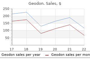
Geodon dosages: 80 mg, 40 mg, 20 mg
Geodon packs: 30 pills, 60 pills, 90 pills, 120 pills, 180 pills, 360 pills

Generic 20mg geodon overnight delivery
Anterior uveitis (or iritis) often manifests with headache anxiety pain 20mg geodon fast delivery, deep eye ache depression era photos generic geodon 20 mg without a prescription, lacrimation, blurred vision, and photophobia. The mostly related respiratory situations are bronchiectasis and chronic bronchitis. Uric acid stones are believed to end result from volume depletion and a hypermetabolic state. Rare intrinsic renal complications embody membranous nephropathy, glomerulonephritis, and renal amyloidosis. Penile and vulvar edema even have been reported, however the mechanism for these occurrences is unknown. Patients might present with venous thromboembolism or, a lot less generally, arterial thrombosis. Circulating immune complexes, elevated ranges of plasminogen activator inhibitors, decreased levels of tissue plasminogen activator, and spontaneous platelet aggregation could additionally be current impartial of bowel inflammation. From symptom to diagnosis: scientific distinctions among varied types of intestinal inflammation. The commonest organisms causing infectious colitis are Salmonella, Shigella, and Campylobacter (See Chapter 110). Patients with this infection, notably children and older adults, often current with bloody diarrhea and might develop associated hemolytic-uremic syndrome or thrombotic thrombocytopenic purpura. Histology is that of ischemic colitis and the Shiga toxins produced by this organism trigger fibrin aggregation inside the vasculature. Development of molecular probes may facilitate the power to set up this analysis. The prognosis is made on the basis of stool tradition or a rising titer of serum antibody. Other, much less widespread bacterial infections inflicting colitis include Aeromonas hydrophila and Listeria monocytogenes; the previous is usually associated with drinking untreated water, and the latter is often related to consumption of unpasteurized milk. In sufferers from endemic areas, sure protozoan and parasitic infections need to be thought of (see Chapters 113 and 114). Schistosomal colitis may be chronic and diffuse, exhibit pseudopolyps, and contain the rectum. Other infectious causes of a bloody diarrhea embrace opportunistic infections of the colon in immunosuppressed sufferers (see Chapters 35 and 36). Endoscopic biopsies ought to be obtained from each the ulcer mattress and adjoining mucosa; careful histologic examination for giant cells with intranuclear inclusion our bodies is essential to verify the analysis. These diagnoses ought to be suspected clinically and confirmed by applicable cultures as properly as histologic look on rectal biopsy specimens. Diverticulitis and ischemic colitis usually present acutely or (less commonly) subacutely, but most noninfectious colitides have extended presentations that can prolong for a quantity of months. When the irritation does lengthen to the rectum, it tends to be patchy and entails solely the proximal rectum. The presence of recurrent oral and genital ulcerations accompanied by uveitis and skin involvement should elevate suspicion of Beh�et illness. The presence of different vasculitic lesions and positive pathergy test supports the prognosis. The classic distribution is segmental involvement within the watershed areas around the splenic flexure or sigmoid colon but any space of the colon may be affected, and isolated involvement of the proper colon and ischemic proctitis also have been described. Symptoms of ischemic colitis normally resolve within 2 weeks, although colonoscopic abnormalities might take as much as 6 months to resolve completely. The location of illness is determined by the sites irradiated however usually involves the rectosigmoid. The onset of signs often temporally corresponds to the radiation remedy but can develop years afterward (see Chapter 41). Though not as yet supported by therapeutic trials, it seems affordable to initiate the evaluation of these sufferers by measuring the extent of calprotectin in a fecal sample. Rather, the analysis is established via a whole assessment of the medical presentation with confirmatory proof from radiologic, endoscopic, and, in most cases, pathologic findings. Initial analysis includes a thorough history-taking, physical examination, and basic laboratory checks. Fever could additionally be associated with the underlying illness or a suppurative complication. A careful examination of the abdomen for signs of obstruction, tenderness, or a mass should be undertaken. Anemic sufferers ought to undergo further analysis to outline the contributions of iron, folate, or vitamin B12 deficiencies. Stool research ought to embrace tradition, examination for ova and parasites, and testing for C. Serology for Entamoeba histolytica must be thought of in vacationers to endemic areas like growing parts of Central and South America, Africa, and Asia. The discontinuous segmental nature of the illness is an important clue to the analysis. Intubation and biopsy of the terminal ileum must be attempted in all sufferers having colonoscopy. In common, the diagnostic accuracy of colonoscopy and histologic interpretation A B. A wide number of findings could also be visualized on endoscopy, partially depending on the length and severity of the irritation. A, Broad primarily based ulceration in the rectum, with surrounding hyperplastic and edematous mucosa, so-called cobblestoning. B, serpiginous ulcerations in the ileum, with effaced villi and proof of fibrostenotic appearance. Care must be taken to keep away from extreme distention throughout colonoscopy in patients with lively illness to minimize danger of perforation. Multiple biopsy specimens ought to be taken from all through the colon and rectum to map the histologic extent of disease and to confirm the analysis. Colonoscopy may be similarly useful, especially in patients whose signs seem out of proportion to the identified extent of disease. With more extreme irritation, the mucosa could also be lined by yellow-brown mucopurulent exudates related to mucosal ulcerations. These inflammatory pseudopolyps may give the colonic mucosa a cobblestoned look. Endoscopically, pseudopolyps usually are small, gentle, pale, fleshy, and glistening; nonetheless, they may be large, sessile, or pedunculated and will have floor ulcerations. In the presence of severe illness, the lumenal margin of the colon-the interface between the colonic mucosa and the lumenal gas-becomes edematous and irregular. Thickening of the colonic wall typically is apparent on a plain movie, and prognostic indicators similar to islands of residual mucosa surrounded by in depth deep ulcerations, distention of the small bowel, and dilatation of the colon may be detected. The infected colon seldom accommodates feces, and no fecal materials is present when the whole colon is inflamed. It is common, nonetheless, for a affected person with left-sided illness to have proximal constipation.
Geodon 40 mg best
This compensates the loss of dorsal nail substance which is brought on by friction and achieves a distal thickening of the nail depression symptoms fever geodon 80 mg generic fast delivery. From the lunule onwards anxiety effects on the body geodon 40 mg with amex, the strips in the space of the sterile matrix serve to stabilise and information the nail plate, which slowly strikes forwards on these. Deficiencies within the area of this portion of the matrix which lead to scarring trigger loss of nail stabilisation and thus, stability. The dense subungual vascularisation, particularly the Hoyer-Grosser organs, are credited with a significance for thermal regulation. In the region of the nail root and on its underside the nail is roofed by an epithelial sheath, which Von Koelliker dubbed the perionychium and which comprises the eponychium, the sterile matrix and the hyponychium. It rests upon the connective tissue of the nail mattress (lectulum or solium unguis), which accommodates vessels, nerves and sensory receptors and which stabilises the epithelial perionychium as far as its distal boundary with the periostum of the distal phalanx and the deep distal phalangeal ligament. Especially if bone supply is absent, a contracting scar within the area of the finger pad can pull the nail mattress towards the palm and provoke formation of a hook nail. Its distal third serves as an abutment for the elastic finger pad and permits the tactile comprehension of fantastic structures and surfaces and the gripping of small objects. The nail itself, too, is an important sensory organ, with which, for instance, the hardness and quality of material surfaces could be examined. The nail complicated consists of the nail itself (unguis) and the hyponychium (nail bed), which is built of germinative and sterile nail matrix. On the nail plate, we distinguish the physique of the nail (corpus unguis), whose dorsal aspect is open, and the foundation of the nail (radix unguis), which is hidden in a pores and skin fold. An intact transition from pores and skin to nail plate also serves as a protection against an infection. If this close connection is affected, as an example as a result of excessive manicuring, infection resistance is lost, which can result in native infection (paronychia). The layer of tissue that dorsally covers the foundation of the nail is recognized as the eponychium. Its free margin extends onto the back of the nail within the shape of a fantastic epithelial skin (the cuticle). Along its shallow proximal boundary fold, this seals off the outer wall of the nail groove, which is rich in capillaries. At the transition from the underside of the free distal margin of the nail (the margo liber) to the hyponychium, the place the nail body detaches itself from the nail bed, the keratin layer of the epidermis sets up a slim seam, which forms a groove towards the finger pad. This portion of the hyponychium, the plantar horn, corresponds to the scutum plantare of clawed animals and is comparably degenerated in humans. Its color is caused by the epithelium of the lunule being loosely connected both with the graceful, papilla-free cutis, whose blood provide is low, and with the nail plate. Dorsally, the matrix reaches to the bottom of the nail groove and, across the free margin of the nail root, partially extends onto the epo- 1 1. The superficial arcade, situated the most proximally, is located on the base of the distal phalanx immediately distally to the attachment of the extensor tendon. It is supplied from several dorsal branches of the right palmar digital arteries which emerge from these at the stage of the median and distal phalanges. The proximal subungular arcade contains the dorsal aspect of the distal phalanx at the stage of its narrowest circumference. The distal subungual arcade is located on the distal margin of the distal phalanx and emits branches to the palmar side of the pad of the finger. Both the proximal and the distal subungual arcade are equipped from distal branches of the proper palmar digital arteries and the distal palmar digital arc, which transfer to the back of the hand throughout the rima unguium, the area between the proper phalangeal ligament and the distal phalanx. Venous drainage from the nail complicated happens primarily via dorsal veins, which converge to larger vessels proximally to the paronychia. By comparability to other regions of the skin, the nail mattress contains a big amount of lymphatic vessels, particularly within the space of the hyponychium. The nail complicated is generously innervated by dorsal branches of the correct palmar digital nerves. Versatile fasciocutaneous flaps based on the medial septocutaneous vessels of the arm. Fasciocutaneous vessels within the upper arm: application to the design of recent fasciocutaneous flaps. The coracobrachialis muscle flap for coverage of exposed axillary vessels: A salvage process. The (elastic) connections between the skin and the underlying tissue, the muscle, as an example, cause rigidity to be transferred to the pores and skin throughout rest and movement. Static rigidity traces exist within the skin and are oriented in particular but variable instructions throughout the physique. They may be determined by noting the furrows and ridges which form when the skin is pinched. Skin pressure strains are usually oriented perpendicularly to the underlying muscles. In elective surgical procedure, cuts should subsequently be made parallel to the pores and skin strains, as skin incisions across the lines will usually depart aesthetically displeasing scars (Kocher 1892, Kraissl 1951, Borges 1973). Repeated stretching of a piece of pores and skin results in a response change, hysteresis, during which the stress-strain curves are shifted to the right. Stress rest Stress relaxation describes the decrease in skin rigidity over time, if a section of skin is stretched to a given size and maintained at that length. The histologic and physiologic changes related to creep are the realignment of collagen fibres to a parallel orientation, fragmentation of elastic fibres, tissue dehydration by the displacement of fluid, and migration of tissue in the direction of the vector of the forces utilized. Excision of small tumors of the skin of the face with particular references to the wrinkle traces. These changes are sometimes described as a stressstrain curve, where stress represents drive per unit area, and strain represents the change in size divided by the unique size. This section of the curve, section 1, corresponds primarily to the deformation of the delicate elastic fibre network. Loss of fibres with age or sun publicity ends in a shift of the curve to the best. In part 2 of the curve, a progressively bigger quantity of drive is required to stretch the skin, which correlates to a progressive change in orientation of the collagen fibres from a comparatively random orientation to one parallel to the course of the drive. In the final part of the curve, section 3, a great amount of force is required to get hold of any increase in size. In the area of the shoulder and the proximal higher arm, we distinguish between (1) the cranial shoulder area (regio supraclavicularis), (2) the lateral shoulder region (regio deltoidea, which extends to the proximal upper arm above the deltoid muscle) and (3) the caudal shoulder area (regio axillaris). On the higher arm we distinguish (1) the ventral area of the higher arm and (2) the dorsal area of the upper arm. Concerning therapy, a division into thirds has proved profitable within the forearm, because it has within the decrease leg. In the proximal third of the forearm, we distinguish 4 subunits according to Masquelet: (a) the dorsal elbow joint surface (olecranon), (b) the ventral elbow joint surface (fossa cubitalis), (c) the lateral elbow joint surface, (d) the medial elbow joint surface.
Discount geodon 40mg line
Because of the wide opening of the 1st commissure during immobilisation organic mood disorder icd 9 order geodon 40mg with visa, the double triangular flap based on depression symptoms quiz geodon 20 mg generic overnight delivery Bonola and Fiocchi, being a variant of the cross-arm flap, has confirmed itself because the remedy of selection. Further therapeutic options embody the groin flap according to McGregor and the belly pores and skin flap based on Zoltan. If attainable, all distant flaps talked about ought to be lifted using the lambeau-greffe (flap graft) method according to Colson and Janvier. Continuous skin growth according to Radovan or continuous gentle tissue extension of the first commissure in accordance with Wenner and Shalvoy, or Epping, characterize new therapeutic procedures which ought to only be employed in highly compliant sufferers. Because of the long period of remedy and the high fee of problems, these procedures are presently reserved as a treatment of final resort. For the treatment of uncommon types of full syndactyly in the region of 15 496 15 Skin and delicate tissue defects of the higher limb indicated. If moreover a muscle-nerve connection have to be established, the missing mobility can, partly, be reconstructed by means of a free useful muscle transplantation. If potential, the axially pedicled groin flap according to McGregor ought to be most well-liked to the abdominal skin flap based on Zoltan. Because of the limited arc of rotation of posterior interosseous artery flaps, the distally pedicled radial artery flap according to Yang represents the one transpositional option from the area of the forearm. Due to the necessity for early mobilisation, the better care and lowered psychological influence, free microvascular flaps are preferred over distant pedicled flaps. Based on the reduced useful donor-site defects, one ought to primarily contemplate the lateral upper arm flap based on Song. In the presence of intensive pores and skin defects and complicated vascular situations, the free microvascular radial artery flap based on Yang ought to be employed as the remedy of alternative. Should defect protection performed with well vascularised flaps not be potential for defects of the dorsal hand involving the first commissure, it should first be examined whether or not or not vascular pedicled flaps from the area of the forearm may be potential. For defects smaller than 6 by 15 cm, the distally pedicled posterior interosseous artery flap in accordance with Penteado or Zancolli has confirmed to be successful. In girls, nonetheless, this flap should solely be employed with caution because of the visible scars left within the dorsal region of the forearm. This is possible when the neurovascular structure of the palm is undamaged and well provided by way of vascularised tissue protection, and when the flexor tendon sheath is undamaged. For defects limited purely to the practical unit of the proximal palmar phalanx, the cerf-volant flap according to Foucher and Braun is is employed. In order to cut back the constraints to the primary ray as far as potential, one should first Table 15. In the presence of co-existing segmental arterial defects, a reconstruction of the vascular defect could be carried out along with delicate tissue coverage, especially when an arterio-arterial variant of venous skin flap has been chosen. For improved venous drainage, nonetheless, a minimum of one venovenous anastomosis should additionally be performed at any rate. This is the case when the peritendineum in the region of the extensor tendon is undamaged. In instances involving additional extensor tendon accidents or within the presence of a fracture, the defect have to be lined using a nicely vascularised flap. For defects restricted to the proximal phalanx, the dorsal metacarpal artery I flap according to Hilgenfeldt of the cerfvolant type based on Foucher and Braun ought to primarily be used. Should contraindications exist for the performance of this method, a distant flap should be used instead. In order to avoid limitations in mobility within the area of the 1st ray as much as attainable, one ought to ideally use free microvascular tissue transplantation. With a skinny subcutaneous fatty tissue layer, one also can make use of the free microvascular lateral higher arm flap in accordance with Song or the free microvascular radial artery flap in accordance with Yang. Due to the unfavourable position of the thumb and because of the appreciable donor-site defects which additionally happen by way of pedicle formation, the groin flap in accordance with McGregor and the stomach skin flap in accordance with Zoltan ought to be used for defect protection as a last resort. In order to limit the mobility of the thumb ray as a lot as possible, one ought to first consider the possibility of a free microvascular venous flap according to Yoshimura. The medial upper arm flap offers a thin skin flap with a well-hidden donor web site and one of the best position for the thumb with a broadly opened 1st commissure. If no free pores and skin transplantation is possible, a nicely vascularised flap should be used. In the dorsal end-phalanx area, normal nail development should be maintained for each practical (counterbearing surface for sensation) and aesthetic reasons. Establishing the differential therapeutic process for distal thumb defects requires a exact topographical description of the defect in addition to data on regional and common elements. Analogous to the distal phalanx regions of the long fingers, the tip portion of the thumb could be divided into 4 totally different levels of amputation. For a slanting course of the defect, the classification is made in accordance with the proximal margin of the incision. Dependent on the course of the dorsopalmar defect line within the transversal aircraft, one can differentiate between a transversal course, mixed palmar and dorsal defects, endphalangeal defects which progress obliquely in a palmar direction, and end-phalangeal defects which slant dorsally. Furthermore, in each of those three teams, and dependent on the course of the defect margin in the sagittal aircraft, subgroups may be distinguished as to being both symmetrical or asymmetrical. With the exception of pure skin accidents to the finger pulp, end-phalangeal accidents at all times encompass a palmar defect of the pulpar soft tissue system and a dorsal defect of the nail advanced, which should be both analysed and treated. Through the curvature within the region of the finger pulp, the actual pores and skin defect is usually discovered to be considerably bigger than was initially assumed. An undersized flap is probably the most frequent error associated with the reconstruction of defects to the finger pulp. From a sensible therapeutic point of view, an extra differentiation within this group with regard to the course of the defect in a transversal and sagittal aircraft is not necessary. The goal of therapy is to reconstruct a functional and aesthetically normal thumb pulp. Independent of the course of the incision line, one should carry out a finger pulp replantation with maintenance of the amputated construction when it comes to a composite graft, especially in younger sufferers and within the presence of a clean finger pulp amputation. If correctly done, secondary wound healing leads to a completely regular aesthetic and functional (sensible) pulp. If defect coverage is ever desired, the finger pulp defect ought to be reconstructed using local flap procedures. The defect coverage with a free skin transplant ought to solely be carried out in exceptional instances. Due to the chance of unstable scar formation and deficient resensibilisation following splitthickness skin transplantation one ought to, if at all, make use of a fullthickness skin transplant in these instances. The major therapeutic objectives are to present sufficient cushioning of the injured bone with sensitive delicate tissue structures, adequate reconstruction and assist of the nail mattress to forestall the event of a claw-like nail deformity and to preserve the best attainable thumb length. For clean amputations which enable one to make use of the amputated construction, particularly in youthful sufferers, one should contemplate the microsurgical reconstruction of arteries and nerves. Otherwise, the defect to the finger pulp should be lined utilizing a vascularised flap. For symmetrical, transversally progressing distal phalanx defects in zone 2, the palmar development flap according to Moberg is the remedy of alternative.
Geodon 80mg cheap visa
With the aid of a Luer rongeur and a reamer depression test for 16 year olds effective 40mg geodon, the necrotic osseus tissues are removed whereas taking care to avoid any injury to the cortical buildings and the lunate bone depression and exercise buy cheap geodon 20 mg on-line. The pisiform bone is now prepared under magnification with a magnifying loupe and its vascular bundle is uncovered from the origin of the ulnodorsal branch or out of the ulnar artery up to its entry into the bone. Subsequently, the pisiform bone is detached on its proximal, ulnar and distal sides from the tendons of the flexor carpi ulnaris muscle and the abductor digiti minimi muscle together with the pisohamate and pisometacarpal ligaments. After release of the tourniquet, the circulation in the bone transponate is evaluated and meticulous haemostasis is carried out. Until the bone has healed utterly, it is recommended that the pisiform bone be fixated in the lunate bone utilizing two Kirschner wires. The tendon of the flexor carpi ulnaris muscle, along with the origin of the abductor digiti minimi muscle, in addition to the pisohamate and pisometacarpal ligaments, are adapted. After elimination of the Kirschner wires, physiotherapeutic remedy might be necessary. Strenuous use of the hand ought to be prevented for three months, for the rationale that initial bone resorption processes in the course of neovascularisation of the lunate bone improve the hazard of suffering a fracture during this period of time. It is fixated in its place by the pisohamate and pisometacarpal ligaments in addition to by the flexor retinaculum. It serves as level of attachment for the tendon of the flexor carpi ulnaris muscle and the abductor digiti minimi muscle as properly acting as an origin for the pisohamate ligament. The dorsopalmar diameter of the pisiform bone is roughly the same as the longitudinal diameter of the lunate bone. One branch courses in a proximal direction as a muscle branch to the flexor carpi ulnaris muscle. The second, center branch passes to the pores and skin and normally divides into an ascending branch to the dorsum of the hand and a descending branch along the ulnar facet of the lower arm. A third branch programs distally and enters the pisiform bone from a radial course. The tendon of the flexor carpi ulnaris muscle is displayed as much as about 4 cm proximal of the pisiform bone. Here, one branch of the ulnar artery progresses to the muscle, while a second department courses alongside the dorsal side up to the pisiform bone. Now, the pisiform bone is indifferent, both from the ulnar aspect and distally, from the abductor digiti minimi muscle, in addition to from the pisohamate and pisometacarpal ligaments, so that it stays pedicled on the tendon of the flexor carpi ulnaris muscle and the branches of the ulnar artery. The loss of operate of this muscle weakens the strength of flexion in the wrist only insignificantly. The dorsal portion of the corticalis of the lunate bone should be maintained due to the dorsal ligamentous structures in order that a luxation of the transplant in a dorsal path is hindered. An S-formed incision is selected which initially runs longitudinally, parallel to the linea vitalis, turns transversely in an ulnar path at the 8. In this stage, alternative of the lunate bone in the variant based on Saffar represents a better therapeutic chance. Contraindications for both flap plastics are current accidents within the ulnar area of the wrist. Hereby, care must be taken to see that no injury occurs to the transitional zone between the fascia and periost, because the vessels offering the blood provide are positioned right here. The level of attachment of the last bone department of the vessel is to be found in the region of the proximal insertion of the radial collateral ligament. With a vascular pedicle of about 5 cm in size, the transplant can simply be re-positioned into the radial carpal bones. In many cases each of those are in touch by method of nice anastomoses so far as the attachment of the radial collateral ligament of the index finger. This major vessel originates either, as a radialis type, immediately from the radial artery or, as metacarpal sort, from the deep palmar arch. One to two concomitant veins accompany the vessels providing a blood provide and ensure the venous drainage of the transplant. The local, pedicled bone transplant is just like the fasciocutaneous pores and skin flap plastic carried out as a cerf-volant flap plastic elevated according to Foucher and Braun. The corticocancellous bone graft is often anchored to the scaphoid bone utilizing a technique modified based on Matti-Russe. An anatomical examine of the first interosseous area in the hand, and an outline of a bony pedicled graft arising from the second metacarpal bone. The vascular blood provide of the second metcarpal bone: anatomic foundation for a brand new vascularized bone graft in hand surgical procedure. For causes of stability, a distance of about 1 cm from the radiocarpal joint surface must be maintained. After scratching the periost with the scalpel, the bone transplant is elevated with a sharp chisel. The vascular pedicle stays on the periost and may now be ready as a lot as the radial artery. If necessary, an area of skin equipped from the blood vessel could be elevated to simplify the postoperative analysis of vitality. Intraoperative X-ray control ought to generally be carried out for documenting the scaphoid correction and the position of the bone graft. After launch of the tourniquet, the circulation is controlled and mild stilling of the bleeding is performed. The vascularised bone transplant is fixated within the region of the recipient with Kirschner wires or a Herbert screw. Subsequently, physiotherapeutic treatment must be initiated in order to improve the mobility and strength. Indications and contraindications the innominate artery flap based on Zaidemberg et al. For avascular necroses of the proximal carpal row, this flap offers a therapeutic choice. In instances involving existent injuries within the radial region of the wrist, nonetheless, the regionally pedicled bone transfer is contraindicated. By method of a dorsoradial entry, the radial artery is exposed and the origin of the innominate artery is dissected. The smart branches of the superficial department of the radial nerve should thereby be preserved. In the same means, the scaphoid bone may be searched for after opening the capsule and the pseudarthrosis can then be eliminated. The correction of the pseudarthrosis may be carried out much more successfully by method of a further palmar entry, a procedure which additionally helps to protect the blood circulation even higher. Conforming with the dimensions of the scaphoid defect, a corti- eight Selected readings Hirase Y, Kojima T. Vascularized bone graft pedicled on the dorsal innominate artery for scaphoid non-union. In its course along the iliac crest, the artery provides off numerous branches to the periost, to the neighbouring muscular tissues and the pores and skin.
Purchase geodon 80 mg
Serratus anterior transplantation for therapy of soppy tissue defects in the hand depression recipes geodon 80mg generic mastercard. Functions of the muscle include extension depression from work 40 mg geodon discount mastercard, adduction, rotation of the arm medially, and drawing the shoulder downward and backward. The latissimus dorsi muscle is a type V muscle based on the classification of Mathes and Nahai (1979) and receives its blood supply from several sources: 1. Dorsal medial and lateral branches of the lumbar arteries the dominant blood provide to the muscle is the thoracodorsal artery, the continuation of the subscapular artery. It lies just anterior to the lateral edge of the latissimus dorsi, working parallel and posterior to the lateral thoracic artery with which it makes a quantity of communications. Near the humeral insertion of the latissimus dorsi muscle a selection of vital vessels connect with the thoracodorsal artery and these play an essential part in the improvement of a collateral circulation following division of the thoracodorsal artery throughout radical mastectomy. The branches to the serratus anterior muscle penetrate the muscle in its midportion alongside the course of the long thoracic nerve. Reversal of flow could happen in these vessels and also in vessels from teres major within the paratenon around the insertion and maybe in different direct vessels entering from axillary and intercostal vessels. The angular branch is 4 to 5 cm long and lies over the serratus anterior muscle within the interval between teres major and the latissimus dorsi. It divides into small branches 1 to 2 cm lateral to the edge of the scapula roughly 2 to three cm above the decrease pole and vascularises the bone through musculoperiosteal vessels. Within the distal a half of the muscle, the lateral branch gives off one or more branches that course parallel the medial branch. The medial department programs parallel to the higher border of the muscle, remaining roughly three. Both branching patterns supply the muscle with long, parallel neurovascular branches which run in the fascia between bundles of muscle fibres and thereby allow the muscle to be break up into unbiased vascularised innervated units. The pores and skin over the upper part of the latissimus dorsi is provided from the intramuscular network by large musculocutaneous perforators which lie about 3 to 5 cm aside. The primary perforators are inclined to lie in a row overlying the course of the descending branch of the thoracodorsal artery. Over the middle third of the muscle the provision is by smaller perforators and likewise via the lateral cutaneous branches of the intercostal and lumbar vessels. This signifies that a pores and skin island iso- lated over the decrease one-third will not be constantly viable, although a portion of pores and skin from the distal third space could additionally be safely carried as an extension of a extra superiorly based skin paddle. Venous drainage is assured by only one concomitant vein, the thoracodorsal vein (external diameter: 2. The lower and medial elements of the muscle preferentially drain by way of the intercostal and lumbar venous system and not by way of the thoracodorsal system. Whereas a reversal of move can easily happen within the arteries and blood can attain the exterior components of the muscle, on the venous side there are issues in draining the inferior finish of the muscle into the thoracodorsal system. By distinction, the myocutaneous flap fares better in its distal part, perhaps as a end result of venous blood from the muscle can discover an additional pathway to return by way of the subcutaneous venous network. It often is positioned three cm medial to the origin of the subscapular artery in the axilla. Variants Bilobed cut up latissimus dorsi muscle flap the flap is raised as has been already described. On the deep floor, the entrance of the neurovascular pedicle is identified and the lateral and medial neurovascular branches are traced from it. The neurovascular pattern may be outlined by transillumination and with the use of a nerve stimulator. Operative technique and postoperative care the operation is carried out beneath common anaesthesia. In a pure muscle flap an incision is made from the posterior axillary fold, running postero-inferiorly toward the loin, three to 5 cm posterior to the lateral margin of the muscle. The size of the incision is in accordance with the scale of the muscle flap required. At instances, a small pores and skin island is designed, remaining on the muscle as a monitor for integrity of the circulation. Anterior and posterior flaps are raised with undermining, to expose the superficial floor of the latissimus dorsi muscle. At its lateral border, dissection proceeds between the latissimus dorsi and serratus anterior muscle tissue. The serratus branch can be seen lying on the thorax and guides the greatest way to the thoracodorsal pedicle on the undersurface of the latissimus dorsi muscle. In the axillary dissection, the thoracodorsal neurovascular pedicle is identified under the latissimus dorsi, roughly 2 to 3 cm medial to the lateral border of the muscle. The branches to the serratus muscle and fascia in addition to the cutaneous branch(es) are divided and ligated and the thoracodorsal neurovascular pedicle is then isolated and marked with a vessel loop. The proximal a part of the muscle is isolated from the teres major muscle and retracted. Next, the muscle is detached from the chest wall depending on the quantity of tissue required at the recipient facet. As the muscle flap is elevated from the chest wall, perforating branches from the lumbar and intercostal segmental arteries are encountered and ligated. The flap is joined to the donor site solely by its proximal insertion and the neurovascular pedicle. The patient is turned within the supine position and dissection in the higher limb region is carried out. When the recipient web site is prepared for transfer, the proximal origin of the muscle is transected and the neurovascular pedicle is ligated at its origin of the subscapular vessels. Lateral cut up latissimus dorsi muscle flap Planning the flap and incision are simply as described above. If a skin paddle is needed it have to be designed on the lateral aspect of the muscle. Taking care that the lateral bundle is included, the lateral part of the muscle is split, and the medial vascular branch is ligated. The lateral branch is separated from the medial department within the thoracodorsal nerve by interfascicular dissection and divided to be so lengthy as attainable, leaving the medial nerve department intact. The lateral split latissimus dorsi flap based mostly on the thoracodorsal-lateral department neurovascular bundle, is isolated. An elliptic skin flap is oriented along the anterolateral border of the muscle as that is the safest area with the most penetrating musculocutaneous perforators. A pores and skin paddle of the required size is printed on the correct location of the muscle. Generally, the myocutaneous perforators from the latissimus dorsi are extra numerous over the proximal two thirds of the muscle. A circumferential incision of the pores and skin flap is carried out and prolonged proximally to the axillary fossa.
Syndromes
- Nipple discharge (may contain pus)
- Excess vitamin D supplements
- Breastfeeding women: 120 mg/day
- 2 months
- Slowed breathing
- Changes in menstrual periods and enlargement of the clitoris
- Is often ill
- Skin biopsy of tumors
- Growth in the eyelid, such as a stye
Geodon 40mg discount line
Owing to its variable course depression myths discount geodon 40mg amex, special consideration has to be paid to the princeps pollicis artery throughout surgical procedure on the ulnar facet mood disorder borderline personality geodon 80 mg purchase on-line. The distal flap pores and skin has to reach slightly beyond the extent of the fingernail to stop secondary formation of a claw nail by scar contraction. The triangular skin defects on either facet, which are attributable to stretching, are closed with rotation flaps. First the rotation flap on the radial side is indifferent along with the muscle fascia and rotated into the defect. The flap is outlined palmarly by two parallel mediolateral lengthwise incisions that extend from the defect to the metacarpophalangeal fold. The process is contraindicated within the case of preexisting circulatory problems and joint stiffening in addition to acute or continual inflammations of the thumb area. There is relative contraindication in patients over 60, as a outcome of incidence of postoperative flexion contractures is highly elevated. To keep away from grafting from the functionally related gripping areas one has to make sure to type a sufficiently giant flap. However, particular caution is required proximally to the sesamoid region owing to quite a few arterial variants. Concerning transposition and fixation of the flap, the same guidelines apply as to the flap in accordance with Moberg. After the bone has been straightened (if necessary), scissors are used to cut the connective tissue septa of the finger pulp to the periosteum and the flexor tendon sheath. At the centre, the flap can be mounted for 2 to three weeks with an interosseous cannula. Suturing the flap into the nail area can cause significant traction pressure inside the flap, which can lead, primarily, to disturbances in perfusion and sensibility, and secondarily, to the formation of a claw nail. The overhanging nail mattress tissue has to be lowered to the length of the underlying bone to stop claw nail formation. With a two-finger splint, the finger is postoperatively stabilised for 5 to 7 days. Rheologic measures are indicated, if ever, solely within the case of impeded blood provide to the flap. Venous drainage happens through the superficial palmar vascular system and the net which is shaped by adventitial veins of the right palmar digital arteries. The finger pad is sensitivised by the distal branches of the correct palmar digital nerves. This means that blood is equipped from the 2 distal branches of the center phalangeal artery. Double dorsal and lateral incisions are made which lengthen to the distal third of the center phalanx (P2) and pass within 2 to three mm of the lateral folds. On the dorsal facet dissection continues up to the distal one third of the center phalanx (P2), taking care not to affect vascularisation. The flap along with the perionychium, the dorsal pores and skin of the middle phalanx (P2) and the underlying soft tissue is pulled again by plication of the bottom until its proximal finish is congruent with the tip of the distal segment. After adjustment, the flap is sutured in place with 4-0 or 5-0 non-resorbable suture. Operative method and postoperative care Surgery is performed whereas the patient is in supine place with the arm on a hand table, with an area anaesthesia and with a tourniquet utilized to the base of the finger. The palmar flap is deliberate as a triangle with the palmar margin of the defect forming the backside line. This must be barely wider than the dorsal margin of the defect on the nail mattress in order to achieve an aesthetically pleasing reconstruction of the finger pad. If the base of the flap is made too wide, although, the finger pad is at danger of turning into too angular. More proximal amputations as a lot as the basal phalanx can be taken care of in the same method. Because the middle (P2) and distal (P3) phalanges obtain their blood supply from palmar vessels, the dorsal branches of the right digital arteries should be protected. Afterwards the neurovascular bundle is dissected as far as possible to the proximal facet, considering possible anatomical variants. Barring the flap, the finger is now rendered cold, and the tourniquet on the higher arm is opened. By slight tugging on the neurovascular bundle and transposing it medially in addition to by flexing the joints proximally to the joint and a further turning motion, the flap can be rotated in the course of the thumb pulp and stuck in place. Afterwards, physiotherapy and, if want be, night time splinting are indicated for complete resolving of the flexion. The distal phalanx of the thumb, for instance, is supplied by each the dorsal and the palmar vascular web. Sensitive provide to the flap is supplied by the dorsal branches of the correct palmar digital nerve. In the lengthy fingers, the dorsal facet of the center and distal phalanges receives sensitive and arterial provide from the branches of the correct palmar digital artery and nerve that rise obliquely to the proper. The flap is planned obliquely from the sting of the defect proximally towards the flexion fold of the median joint and the mediolateral line. Also, a safety distance of approximately 3 mm to the nail mattress has to be stored within the long fingers. On the dorsal aspect, the incision is first made parallel to the median line and then obliquely towards the flexor fold of the median joint on the proximal facet. The mediolateral incision is extended proximally, and the neurovascular pedicle is recognized. Before the dorsal skin flap is severed, the circulation must be restored and the blood provide to the flap verified. After transversal amputations of the distal phalanx of the thumb in the area of the nail root, delicate reconstruction of the thumb pad may be achieved without losing size. The pores and skin flap can lengthen from the basal joint to about three mm proximally to the nail bed. A mediolateral incision on the degree of the basal joint of the thumb permits securing of the neurovascular pedicle, which must be raised together with as a lot adipose tissue as attainable to avoid endangering venous drainage. When the flap has been minimize out, preparation begins at the distal facet and proceeds proximally. When the flap is loosened, the peritendineum of the extensor aponeurosis and the flexor tendon sheath need to be preserved on all accounts. First, a pores and skin bridge is preserved on the proximal and dorsal margin of the flap, which can maintain blood supply to the flap in case of an injury to or thrombosis in the neurovascular pedicle or of a congenital aplasia. A local dorsolateral island flap for restoration of sensation after avulsion damage of fingertip pulp. Restoration of sensation using an area neurovascular island flap as a major procedure in extensive pulp lack of the fingertip. Local composite neurovascular island flap for skin cover in pulp loss of the thumb. For overlaying the defect, two lateral triangular pores and skin flaps are pulled to a medial-distal place.
Geodon 40 mg purchase fast delivery
Sinuses and fistulas symbolize extensions of fissures; sinus tracts end blindly anxiety 24 hour hotline 80mg geodon best, and fistulas enter epithelial-lined organs similar to bowel anxiety quotes and sayings 80mg geodon order with amex, pores and skin, bladder, or vagina. With penetration of inflammation to the serosa, serositis can happen, leading to adhesion of bowel to loops of small gut, colon, or different adjoining organs. As a result of the chronicity of the inflammatory process and adhesions, free perforation is far less frequent than walledoff or contained intra-abdominal abscesses. Fissures and fistulas are lined by neutrophils and surrounded by histiocytes and a mononuclear cell infiltrate; partial epithelialization also is commonly observed, maybe reflecting incomplete healing. Fibrosis could additionally be evident grossly as irregular thickening of the bowel wall and, together with hypertrophy of the muscularis mucosa, can contribute to the event of strictures. Note the loosely fashioned collection of cells, consisting of multinucleated big cells (not all the time observed) and mononuclear cells, including T cells and epithelioid macrophages. Granulomas may be discovered in involved and uninvolved bowel, in any layer of the intestine, and in mesenteric lymph nodes. Surgeons have long taken fat wrapping as a dependable indicator of the presence of diseased tissue. Mesenteric adipose tissue hypertrophy and fat wrapping are acknowledged early in the course of illness at laparotomy or laparoscopy. These blunt or finger-like lesions develop as byproducts of ulcers that penetrate into the submucosa, leaving islands of adjacent regenerative mucosa. Although the intervening areas of colonic mucosa are ulcerated, pseudopolyps can persist even when inflammation has abated and the mucosa has healed. It is intriguing that sufferers with an elevated ratio of visceral to subcutaneous fat are at significantly elevated risk for classy disease conduct. Careful descriptive immunopathology of areas of pyloric metaplasia reveals the presence of an ulcer-associated cell lineage. As disease progresses, the mucosa becomes hemorrhagic, with seen punctate ulcers. They are often irregular in shape with overhanging edges or may be linear alongside the road of the teniae coli. Patients with severe illness can develop acute dilatation of the colon, additionally characterised by thin bowel wall and grossly ulcerated mucosa with solely small fragments or islands of mucosa remaining. With perforation of the colon, a fibrinopurulent exudate may be seen on the serosal floor of the bowel. These findings are adopted by an acute inflammatory cell infiltrate of neutrophils, lymphocytes, plasma cells, and macrophages, often accompanied by increased numbers of eosinophils and mast cells. Neutrophilic infiltration of colonic crypts gives rise to cryptitis and in the end to crypt abscesses with neutrophilic accumulations within the crypt lumens. Cryptitis is associated with discharge of mucus from goblet cells and elevated epithelial cell turnover. Thus the acute inflammatory infiltration leads to the attribute histopathology of goblet cell mucin depletion, formation of exudates, and epithelial cell necrosis. The inflammatory changes usually finish on the lumenal side of the muscularis mucosa. With growing irritation, nonetheless, the surface epithelial cells turn out to be flattened, ultimately ulcerate, and may turn out to be undermined if the ulcers are deep. At this stage of the illness, some irritation and vascular congestion could also be current within the submucosa, and ulceration can lengthen into the muscularis mucosa. Epithelial cells present process regenerative adjustments turn into cuboidal with eccentric, giant nuclei, and distinguished nucleoli; these features may be confused with dysplasia. Accordingly, surveillance colonoscopy must be carried out, when potential, throughout a interval of remission. Varying levels of acute or persistent irritation of the lamina propria could additionally be current in chronic quiescent disease. Features that replicate chronicity and thus argue towards a analysis of infectious or acute self-limited colitis include distorted crypt structure, crypt atrophy, increased intercryptal spacing to fewer than 6 crypts per millimeter, an irregular mucosal surface, basal lymphoid aggregates, and a chronic inflammatory infiltrate. A, Diffuse chronic inflammation of the lamina propria and crypt distortion are present. There are many plasma cells between the crypt and the muscularis mucosae, another necessary discovering that helps differentiate acute from persistent colitis. The bottom of this distorted crypt has been destroyed by an aggregate of polymorphonuclear neutrophils. The colon exhibits diffuse mucosal irritation that extends proximally from the rectum without interruption to the transverse colon. The distal mucosa is erythematous and friable, with many ulcers, erosions and pseudopolyps. One third have illness confined to the small intestine, primarily the terminal ileum, and there may be an increasing group with isolated colonic disease. Gross involvement of the esophagus, stomach, or duodenum also is rare and virtually always is seen in association with disease of the distal small intestine or colon. Factors contributing to this variability embody the situation of disease, the depth of inflammation, and presence of particular intestinal and extraintestinal complications. Pain is attributable to irritation, abscess, or obstruction and may be intermittent and colicky or sustained and extreme. Some sufferers expertise symptoms that are delicate and longstanding or which may be atypical. Such patients are extra doubtless to expertise a delay in prognosis that exceeds one year. Perianal findings could additionally be categorized as pores and skin lesions, anal canal lesions, and perianal fistulas. Skin tags are usually of 2 varieties: type 1 ("elephant ears") are characteristically soft, nontender, could be quite giant, and typically not associated with underlying anal pathology; and kind 2, which regularly arise from healed fissures, ulcers, or hemorrhoids, are sometimes edematous, exhausting, and tender. In most circumstances, an anal stricture is asymptomatic, however pain and sometimes obstruction happens, particularly if stool consistency improves in the midst of treatment. Deeper abscesses can arise secondary to fistulas, particularly when the internal opening is positioned high in the rectum. When outflow obstruction occurs due to stricture formation or edema, early satiety, nausea, vomiting, and weight loss can predominate. Presenting symptoms can embrace dysphagia, odynophagia, substernal chest pain, and heartburn; these signs may be progressive and result in profound weight loss. Esophageal stricture and even esophagobronchial fistula can complicate the course of illness. When idiopathic granulomatous inflammation is confined to the appendix, the presentation most often resembles that of acute appendicitis and infrequently peri-appendiceal abscess. The condition is uncommon, and the lack of illness in different places of bowel portends a good prognosis, with a postoperative recurrence price as little as 6%. Many years of subclinical inflammation can allow development to fibrotic stenosis, with the next onset of intermittent colicky pain, generally accompanied by nausea and vomiting. Physical examination can reveal fullness or a tender mass in the best hypogastrium, which may be more outstanding during obstructive episodes and displays matted loops of thickened bowel, normally ileum. Patients with an lively inflammatory component to their illness more usually current with anorexia, unfastened or frequent stools, and weight reduction; their examination may reveal fever or evidence of malnutrition.
Geodon 80 mg purchase amex
Malabsorption plays solely a minor position in lowering average web intestinal power absorption after long-limb Roux-en-Y gastric bypass anxiety home remedies generic geodon 80 mg overnight delivery. In a examine of 9 severely overweight sufferers anxiety journal order geodon 80mg online, gastric bypass reduced fat absorption in each patient, though the severity of malabsorption various widely and was correlated with the size of the biliopancreatic limb. Intestinal absorption of combustible vitality averaged 3505 kcal/day earlier than bypass, 1318 kcal/day 5 months after bypass, and 1914 kcal/day 14 months after bypass. The vast majority of the discount in power absorption after bypass was defined by lowered intake somewhat than malabsorption Early pathologic changes are characterized by vasculopathy, which results in ischemia and progressive organ dysfunction. Symptoms and signs include osmotic diarrhea, failure to thrive, excess flatus, and occasional vomiting. Diagnosis could be established by an oral sucrose absorption take a look at or the absence or markedly decreased sucrose exercise in duodenal biopsies. Treatment includes avoidance of dietary starch and sucrose and enzyme alternative remedy with oral administration of sacroidase. Older youngsters and adults tolerate the offending carbohydrates better, however the transport defect is lifelong. Diagnosis may be established with an oral glucose tolerance take a look at or by in vitro glucose absorption exams performed on intestinal biopsy specimens. After the age of three months, addition of meals containing low quantities of glucose or galactose similar to greens, fruits, and cheese is taken into account safe. This illness appears to be brought on by mutations of the apolipoprotein B gene generally. General therapy measures in abetalipoproteinemia, hypobetalipoproteinemia, chylomicron retention disease, and Anderson illness include alternative of triglycerides that contain long-chain fatty acids with medium-chain triglycerides and dietary supplementation with tocopherol. In Hartnup dysfunction, oral administration of nicotinamide and a high-protein food plan have been proven to relieve signs to some extent. Congenital Disorders of Cobalamin Absorption Several congenital ailments may find yourself in vitamin B12 malabsorption. Intestinal Enterokinase Deficiency Enterokinase, an enzyme secreted by the intestinal mucosa, initiates activation of pancreatic proenzymes. Several sufferers have been reported to have an inborn deficiency of this enzyme, with resultant diarrhea, failure to thrive, and hypoproteinemia primarily from protein malabsorption. These sufferers respond properly to pancreatic enzyme replacement, and a few patients show an inclination to improve with age. Secondary enterokinase deficiency also has been reported in sufferers with villus atrophy, although patients with celiac disease appear not to be affected. Autoimmune and allergic diseases are also generally seen in patients with this dysfunction. A 10- to 16-fold elevated incidence of gluten-sensitive enteropathy has been reported in patients with IgA deficiency,242 and a subgroup of patients with selective IgA deficiency have sprue-like small intestinal lesions that lead to extreme diarrhea and malabsorption but are unresponsive to a gluten-free diet. Successful bone marrow transplantation with amelioration of enteropathy has been reported in some cases. Affected patients are additionally at increased risk for autoimmune and neoplastic ailments. Symptoms are associated with stunting of intestinal villi or their full absence. The pathophysiology of malabsorption is unknown, and the syndrome often fails to reply to antimicrobial therapy. Severe diarrhea and malabsorption happen on account of diffuse villus atrophy, and ulcerations may be current within the small and large gut. Although the name autoimmune enteropathy implies a causative autoimmune course of, little or no is understood in regards to the pathophysiology of this disease. Proposed diagnostic criteria require the presence of continual diarrhea and malabsorption; exclusion of other small intestinal ailments, corresponding to celiac illness; histologic modifications on intestinal biopsies corresponding to partial or full villus atrophy, deep crypt lymphocytosis, increased crypt apoptotic our bodies, and minimal intra-epithelial lymphocytosis; and the presence of antienterocyte antibodies and antigoblet cell antibodies. Different histologic patterns are described, either as continual lively enteritis, celiac disease-like, graft-versus-host disease�like, or as combined image. Therapy of autoimmune enteropathy is challenging, and some sufferers have been handled efficiently with glucocorticoids and immunosuppressive medication, such as cyclosporine A and tacrolimus. A current case series reported an 85% response rate to open-capsule budesonide even in patients who had not responded to systemic steroids. A, Histopathologic features embody villus atrophy, diffuse infiltration of lamina propria with inflammatory cells, and crypt abscesses (arrow). C, H&E stain D, immunohistochemical staining for caspase 3 highlighting apoptosis. Some of the steatorrhea in hyperthyroid sufferers might outcome from hyperphagia with elevated dietary consumption of fat. The prevalence of celiac disease in sufferers with autoimmune thyroid disease is approximately 2% to 4%. Although the pathophysiologic mechanism of malabsorption and diarrhea in sufferers with diabetes mellitus is unknown, poor glycemic control is a crucial cofactor. In sufferers with diabetes mellitus type 1, a high prevalence (3% to 8%) of celiac illness has been reported from screening research, but most of those patients are asymptomatic. In 40% of diabetic patients with lowered fecal elastase ranges, fats malabsorption with fecal fats output of larger than 10 g/day was detected. Foods beneficial to diabetics because they include poorly absorbable carbohydrates, such as fructose or sorbitol, can lead to bloating and diarrhea. Although as a lot as one half of patients on a gluten-free diet have osteoporosis,298 some research have proven significant enchancment in bone mineral density 1 12 months after starting a gluten-free diet. In addition to treating the underlying explanation for malabsorption, calcium supplementation is needed to ensure a day by day intake of 1500 mg of calcium. The reader is referred to the related chapters of this e-book for discussion about therapy of specific diseases and their nutritional administration. In sufferers with belly bloating and gas-related complaints because of sugar malabsorption, a diet with decreased content material of poorly absorbable carbohydrates In patients with short bowel syndrome and some remaining colon, colonic salvage capability can be used to regain energy from carbohydrates;302 these patients should eat a food regimen wealthy in carbohydrates and medium-chain triglycerides. Teduglutide, a glucagon-like peptide 2 analog, has been proven to scale back the amount of malabsorbed calories and the need for parenteral quantity supplementation. Reduced bone mineral density is a common finding in patients with gastric resection,296 celiac illness,297 and lactose malabsorption. In sufferers with diarrhea, symptomatic remedy with opiates or loperamide can improve the time out there for absorption of vitamins. In sufferers on house parenteral diet, catheter-related bloodstream infections stay the main risk. A prevention strategy using taurolidine, which is a potent antimicrobial agent, has been shown to reduce the risk of these infections. Bile salt and micellar fat focus in proximal small bowel contents of ileectomy patients. Colipase and maximally activated pancreatic lipase in normal subjects and patients with steatorrhea. Disorders of the biogenesis and secretion of lipoproteins containing the B apolipoproteins. First evidence of a possible affiliation between gastric acid suppression throughout pregnancy and childhood asthma: a population-based register research. Clin Exp Allergy: Journal of the British Society for Allergy and Clinical Immunology 2009;39:246�53.
Buy 80mg geodon amex
They normally emerge from the princeps pollicis artery proximally to the sesamoid bones or instantly from the superficial palmar arch or the deep palmar branch of the ulnar artery depression symptoms patient uk generic geodon 80 mg with mastercard. There are anastomoses between each palmar digital arteries on the degree of the basal joint (the proximal digitopalmar arch) and distally to the attachment of the flexor tendon (the distal digitopalmar arch) depression definition us history geodon 80mg with amex. In contrast to the lengthy fingers, the dorsal side of the thumb is sufficiently equipped by the dorsal digital arteries. Additionally, the proper palmar digital arteries have very delicate concomitant veins which may guarantee venous drainage of a pores and skin flap plasty along with an internet of adventitial veins. Innervation of the palmar side of the thumb is achieved by the correct palmar digital nerves, which commonly emerge from the radial portion of the median nerve. They continually accompany the eponymous arteries at their inner aspect distally to the sigmoid bones. For this the mediolateral incision is continued to the base of the first metacarpal bone. On the ulnar aspect, the mediolateral incision is sustained through the 1st commissure approximately to the extent of the head of the 2nd metacarpal bone. The skin flap is then cut round and detached from the fibrous peritenon along with the correct palmar digital arteries and nerves from the distal to the proximal facet and laterally from the finger skeleton and the thenar fascia. The distal basis, which borders instantly on the defect and which must be about 7 mm broad at the level of the nail groove, has to be of equal measurement for each triangles. The two palmar sides of the triangle should converge on the cusp of the distal flexion fold to guarantee maximum mobilisation. With larger finger pulp defects, this point could also be moved proximally to the mediolateral line of the median phalanx. The skin incisions should fully sever the cutis however must go away the subcutaneous tissue and the neurovascular supply intact. The rotation radius of the flap is restricted especially on the dorsal facet of the two triangular pores and skin flaps, and neurovascular provide to the finger pulp happens primarily from the palmar facet. Both skin flaps at the moment are converged to kind the pulp, both symmetrically or barely laterally, comparable to the configuration of the defect. Especially in laterally indirect defects, a relatively larger mobilisation (distalisation) of the longer facet of the flap results in an improved anatomical shape of the finger pad. A slight shortening of the bone may be necessary to enable for tension-free closure of the defect. The two pores and skin flaps are then sutured collectively in the area of the centre of the nail groove. An various is to repair each skin flap at the finger pulp with an intraosseous cannula. Selected readings: Surgical anatomy As a rule, complete vascular provide of the fingers is achieved by the 2 proper palmar digital arteries. Depending on the depth of the indirect palmar incision, further collateral branches of the contralateral palmar neurovascular bundle can contribute to the availability of the lateral enlargement flap based on Venkatraswami and Subramanian. The finger pulp is supplied by the distal branches of the proper palmar digital arteries, which converge into the pulpar arcade on the centre of the distal phalanx, which emits particular person distal and dorsal branches. The correct palmar digital arteries have very delicate concomitant veins, which may, along with the net of adventitial veins of the proper palmar digital artery, guarantee sufficient venous drainage. The finger pulp is innervated by the distal branches of the proper palmar digital nerves. Distally to the joint, they branch out into quite a few delicate branches lying deeper in the pulp. Postoperative therapy with a night splint is really helpful for stopping finger contracture. With small indirect defects of the finger pads, a local infiltration anaesthetic with a bloodless area in the finger (surgical glove) will suffice. The tip of the triangle is centred dorsally on the neurovascular bundle alongside the mediolateral line. The flap is barely raised distally with two skin hooks and severed from the sheath of the flexor tendon with scissors, starting at the distal finish and moving in direction of the proximal aspect. Afterwards, the vascular pedicle is ready from the dorsal facet by way of lateral incision and left hooked up to the graft. Then the side of the triangle that obliquely crosses the palmar facet of the finger is ready. Once the tourniquet is launched, blood provide to the flap is examined, complete haemostasis obtained and the flap sutured into the defect without pressure. The nail should be sutured at the finger pad only within the region of the nail groove. Fixation throughout the nail ought to be prevented so as to prevent formation of a claw nail. The finger is immobilised postoperatively on a two-finger splint on the palmar aspect. However, the corresponding triangles on the other facet of the digit are raised thinly so that a longitudinal mid-line dissection could be made all the way down to the sheath and then prolonged beneath the flap. It is advanced by participating each triangular flap one step distally and by immediately closing the proximal triangle and the hole above the pedicle. Bilateral V-Y flap according to Kutler is indicated solely in choose instances for primary reconstruction of an amputated finger pad. A lateral expansion flap in accordance with Venkataswami and Subramanian is indicated for indirect defects of the distal third of the finger pad that stretch palmarly and transversally, respectively. This may also be utilized for transversal finger pad amputations with exposed bone. All flap varieties are contraindicated after vascular trauma of the fingers and a recognized tendency to acral ischaemia. If the choice is made during surgery for a neurovascular-pedicled homodigital flap, the configuration is predetermined. With a strictly mediolateral or a hemi-Bruner incision the neurovascular bundle may be recognized and prepared within the proximal region of the finger. The neurovascular pedicle ought to be left hooked up to its subcutaneous base wherever attainable, as a result of it contains the fragile concomitant veins that guarantee venous drainage from the graft. Slight flexing of the joints above the graft and slight traction allows distal transposition of the flap by about 15 mm. Dissection of the neurovascular bundle, which forms a laterally convex arch within the metacarpal region, and subsequent medialisation can allow transposition as a lot as 22 mm in the area of the index finger. After the tourniquet is launched and full haemostasis obtained on the dorsal and retrotendinous palmar branches, the flap is fixated with- 5. Oblique Triangular Flap: A New Method for Repair for Oblique Amputations of the Fingertip and Thumb. Couverture des amputations distales des doigts par lambeau neurovasculaire homodigital en ilot. If a mediolateral incision is chosen, small cusps ought to be laid out (arrow principle according to Rubin et al. Preferably, the neurovascular bundle remains in its subcutaneous base because it accommodates the fairly delicate concomitant veins which guarantee venous drainage from the graft.
Geodon 20mg buy online
Likewise anxiety 24 hours 80 mg geodon discount with mastercard, binding to progress receptors or their ligands may abrogate important alerts required for cell proliferation anxiety in spanish generic geodon 40 mg without prescription. Depending on the isotype of therapeutic antibody complement part, C1q is activated and triggers a cascade of enzymatic reactions resulting in a recruitment of phagocytes and formation of a membrane-attack complex that finally leads to the lysis of tumor cells [83]. Upon activation, these effector cells release cytotoxic granules from the cytosol delivering a kiss of death to the tumor goal. Unfortunately, many antibodies elicit neither direct nor oblique effects, this holding especially true for murine antibodies. However, these reagents can be efficiently used as carriers for toxins, radionuclides, or chemotherapeutic substances. Recently, a novel strategy for cancer remedy has been explored by modulating the amplitude of immune responses. Finally, bispecific antibodies are man-made molecules that carry two totally different antigen binding websites. As already talked about, in vitro immunization was not in a position to solve the problem owing to predominant IgM responses. This technique, nonetheless, is advanced and infrequently results in instable hybridomas that require repeated recloning. Lacking a human nonsecretor myeloma cell line with high fusion frequency, the production of human monoclonal antibodies by the hybridoma approach has no longer been pursued for fairly a few years. Neutralizing monoclonal antibodies of high affinity towards the virus conferring protection in a mouse mannequin had been efficiently isolated [96]. At present, there are at least three alternative core technologies obtainable that permit for the creation of human monoclonal antibodies. Screening of large recombinant antibody libraries is exploited to construct human antibodies with excessive specificity and affinity [98]. Transgenic mice carrying human immunoglobulin genes will reply to immunization with the manufacturing of completely human antibodies. After fusion with mouse myeloma cells, these human antibodies are secreted by the resulting hybridomas [99]. In addition, recombinant antibodies containing minimal binding fragments can be reconstructed to multivalent excessive affinity reagents [100]. After a first wave of innovation based mostly on mouse monoclonals, molecular biology has offered instruments for reshaping the antibody molecule to get hold of chimeric, humanized, and totally human antibodies in addition to recombinant antibody fragments. Therapeutic antibodies have evolved as efficient pharmaceutical compounds not just for the therapy of malignant tumors but additionally of autoimmune ailments and infections. Purification and some properties of streptococcal protein G, a novel IgGbinding reagent. The first antibody serum was directed against diphtheria and produced in horses [1]. Hybridoma technology was the following milestone, allowing the manufacturing of monoclonal antibodies by fusion of an immortal myeloma cell with an antibody-producing spleen cell [2]. There are two potentialities for the generation of human antibodies utilizing the hybridoma know-how. This expertise could be very tough in the experimental way and depends on the supply of B-lymphocytes from patients [9ͱ2]. An alternative is the antibody era utilizing transgenic mice or cows, which comprise human immunoglobulin loci instead of the murine Ig loci. By using the hybridoma technology, monoclonal human antibodies could be generated [13ͱ8]. Because of the immunization of mammals, this in vivo expertise is proscribed regarding poisonous substances or extremely conserved mammalian proteins [11, 19, 20]. For this objective, a panel of applied sciences were developed such as bacterial surface show [21Ͳ3], yeast floor show [24Ͳ6], ribosomal display [27ͳ0], or puromycin show [31] (Table three. Despite these manifold technologies, phage display became the most extensively used choice technique. Advantages Somatic hypermutation Disadvantages Selection system 3 Antibody Phage Display Transgenic mice Hybridoma technology Cellular display Bacteria Yeast Immunization required, not freely available Not matured, requires particular person sorting Requires individual sorting Intracelluar show Yeast two hybrid Molecular show Genomic Puromycin/ribosomal Cytoplasm not optimum for antibody folding Finicky method Phage show Filamentous N- and C-terminal and sandwich fusion Display of bigger proteins, N- and C-terminal and sandwich fusion Screening library versus library attainable Largest achievable library measurement in vitro Robust, multivalent show Phagemid Prone to mutation, phage manufacturing and propagation are coupled, solely C-terminal fusion Only C-terminal fusion T7 Robust, monovalent, and multivalent show by choice of helperphage Well suited to peptide show Robust, easy Arrays Gridded clones No show of antibody fragments, lytic phage Small library sizes Modified from Ref. Display methods employing insertion of antibody genes into the phage genome have been developed for phage T7 [33] and phage Lambda [34ͳ6]. For antibody phage display it was important that filamentous phage assembly occurred in the periplasm. Usually, only the oxidizing milieu of the bacterial periplasm permits folding of useful antibody fragments [38]. Antibody phage display know-how was developed by three completely different teams in Heidelberg, Cambridge, and La Jolla in 1990/1991 utilizing filamentous M13 phage [39ʹ3]. The phagemid contains a phage morphogenetic sign for packaging of the vector into phage particles throughout assembly. Hence, the antibody gene replication and expression is uncoupled from the phage replication cycle, resulting in a higher genetic stability because of lack of choice strain throughout phagemid propagation. Using the phagemid system, a helperphage is needed for the production of antibody phage particles [39ʹ2]. The few antibody phage particles in these mixtures are mainly monovalent, with phage carrying two or more antibody fragments being extraordinarily rare. This permits selecting for antibodies with a high monovalent affinity, because avidity ends in decreasing the dissociation rate. However, in the first panning spherical, when a couple of binders have to be fished out of a library with large excess of undesirable antibody phage, the fact that solely a small proportion of the phage carries antibodies hampers the efficiency of the system. In addition, this technique permits switching between polyvalent display in the first panning spherical utilizing the library and the monovalent display in the following panning rounds using M13K07 to select for higher affinities [56, 57]. The antigen is immobilized to a strong floor corresponding to nitrocellulose [59], magnetic beads [60], column matrices [40], plastic surfaces like polystyrene tubes [61], or 96-well microtitre plates [39] and incubated with antibody phage particles of the antibody gene library. Antibody phage particles that bind weakly to the antigen and the huge excess of nonbinding antibody phage are eliminated by stringent washing. Usually, two or three, generally as a lot as six, panning rounds are essential to choose particularly binding antibody fragments. Afterwards, these particular person binders could be sequenced and additional characterized biochemically [17, 20, 56]. Owing to the provision of the gene sequence of the binder, the antibody depending on the desired software and the end-user requirements could be reconverted into totally different antibody formats such as scFv-Fc fusion or IgG and produced in different manufacturing hosts [56, 68ͷ1]. Using additional in vitro affinity maturation steps, the affinity of the antibodies selected by phage display could be increased [72ͷ4]. A choice of phage display vectors, without pretending to be complete, is given in Table 3. A variety of completely different promoters have been employed for antibody fragment show on the surface of phage particles. For scFvs, two different sorts of linkers are used: the Yol linker [56, 116, 124, 125] and the Gly-Ser linker [99, 111, 126]. The bicistronic system is more adequate for the Fab phage show and expression [118].