

Endep dosages: 75 mg, 50 mg, 25 mg, 10 mg
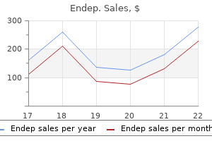
Endep packs: 30 pills, 60 pills, 90 pills, 120 pills, 180 pills, 270 pills, 360 pills
10 mg endep discount visa
The decrease limb rotates in utero medially 90 degrees treatment zone lasik endep 50 mg on line, while the higher limb rotates laterally ninety degrees symptoms 5 days after iui endep 25 mg cheap online. Thus, the limbs are a hundred and eighty diploma out of part with one another (knee anterior and large toe medial versus elbow posterior the extensor compartment in decrease limb comes anterior and the flexor compartment becomes posterior. The dorsal and ventral axial strains each reach the ankle joint, ventral reaches the medial facet. The blood provide to the decrease limb is derived from the lateral department of the fifth lumbar intersegmental artery, which the preaxial vein turns into the long saphenous vein, which drains into the femoral vein on the saphenous opening. Lower Limb the iliac crest terminates posteriorly because the posterior superior iliac spine, which is marked superficially by the gluteal Palpation places the posterior superior iliac spines in the vary of the L5�S1 vertebral junction to the S2 spinous course of the caudal restrict of the dura mater ranges from the L5�S1 junction to S4, with the bulk sitting at the decrease S1 to the S2 the termination of the dural sac is situated at imply of 31. Thigh: the inguinal ligament could be felt operating between the anterior superior iliac backbone and the pubic tubercle. Femoral Pulse Femoral artery pulse can be taken at mid-inguinal level (� 1 cm both side). Note: the midinguinal level is midway between the anterior superior iliac spine and the pubic symphysis. It is represented by the upper two third of a line joining the mid inguinal point to adductor tubercle. Note: Femoral vein has same markings as femoral artery except that the upper point is taken 1 cm medial to mid inguinal level and lower level 1 cm lateral to adductor tubercle. The third is a horizontal line from the tip of greater trochanter to the first line. Femoral artery � Halfway between the anterior superior iliac spine and the pubic symphysis lies the midinguinal level. Femoral artery � In slight flexion, abduction and lateral rotation of hip joint, a line drawn from the midinguinal level to the adductor tubercle represents the femoral artery. Bones Bones of the decrease limb embrace Hip (coxal/innominate) bone fashioned by the fusion of the ilium, ischium, and pubis. Foot region: Tarsal bones (talus, calcaneus, navicular, cuboid, and three cuneiform bones); metatarsals and phalanges (proximal, center, and distal). Hip Bone Hip bone Is formed by the fusion of the ilium, pubis, and ischiumin the acetabulum. Ilium forms the lateral part of the hip bone and consists of the physique, which joins the pubis and ischium to kind the acetabulum, and the ala or wing, which varieties the iliac crest. It comprises also the anterior superior iliac backbone, anterior inferior iliac spine, posterior iliac spine, higher sciatic notch, iliac fossa, and gluteal strains. Superior ramus which enters the formation of the acetabulum Inferior ramus which joins the ramus of the ischium, a part of the obturator foramen (formed by fusion of the ischium Ischium varieties the posteroinferior part of the acetabulum and the decrease posterior part of the hip bone. It consists of the Body which joins the ilium and superior ramus of the pubis to form the acetabulum Ramus which joins the inferior pubic ramus to kind the ischiopubic ramus. Acetabulum is an incomplete cup-shaped cavity on the lateral aspect of the hip bone during which the head of the femur articulates. It is fashioned by the ilium superiorly, the ischium posteroinferiorly, and the pubis anteromedially. It consists of the acetabular notch, which is bridged by the transverse acetabular ligament. The higher space of the tuberosity is further subdivided by an oblique line right into a superolateral half for semimembranosus and an inferomedial half for the lengthy head of biceps femoris and semitendinosus. The lower area is subdivided by an irregular vertical ridge into lateral and medial areas. The medial area is covered by fibroadipose tissue that normally contains the sciatic bursa of gluteus maximus, which helps the physique in sitting. The higher and decrease borders of gluteus maximus muscle are indicated by thick purple lines Sacrotuberous ligament (runs from the sacrum to the ischial tuberosity) and sacrospinous ligaments (runs from the sacrum to the ischial spine) convert the higher and lesser sciatic notches of the hip bone into larger and lesser sciatic foramina, the two necessary exits from the pelvis. Sacrotuberous ligament is a broad band of fibrous tissue which extends from sides of the sacrum and coccyx to the medial facet of the ischial tuberosity. The lowest fibres of gluteus maximus are attached to it and the decrease a half of the ligament continue into the tendon of biceps femoris. The coccygeal branches of the inferior gluteal artery, the perforating cutaneous nerve and filaments of the coccygeal plexus pierce the ligament. Sacrospinous ligament extends from the ischial spine to the lateral margins of the sacrum and coccyx anterior to the sacrotuberous ligament. Greater sciatic foramen is bounded anterosuperiorly by the larger sciatic notch, posteriorly by the sacrotuberous ligament and inferiorly by the sacrospinous ligament and ischial backbone. Piriformis muscle cross through it, above which the superior gluteal vessels and nerve leave the pelvis. Other necessary structures as they exit the pelvic cavity to enter the gluteal and thigh areas: Superior gluteal vein, artery, and nerve; inferior gluteal vein, artery, and nerve; sciatic nerve. Adductor magnus � Posterior (hamstring) a half of adductor magnus takes origin from the ischial tuberosity. Origin of semimembranosus from superolateral area � Semi-membranosus arises by an extended, flat tendon from a superolateral impression on the ischial tuberosity. Obturator internus muscle � It is the tendon (not muscle) of obturator internus, which passes by way of lesser sciatic notch. Nerve to obturator internus � Pyriformis muscle move through higher sciatic notch. Coccygeal nerve � the sacrotuberous ligament is pierced by the coccygeal branches of the inferior gluteal artery, the perforating cutaneous nerve and filaments of the coccygeal plexus. It reveals the following options: Head varieties about two-thirds of a sphere and is directed medially, upward, and barely ahead to match into the acetabulum. It has a despair in its articular surface, the fovea capitis femoris, to which the ligamentum capitis femoris is connected. It is separated from the shaft in entrance by the intertrochanteric line, to which the iliofemoral ligament is attached. It supplies an insertion for the gluteus medius and minimus, piriformis, and obturator internus muscular tissues. It receives the obturator externus tendon on the medial side of the trochanteric fossa. It exhibits lateral and medial lips that present attachments for many muscular tissues and the three intermuscular septa. It is perforated slightly below its middle by the nutrient canal, which is directed obliquely upward. Pectineal Line runs from the lesser trochanter to the medial lip of the linea aspera. Adductor Tubercle is a small prominence at the uppermost part of the medial femoral condyle. Nutrient foramina are located within the area of in the linea aspera (are directed proximally) one is usually close to its proximal end and a second usually close to its distal end. The main nutrient artery to femur is normally derived from the second perforating artery (branch of profunda femoris If two nutrient arteries happen, they may branch from the primary and third perforators. Linea aspera is a outstanding longitudinal ridge or crest, on the middle third of the bone, presenting a medial and a lateral lip. It is a vital insertion level for the adductors and the intermuscular septa that divides the thigh into three compartments.
Buy endep 75 mg amex
Left border:- Left free margin of greater omentum;gastrosplenic medicine wheel colors purchase endep 25 mg overnight delivery, linorenal and gastrophrenic ligaments treatment 5th toe fracture endep 10 mg order online. Omental bursa � A posterior gastric ulcer may perforate into the lesser sac (omental bursa). Inferior liver � Epiploic foramen (foramen of winslow or aditus to lesser sac) is a slit like opening by way of which lesser sac communicates with higher sac. Its boundaries are:� Anterior:- Right free margin of lesser omentum (contains portal vein,hepatic artery proper and bile duct). Left subhepatic space � Lesser sac (Omental bursa) is left posterior intraperitoneal space, also known as left subhepatic house. Uterus and rectum � In females rectouterine pouch (pouch of Douglas) lies between rectum (posteriorly) and uterus and posterior fornix of vagina (anteriorly). Inferior mesenteric vein � Inferior mesenteric vein lies in the free edge of the peritoneal fold of paraduodenal recess. Intersigmoid recess � Intersigmoid recess is continually present in the foetus and in early infancy, however may disappear with age. Left ureter � Left ureter crossing the bifurcation of left widespread iliac artery lies behind intersigned recess, which is a surgical guide for locating left ureter. Middle colic artery Mesentery of gut Mesentery proper (Mesentery of small intestine) Transverse mesocolon Mesoappendix Sigmoid mesocolon Vessels contained by mesentery Jejunal and ileal branches of superior mesenteric vessels Middle colic vessel Appendicular vessels Sigmoid vessels 15. Fascia between the rectal ampulla and the prostate and the seminal vesicles � Posterior surface of prostate is separated from rectum by the fascia of Denonvilliers which is the obliterated rectovesical pouch of peritoneum. Right and left gastric arteries which provide the lesser curvature (abdominal aorta celiac trunk widespread hepatic Right and left gastroepiploic arteries which provide the larger curvature (abdominal aorta celiac trunk common artery right gastric artery; abdominal aorta celiac trunk left gastric artery). Short gastric arteries which provide the fundus (abdominal aorta celiac trunk splenic artery quick gastric arteries). Right and left gastric veins (right and left gastric veins portal vein hepatic sinusoids central veins hepatic veins Left gastroepiploic vein and short gastric veins (left gastroepiploic vein and short gastric veins splenic vein portal Right gastroepiploic vein (right gastroepiploic vein superior mesenteric vein portal vein hepatic sinusoids central veins hepatic veins inferior vena cava). Innervation the innervation of the stomach is by the enteric nervous system which in the abdomen consists of the myenteric plexus of Parasympathetic Preganglionic neuronal cell our bodies are located in the dorsal nucleus of the vagus. The enteric nervous system is modulated by the parasympathetic and sympathetic nervous systems. Sympathetic Preganglionic neuronal cell bodies are situated within the intermediolateral cell column of the spinal twine (T5 to T9). Postganglionic axons synapse within the advanced circuitry of the enteric nervous system. Gastric ulcers most often occur throughout the physique of the abdomen alongside the lesser curvature above the incisura angularis. Carcinomas of the stomach are most commonly found within the pylorus of the abdomen and will metastasize to supraclavicular lymph nodes (Virchow nodes) on the left facet which can be palpated within the posterior triangle of the neck. The quick gastric arteries, left gastroepiploic artery and, when current, the posterior gastric artery are branches of the splenic artery. The right gastric artery and proper gastroepiploic artery arise from the hepatic artery and its gastroduodenal branch, respectively. Stomach � Main gastric nerve of Latarjet is branch of vagus and supplies the abdomen. All of the above � Arterial provide of abdomen is as follows � Along lesser curvature: Left gastric artery (branch of coeliac trunk) and proper gastric artery (branch of correct hepatic artery). Left gastric artery � he persistently largest artery to the abdomen is left gastric artery. These structures forming abdomen bed are (i) Diaphragm,(ii) left kidney, (iii) left suprarenal (adrenal) gland, (iv) pancreas (body), (v) transverse colon, (vi) splenic flexure of colon and (vii) splenic artery. Pre aortic nodes Lymphatic drainage of stomach � the abdomen can be divided into 4 lymphatic territories. Lymph vessels from these nodes travel along the splenic artery to reach the coeliac nodes. Lymph vessels arising in these nodes drain into the primary and second elements of the duodenum. From right here the lymph is drained additional into the hepatic nodes that lie alongside the hepatic artery; and eventually into the coeliac nodes. From here it passes via the intestinal lymph trunk to attain the cisterna chyli. Superior Part (First Part) the primary 2 cm of the superior half is intraperitoneal and due to this fact has a mesentery and is mobile; the remaining distal 3 cm of the superior part is retroperitoneal. Radiologists check with the primary 2 cm of the superior part of the duodenum because the duodenal cap or bulb. The superior half begins at the pylorus of the stomach (gastroduodenal junction) which is marked by the prepyloric vein. The hepatoduodenal ligament attaches superiorly and the greater omentum attaches inferiorly. Descending Part (Second Part) the descending half is retroperitoneal and receives the common bile duct and primary pancreatic duct on its posterior/ medial wall on the hepatopancreatic ampulla (ampulla of Vater). In extreme belly accidents, this part of the duodenum could additionally be crushed in opposition to the L3 vertebra. Ascending Part (Fourth Part) the ascending part is intraperitoneal and ascends to meet the jejunum on the duodenojejunal junction which occurs roughly on the L2 vertebral level about 2 to 3 cm to the left of the midline. This junction usually types an acute angle which is known as the duodenojejunal flexure which is supported by the ligament of Treitz (represents the cranial end of the dorsal mesentery). The arterial provide of the duodenum is from the next: Supraduodenal artery which provides the upper portion of the duodenum (abdominal aorta celiac trunk frequent hepatic artery gastroduodenal artery supraduodenal artery). Anterior and posterior superior pancreaticoduodenal arteries (abdominal aorta celiac trunk frequent hepatic artery gastroduodenal artery anterior and posterior superior pancreaticoduodenal arteries). Anterior and posterior inferior pancreaticoduodenal arteries (abdominal aorta superior mesenteric artery anterior and posterior inferior pancreaticoduodenal arteries). The venous drainage of the duodenum is to the next: Anterior and posterior superior pancreaticoduodenal veins (anterior and posterior superior pancreaticoduodenal veins portal vein hepatic sinusoids central veins hepatic veins inferior vena cava). Anterior and posterior inferior pancreaticoduodenal veins (anterior and posterior inferior pancreaticoduodenal veins superior mesenteric vein portal vein hepatic sinusoids central veins hepatic veins inferior vena cava). Clinical Considerations Duodenal ulcers most often happen on the anterior wall of the first a half of the duodenum. Perforations of the duodenum happen most often with ulcers on the anterior wall of the duodenum. However, posterior wall perforations may erode the gastroduodenal artery causing extreme hemorrhage and prolong into the pancreas. Different arteries of the duodenum derived instantly or not directly from the above two arteries are: 1.
25 mg endep generic free shipping
Spinal twine harm is an infrequent but devastating complication of anterior cervical spine surgical procedure with a reported incidence of 0 symptoms 4 weeks 75 mg endep buy overnight delivery. A lately revealed European study with as a lot as 97110 treatment code 50 mg endep generic otc 8-year follow-up demonstrated no instances of anteroposterior migration higher than three mm or subsidence greater than 2 mm at any time level. During the anterior cervical strategy, the cervical sympathetic trunk lies at an average distance of 11. Reported charges of secondary surgical procedure on the index level following cervical disc arthroplasty at 2-year follow-up vary from 1. There have been two known circumstances of aseptic osteolysis; one case was in an asymptomatic patient in whom osteolysis of the adjoining vertebral our bodies was detected throughout routine follow-up radiographs three years postoperatively, and the second case was in a patient who developed a recurrent radiculopathy 5 years postoperatively secondary to osteolysis with associated reactive hypertrophic bone formation that resulted in nerve root compression. Histological studies of explanted CoCr alloy metal-onmetal lumbar units present tissue necrosis and lymphocytedominated inflammatory responses with no proof of osteolysis. Histologically, the tissue response is characterised by a mononuclear persistent inflammatory response just like that observed with other stainless steel surgical devices and has not been associated with tissue necrosis, osteolysis, or tissue degeneration. A case report has been printed describing rupture of the polyurethane sheath eight years after implantation of a Bryan cervical disc. When this was explored surgically, a crack was discovered within the polyurethane sheath and metal particles was current within the surrounding tissues along with vital soft-tissue irritation. Arthroplasty with intercorporal endoprosthesis in herniated disc and in painful disc. A medical evaluation of 4and 6-year follow-up results after cervical disc replacement surgical procedure using the Bryan Cervical Disc Prosthesis. With that mentioned, cervical disc arthroplasty has particular problems inherent to the design 134 Kim et al. Anterior discectomy without fusion for remedy of cervical lateral gentle disc extrusion: a follow-up of a hundred and twenty cases. Stretch-induced nerve harm as a reason for paralysis secondary to the anterior cervical method. Incidental durotomy during spine surgical procedure: incidence, management and problems. Airway obstruction attributable to cerebrospinal fluid leakage after anterior cervical backbone surgery. Cerebrospinal fluid fistula secondary to dural tear in anterior cervical discectomy and fusion: case report. Can airway problems following multilevel anterior cervical surgical procedure be avoided Cervical myelopathy after cervical whole disc arthroplasty: case report and literature evaluation. Vertebral physique cut up fracture after a single-level cervical whole disc substitute. Posterior avulsion fracture at adjoining vertebral physique during cervical disc substitute with ProDisc-C: a case report. Part 2 Thoracolumbar 21 Lumbar Pedicle Screw Complications 22 Junctional Breakdown in Pedicle Screw Constructs 23 Hook Complications within the Thoracic and Lumbar Spine 24 Sublaminar Wiring Complications 25 Complications of Percutaneous Vertebral Cement Augmentation 26 Complications of Vertebral Body Implants Introduced via the Posterolateral Approach 27 Complications of Vertebral Body Replacement Cages 28 Complications of Anterior Thoracic Instrumentation Systems 29 Interspinous Spinous Process Fusion Plate Complications one hundred forty 146 152 156 158 one hundred sixty five 169 173 183 30 Complications of Cortical Screw Fixation 188 31 Complications of Posterior Screw Fixation in Spine Surgery 32 Interspinous Spacer Complications 33 Complications of PresacralApproach�Based Fusion Devices 194 203 208 34 Complications of Posterior and Transforaminal Lumbar Interbody Fusion 214 35 Complications of Open Transforaminal Lumbar Interbody Fusion 230 Kim et al. In 2001, a total of 122,000 lumbar fusions were carried out within the United States,6 which represented a 220% enhance in variety of fusion per a hundred,000 in 1990. This article will evaluate the related lumbar anatomy, techniques for screw insertion, and complications related to their placement. Image-guided pedicle screw methods have also emerged as a means for correct placement of instrumentation within the lumbar spine. The pars interarticularis is a vital osseous landmark, because the lateral pars provide a tough estimate to the medial wall of the corresponding pedicle. The internal diameter of the pedicle can vary significantly from L1 to L5, with diameter sometimes growing from proximal to distal. Different beginning points have been advised for the rationale that introduction of transpedicular lumbar fixation. Roy-Camille et al recognized the start line because the intersection of the middle parts of the transverse course of and facet joint. Variations in pedicle morphology, obesity, degenerative modifications, and three-dimensional deformity can all complicate pedicle screw placement. Despite improvements in surgical approach, pedicle screw malposition is a relatively widespread incidence even in the hands of experienced surgeons. Reported rates of screw malposition differ considerably in the in vitro cadaveric versus the in vivo medical setting starting from 5. The relative risk of screw malposition was statistically larger with anatomic insertion techniques when in comparability with image-guided strategies. There are presently a complete of 5 potential in vivo research evaluating the accuracy of pedicle screw placement within the lumbar spine. Lumbar Pedicle Screw Complications Laine et al prospectively evaluated the accuracy of lumbar pedicle screw placement in 30 consecutive sufferers using a computer-assisted navigation technique. All of the pedicle perforations utilizing the computer-assisted technique were lateral, while freehand screw placement resulted in three medial perforations, one lateral, and one inferior. This finding is in preserving with that of Gelalis et al, who reported an inclination towards medial breach with freehand methods compared to lateral breach with image-guided methods. No surprise that probably the most tough screws to place had the next fee of cortical breach. Laine et al performed one other potential randomized in vivo management trial, evaluating the accuracy of pedicle screw insertion with and with out computer-assisted navigation in a consecutive series of ninety one sufferers. This contrasts the 10 misplaced screws using the computer-assisted approach with 9 screws lateral (90%) and 1 medial breach. Castro et al prospectively evaluated 30 consecutive patients who obtained instrumented lumbar fusion. Image-guided methods do persistently exhibit a decrease rate of misplaced screws when compared to freehand conventional strategies. Factors that influence the scientific consequence of screw misplacement embody pedicle location, course, and diploma of cortical violation. Direct dural repair in this setting may be difficult due to the ventral lateral location of the durotomy. Direct restore may be performed by a selection of methods with nobody particular method proven superior. Improvement within the locking mechanisms on the screw�rod interface has resulted in much less rod migration. Technically, the surgeon should remember to finally tighten the screws using an anti-torque system. Larger diameter screws and anterior column help are two other components surgeons should consider when performing multilevel fusion procedures, especially when crossing the lumbosacral junction. The use of a cortical trajectory for posterior instrumentation has also be advocated as a method to increase pullout strength in osteoporotic sufferers.
Endep 50 mg proven
Endovascular treatment of an iatrogenic thoracic aortic harm after spinal instrumentation: case report medicine logo 50 mg endep buy with mastercard. The pearls and tips help the surgeon with a global image medications with dextromethorphan cheap endep 10 mg overnight delivery, on condition that limiting complication danger for instrumentation failure includes the necessity to perceive both anatomy and approach info. Stackable carbon fiber cages for thoracolumbar interbody fusion after corpectomy: long-term consequence evaluation. Minimally invasive, extracavitary strategy for thoracic disc herniation: technical report and preliminary results. Minimally invasive extracavitary approach for thoracic discectomy and interbody fusion: 1-year medical and radiographic outcomes in 13 patients in contrast with a cohort of conventional anterior transthoracic approaches. Thoracic aortic dissection and mycotic pseudoaneurysm within the setting of an unstable higher thoracic sort B2 fracture case report. Interspinous Spinous Process Fusion Plate Complications 29 Interspinous Spinous Process Fusion Plate Complications Andrew H. Advances in materials and minimally invasive surgical strategies have renewed curiosity in implantable devices for interspinous fusion. Such constructs may be used alone to facilitate posterolateral fusion or could additionally be used at the aspect of interbody techniques for achievement of anterior and posterior fusion. In addition, the midline paraspinal aponeurosis is a comparatively avascular plane that decreases the probability of serious blood loss or vascular injury in the course of the procedure. A variety of different potential benefits to interspinous distraction have been posited, together with elevated disc house height and reduced side contact strain. The spinous processes and medial borders of the side joints are uncovered and decorticated. Risk factors for certain potential complications may be extrapolated from biomechanical testing knowledge, such because the potential for acute fracture during implantation. To harness the potential for interspinous distraction with gadget implantation, a variable quantity of resistance is encountered throughout device insertion and seating. A related trend towards elevated rates of spinous fracture in sufferers with low bone mineral density has additionally been observed clinically following interspinous spacer implantation. Lindsey et al found that dynamic interspinous spacer implantation produced an isolated reduction of flexion�extension at the instrumented level with out important effects on vary of motion at the cranial or caudal adjacent ranges. An equivalent and statistically vital improvement in medical outcome measures was seen in both groups at the ultimate follow-up. Both methods had an excellent safety profile, as no circumstances of major surgical complications, implant failure, or pseudoarthrosis occurred in either group over the 1- to 12-month follow-up period. For an isolated asymptomatic spinous course of fracture recognized by the way on follow-up imaging with out evidence of gadget migration or impending failure, no immediate intervention is required. Painful fractures of the posterior parts might benefit from preliminary therapy with analgesia and bracing as needed for consolation, as a major proportion of such fractures will heal spontaneously. For painful nonunions of the posterior components, surgical options range from fragment excision to device removing and revision instrumentation as needed to obtain enough stability to allow fusion. Any progressive neurologic deficit prompts quick analysis and pressing decompression with instrumentation and fusion as wanted to restore stability. Historic approaches to interspinous process fusion have been surpassed in reputation by the supply of posterior pedicle screw�rod instrumentation. Biomechanics of a lumbar interspinous anchor with transforaminal lumbar interbody fixation. Biomechanical effect of various interspinous gadgets on lumbar spinal range of movement beneath preload situations. Biomechanics of a lumbar interspinous anchor with anterior lumbar interbody fusion. Invited submission from the Joint Section Meeting on Disorders of the Spine and Peripheral Nerves, March 2005. The positional magnetic resonance imaging modifications within the lumbar backbone following insertion of a novel interspinous process distraction gadget. Biomechanical comparison of an interspinous gadget and a inflexible stabilization on lumbar adjacent segment vary of movement. Posterior interspinous fusion system for one-level fusion in degenerative lumbar spine disease: comparison with pedicle screw fixation - preliminary report of at least one 12 months follow up. Invited submission from the Joint Section Meeting On Disorders of the Spine and Peripheral Nerves, March 2005. Because the starting point for cortical screws is positioned inside this area, the anatomic landmarks for cortical screws are usually extra consistent than these used for pedicle screws, significantly in sufferers with important facet hypertrophy. With their lateral-to-medial angulation, pedicle screws inserted via an open strategy require an extensile exposure, which produces a significant iatrogenic insult to the surrounding soft tissues, increases blood loss, and prolongs the operative time such that these people could often expertise severe postoperative pain and useful disability. There can be an inherent risk of neurologic harm with misdirected implants, significantly since their trajectory places them in close proximity to each the nerve roots in the foramina in addition to the thecal sac within the central canal. The authors demonstrated that cortical screws exhibited larger pullout strength which they attributed to the upper density of the bone surrounding these implants. Mobbs et al, in a clinical review of cortical screws, detailed the less invasive nature of this surgical technique which makes use of a novel medial-to-lateral trajectory through the vertebra. The biomechanical characteristics of cortical instrumentation has been elucidated by a quantity of in vitro cadaveric studies and their higher pullout strength relative to pedicle screws was attributed to the upper mineral density of the encircling cortical bone across the implants. Cortical screws may confer numerous other advantages on account of their distinctive trajectory. For occasion, the superolateral angulation of those screws away from the thecal sac and exiting nerve root doubtless reduces the risk of neural injury in comparison with the usage of transpedicular fixation. The cortical pedicle fixation (b) makes use of a caudal to cranial upward angulation to optimize buy in the dense cortical bone of the pars, inferior pedicle, and vertebral endplate. The typical starting point for cortical screws is at the intersection of a horizontal line along the inferior border of the transverse process and a vertical line bisecting the pars interarticularis, no more than 3 mm from its Kim et al. The cortical screws (a) have a novel pitch, thread design, and tapered root diameter taper to maximize cortical bone purchase at the posterior facet of the screw and cancellous buy on the distal tip of the screw. Once the trajectory has been verified in a number of planes, the drill is advanced via the posterior elements in a superior and lateral path till it reaches the right depth within the cancellous bone of the vertebral physique, immediately below the extent of the endplate. Because these screws should traverse dense cortical bone, it is suggested that the holes be tapped "line to line". In general, less exposure of the lateral compartment and intertransverse region is required with cortical screws in comparability with conventional pedicle screws. The favorable biomechanical profile of cortical fixation has been established by a quantity of studies, however these constructs may still be subject to implant loosening or catastrophic failure indicative of a pseudarthrosis. As with all spinal procedures, most circumstances of infection or different wound issues might be evident upon inspection of the incision, however within the absence of any obvious indicators or symptoms, laboratory testing or further imaging can also serve to establish the diagnosis. Likewise, patients who develop medical or radiographic findings according to a symptomatic nonunion may be candidates for revision fusion procedures with removal of the loose hardware, replacement 192 Kim et al. Complications of Cortical Screw Fixation with pedicle screws, and probably even supplementation of the assemble with anterior column help or different stabilizing techniques if indicated.

Buy endep 75 mg mastercard
Difference within the top of the best and left ethmoidal roofs: a potential danger issue for ethmoidal surgical procedure medicine 5658 order endep 25 mg visa. Height and shape of the cranium base as danger factors for cranium base penetration throughout endoscopic sinus surgical procedure treatment 101 purchase 25 mg endep. Lateral retropharyngeal node metastasis from carcinoma of the upper gingiva and maxillary sinus. Anatomical variation and morphology in the place of the palatine foramina in adult human skulls from Greece. Bilateral nasal squamous carcinoma arising in papillomatosis: report of a case growing after chemotherapy for leukemia. Craniofacial resection for malignant paranasal sinus tumors: report of a global collaborative examine. Patterns of dural involvement in sinonasal tumors: prospective correlation of magnetic resonance imaging and histopathologic findings. European Rhinologic Society Advisory Board on Endoscopic Techniques within the Management of Nose, Paranasal Sinus and Skull Base Tumours. European place paper on endoscopic administration of tumours of the nostril, paranasal sinuses and skull base. The sinonasal tract: one other potential "hot spot" for carcinomas with transcriptionally-active human papillomavirus. Endonasal endoscopic surgery for squamous cell carcinoma of the sinonasal cavities and skull base: oncologic outcomes primarily based on remedy strategy and tumor etiology. Incidence and survival in patients with sinonasal most cancers: a historic evaluation of population-based knowledge. Update on sinonasal adenocarcinoma: classification and advances in immunophenotype and molecular genetic make-up. Sinonasal undifferentiated carcinoma: clinical and pathologic features and a dialogue on classification, cellular differentiation, and differential analysis. Promising outcomes with chemoradiation in sufferers with sinonasal undifferentiated carcinoma. Salivary adenoid cystic carcinoma in Denmark 1990�2005: consequence and impartial prognostic elements together with the good thing about radiotherapy. Sinonasal tract mucoepidermoid carcinoma: a clinicopathologic and immunophenotypic examine of 19 cases mixed with a comprehensive review of the literature. Multidisciplinary treatment of olfactory neuroblastoma: patterns of failure and administration of recurrence. Induction of olfactory neuroepithelial tumors in Syrian hamsters by diethylnitrosamine. Activation of retrovirus in transgenic mice: affiliation with development of olfactory neuroblastoma. Perineural unfold of malignant melanoma of the pinnacle and neck: scientific and imaging options. Treatment outcome and sample of failure in seventy seven sufferers with sinonasal pure killer/T-cell or T-cell lymphoma. Osteosarcoma of the jaw/craniofacial area: outcomes after multimodality therapy. Head and neck osteosarcoma in adults: the province of Alberta expertise over 26 years. The role of the human papillomavirus within the pathogenesis of Schneiderian inverted papillomas: an analytic overview of the proof. Invasive development patterns of juvenile nasopharyngeal angiofibroma: radiological imaging and medical implications. Primary intraosseous meningioma of orbit and anterior cranial fossa: a case report and literature evaluate. Hyperostosis related to meningioma of the cranial base: secondary changes or tumor invasion. Endoscopic endonasal versus open transcranial resection of anterior midline skull base meningiomas. Long-term tumor control of benign intracranial meningiomas after radiosurgery in a collection of 4565 sufferers. Inducible cyclooxygenase and interleukin 6 gene expressions in nasal polyp fibroblasts: possible implication within the pathogenesis of nasal polyposis. Computed tomography and magnetic resonance diagnosis of allergic fungal sinusitis. An strategy to fulminant invasive fungal rhinosinusitis in the immunocompromised host. Fibrous dysplasia and aneurysmal bone cyst of the cranium base presenting with blindness: a report of a rare regionally aggressive instance. Surgery versus watchful waiting in sufferers with craniofacial fibrous dysplasia�a metaanalysis. The many faces of granulomatosis with polyangiitis: a evaluate of the top and neck imaging manifestations. Ocular manifestations of systemic illness: antineutrophil cytoplasmic antibody-associated vasculitis. Otolaryngological development of granulomatosis with polyangiitis after systemic therapy with rituximab. Conversion of the mesenchyme into cartilage at the cranium base begins across the fortieth day of gestation. The various foramina of the cranium base are current within this cartilaginous formation because the primitive nerves develop previous to chondrification and the cartilage develops around them. During the 5th week of gestation, the notochord passes into the basiocciput from the upper cervical vertebral bodies. It then passes obliquely through the basiocciput, exiting ventrally to are out there in contact with the primitive pharynx, and then again into the basisphenoid and terminates just caudal to the pituitary fossa at the dural margin. It passes by way of the basal mesoderm just cephalad to the tip of the notochord to meet the precursor of the posterior pituitary, which forms from a diverticulum of the diencephalon. More rostrally, the paired presphenoid cartilages fuse to turn into probably the most anterior portion of the sphenoid bone. However, remaining cartilage may be found in numerous synchondroses and persists into adult life, mostly in the foramen lacerum and petroclival junction. Skull base chondrosarcomas are thought to largely come up from remnants of embryonal cartilage. It is bounded anteriorly by the tuberculum sellae and posteriorly by the dorsum sellae. Anterior to the tuberculum sellae is the prechiasmatic sulcus and the planum sphenoidale. The sella turcica is surrounded by four bony projections, the anterior and posterior clinoid processes. The dorsum sellae is contiguous posteroinferiorly with the clivus through the spheno-occipital synchondrosis. The optic canal is located above and is separated from the superior orbital fissure by the optic strut, and transmits the optic nerve and the ophthalmic artery.
Endep 75 mg generic free shipping
Note the very heterogeneous appearance of the mass and the considerable adjacent brain edema treatment enlarged prostate cheap endep 10 mg amex. Giving the surgeon an estimate as to the degree of tumor vascularity as evidenced by the diploma of distinction avidly and enhancement could predict the next likelihood of excessive bleeding at surgical procedure medicine tablets cheap endep 10 mg fast delivery. Other options to notice are the presence of brainstem compression and secondary obstructive hydrocephalus. During surgical procedure you will want to excise the tumor, dural tail, and any concerned bone to decrease the danger of a recurrence. The mass shows avid enhancement and each images also reveal the presence of large intralesional vessels indicating a very well-vascularized tumor. More recently, 5 fraction approaches are more and more practiced (25�30 Gy) as a method to allow for the benefits of fractionation in a shorter time course to enhance patient convenience. A margin extending into the parenchymal tissue is needed to cut back the chance of recurrence. They are more predominant in females (male-to-female ratio is 1:2) and usually current within the fifth to seventh a long time of life. Despite this, the bony foraminal margins are still higher defined than with paraganglioma. These meningiomas will show minimal vascularity and can lack the standard extended cloudlike blush of enhancement. One also needs to assess for possible extension to involve the mastoid section of the facial nerve. The presence of serious brainstem compression must be communicated urgently, as it might necessitate emergent surgery. Inferior extension into the nasopharyngeal carotid area with a dumbbell morphology. These lesions can grow into the hypotympanum, via the jugular vein and likewise into the posterior fossa. Injury to a dominant sigmoid sinus/jugular bulb throughout surgical procedure would possibly end in thrombosis and cerebral venous hypertension. The affected person has to be well informed concerning the potential complications of the surgical procedure which could possibly be worse than nonresection. Other treatment options include radiation or surgical debulking with a deliberate subtotal tumor removal for any compressive symptoms. This jugular fossa meningioma mass extends inferiorly along the post-styloid parapharyngeal space and protrudes intracranially into the posterior fossa causing mass impact on the cerebellum. This can mirror a mix of reactive hyperostosis and/or tumor infiltration. The sagittal image shows the tumor monitoring along the dura in the posterior fossa. Radiation is related to long-term native tumor management with out the related morbidities of surgical procedure. Atypical and malignant meningiomas require higher doses and are treated with stereotactic fractionated radiotherapy (60�70 Gy over 6�7 weeks). In those presenting within the subacute stage of denervation, the presence of tongue fasciculation may be a dominant symptom. There is an expansile transforming of the right lateral clivus and the adjacent occipital bone. Axial (d), coronal (e), and sagittal (f) enhanced T1-weighted photographs show a heterogeneously enhancing mass. The giant extracranial part extends down the proper poststyloid parapharyngeal area with displacement of the interior carotid artery anteriorly. Large hypoglossal schwannomas can have sizable intracranial and extracranial components. Care ought to be taken when assessing for putative hypoglossal schwannoma, as uneven enhancement in the hypoglossal canal can be seen as a end result of the presence of regular sturdy venous drainage or rarely a persistent hypoglossal artery. Typically, that is delivered as stereotactic fractionated radiotherapy (50�54 Gy in 5�6 weeks of 1. Fractionated stereotactic radiation gives rise to a low risk of cranial neuropathy (~5% or less) and achieves longterm tumor control of roughly 80�90% at 10 years. Other signs embody facial nerve palsy (8%), pulsatile tinnitus (50�92%), and vertigo (20�62%). Gradient-weighted or susceptibility-weighted imaging will present areas of internal low signal comparable to the presence of residual bone fragments and or blood merchandise. One should also assess for the presence of other lesions concerned in the context of von HippelLindau syndrome (such as cerebellar, spinal, or retinal hemangioblastomas and choroid plexus papillomas). Intratumoral calcific spicules and posterior rim calcification are reported to be present in one hundred pc of the circumstances. Foci of high sign Prognosis and Treatment If the tumor is sluggish rising, then watchful waiting can be adopted. If the tumor is quickly growing, then intervention ought to be considered to prevent further listening to loss. Axial T1- (b), T2- (c), and postcontrast T1-weighted (d) photographs show a corresponding enhancing lesion causing mass effect on the adjoining cerebellum and in addition extending into the vicinity of the left dural sigmoid sinus. Imaging Imaging of metastatic disease can be fairly variable leading to solitary, multifocal, or diffuse illness. If the metastasis provides rise to a sclerotic bony response, the lesions will present very low T1 and T2 signal. Fat-saturated contrast-enhanced T1-weighted images will present an enhancing bone marrow mass with or with out extraosseous gentle tissue part. The location of the metastatic disease may be bony, dural primarily based, leptomeningeal, or a mixture of any of those places. Skull base bony metastases from extracranial primaries happen in 4% of cancer sufferers. In kids and younger adults, histiocytosis and embryonal rhabdomyosarcoma also need to be thought-about. Of observe, malignant otitis externa and primary cranium base osteomyelitis can have an aggressive appearance and mimic a malignancy. Leptomeningeal carcinomatosis is the malignant infiltration of the pia, arachnoid, or each. The leptomeninges (arachnoid) extend alongside the cisternal and canalicular segments of the facial and vestibulocochlear nerves and metastases can seed into these places. Intracranial metastases have gotten more and more common as the survival price of cancer patients is bettering. There are metastatic lesions located in the left jugular fossa and laterally in the left petrous bone (long arrow; a). There are in depth permeative erosive adjustments within the left petroclival junction with erosion of the left jugular tubercle and extension into the left hypoglossal canal (short arrow; b). Multilevel sclerotic metastases to the cervical and higher thoracic vertebral bodies are additionally current (red arrows; c).
Order endep 75 mg without prescription
Table 19: Arrangement of structures at the lung hilum Right side Left aspect Pulmonary artery Left principal bronchus Inferior pulmonary vein � Eparterial � Pulmonary artery � Hyparterial bronchus � Inferior pulmonary vein In situs solitus symptoms 0f pneumonia endep 75 mg generic on-line, the right principal bronchus is short and eparterial (its department for the proper higher lobe lies over the second branch of the right pulmonary artery) and the left principal bronchus is longer and hyparterial (it programs beneath the left pulmonary artery) medical treatment 80ddb generic endep 25 mg online. In situs ambiguus, the bronchi and lungs can display both a bilateral proper morphology with bilateral trilobed lungs and bilateral eparterial bronchi (heterotaxy syndrome: incomplete or inappropriate lateralization of the thoracic and stomach viscera, and asplenia) or a bilateral left morphology with bilateral bilobed lungs and bilateral hyparterial bronchi (heterotaxy syndrome and polysplenia). Bronchopulmonary Segments 596 the bronchopulmonary section is the anatomical, practical, and surgical unit of the lungs. Thorax It is the wedge shaped largest subdivision of a lobe, named based on the segmental bronchus supplying it, and is surgically resectable. It incorporates a segmental (tertiary or lobular) bronchus, a department of the pulmonary artery, and a branch of the bronchial artery, which run collectively through the central part of the phase, surrounded by a delicate connective tissue (intersegmental) septum. The tributaries of pulmonary veins are intersegmental and lie at the margins of bronchopulmonary segments. They create surgical planes, which a surgeon can observe for segmental resection, with minimal tissue harm. Apical and posterior of left upper lobe usually mix into apicoposterior section. Anterior and medial basal of left lower lobe often combine into anteromedial basal phase. Clinical Correlations � Foreign physique aspiration: � Location of the foreign body aspirate depends upon the posture of the patient and decided by the gravity factor as nicely (most dependent location). Pulmonary System of Arteries Pulmonary Trunk extends upward from the conus arteriosus of the best ventricle of the guts and carries deoxygenated blood to the lungs for oxygenation. It passes superiorly and posteriorly from the entrance of the ascending aorta to its left aspect for and bifurcates into the best and left pulmonary arteries throughout the concavity of the aortic arch at the degree of the sternal angle (approximately). Left pulmonary artery is shorter and narrower than the right and is connected to the arch of the aorta by the ligamentum arteriosum. Pulmonary arteries department to follow the airways to the level of the terminal bronchioles, at which point they kind a pulmonary capillary plexus. Bronchial System of Arteries Bronchial arteries carry oxygenated blood to the lung and the visceral pleura. Bronchial arteries branch to observe the airways until the extent of the respiratory bronchioles, at which point they drain into the pulmonary capillary plexus. Pulmonary System of Veins Pulmonary veins carry oxygenated blood from the pulmonary capillary plexus and deoxygenated bronchial blood to the left atrium. Within the lung pulmonary veins are intersegmental (found on the periphery of the bronchopulmonary segments), not accompanied by branches of bronchi, pulmonary arteries, or bronchial arteries. Bronchial System of Veins Bronchial veins carry deoxygenated blood from the bronchial arteries that supply giant bronchi. Right bronchial veins drain into the azygos vein, left into the accent hemiazygos vein (or the left superior intercostal vein). Thorax They run along the bronchiole and bronchi toward the hilum to drain into the pulmonary after which bronchopulmonary nodes, which in flip drain to the inferior (carinal) and superior tracheobronchial nodes, the tracheal (paratracheal) nodes, bronchomediastinal nodes and trunks, and finally to the thoracic duct (on the left) and proper lymphatic duct (on the right). Nerve Supply Pulmonary plexuses are anterior and posterior to the opposite buildings at the hila of the lungs. They are fashioned by cardiac branches from the second to fifth (or sixth) thoracic sympathetic ganglia and from the vagus and cervical sympathetic cardiac nerves. Vagus nerve carrying parasympathetic fibers that innervate the graceful muscle and glands of the bronchial tree are excitatory (bronchoconstrictor and secretomotor). Vagus additionally carry sensation of stretching of the lung during inspiration and is anxious within the reflex control of respiration. Sympathetic fibers innervate blood vessels, clean muscle, and glands of the bronchial tree are inhibitory (bronchodilator and vasoconstrictor). Breathing Breathing involves altering the thoracic volume by altering the vertical, transverse and anteroposterior diameters of the thorax. The external and internal intercostals, transversus thoracis, subcostalis, levator costarum, serratus posterior superior and serratus posterior inferior can elevate or depress the ribs, and hence can act as accent muscles of ventilation. Inspiration entails muscular tissues (table) that elevate the thoracic cage (ribs and sternum) and accordingly, expiration includes the melancholy of the cage. Diaphragm is the chief muscle of inspiration, pulls the dome inferiorly into the stomach, thereby rising the vertical diameter of the thorax and expansion of lung (air moves in). Enlargement of the pleural cavities and lungs reduces the intrapulmonary pressure (creates a unfavorable pressure), thus allowing air to rush into the lungs passively due to atmospheric strain. Inspiration involves varied muscular tissues causing elevation of the ribs (superolateral movement), with the sternum transferring anteriorly. It leads to elevated transverse and anteroposterior diameters of the thoracic cavity and belly quantity is decreased with an elevated belly pressure. Elevation of upper 6 ribs causes sternum to be pushed ahead and upward, which increases the anteroposterior diameter of the thorax and lungs broaden (inspiration). The lower ribs elevate by swinging upward and laterally leading to an increase in the transverse (lateral) diameter of the thorax for lung expansion (inspiration). It involves leisure of the diaphragm and other muscle tissue; lower in thoracic volume; and enhance in the intrathoracic stress. Elastic recoil of the lungs produces a subatmospheric pressure within the pleural cavities and the air is expelled. Forced expiration requires contraction of the anterior stomach muscle tissue and few others Table 22: Respiratory muscles Type of respiration Inspiration (rib elevators) Quiet respiration � Diaphragm (chief muscle) � External intercostal muscles Deep inspiration � External intercostal muscular tissues � Scalene muscles � Sternocleidomastoid � Levator costarum � Serratus posterior superior � Innermost intercostal Forced expiration � Levator scapulae � Trapezius � Rhomboids � Pectoral muscle tissue � Serratus anterior Expiration (rib depressors) Passive Passive � � � � � Anterior abdominals* Quadratus lumborum Internal intercostal (costal part) muscles** Transverse thoracis Serratus posterior inferior Anterior abdominals are: External and inner oblique muscle tissue along with transversus abdominis. Latissimus dorsi Accessory muscular tissues of inspiration are elevators of rib and improve the diameters of thoracic cavity. Internal intercostal Muscles of expiration are depressors of rib and decrease the diameters of thoracic cavity. Internal intercostal (costal part) helps in expiration, whereas the interchondral half participates in inspiration. Diaphragm is the chief muscle of inspiration, assisted by external intercostal and serratus anterior. Serratus posterior inferior Muscles that elevate the ribs work for inspiration, whereas serratus posterior inferior depress the ribs (for expiration). The line lies 3�4 cm above the anterior finish of the primary rib; the surface marking is posterior to the medial third of the Anterior border of pleura: the proper and left pleurae are in contact retrosternally in the midline from the second to the fourth costal cartilages, at which point they diverge. The pleura on the proper descends vertically to the xiphisternum, whereas the pleura on the left deviates laterally by 3�5 cm after which passes inferiorly to cross the anterior finish of the sixth rib. This deviation produces an area between the center and the sternum that is free of pleura; a needle puncture Inferior border of pleura: the costodiaphragmatic reflections of the best pleurae follows across the chest wall from the midpoint of xiphisternal angle anteriorly to the eighth rib within the mid-clavicular line, tenth rib within the mid-axillary line and twelfth rib at paravertebral line. It is type of the same for left lung but with one exception: It begins from the sixth rib/costal cartilage anteriorly to the eighth rib in the mid-clavicular line, tenth rib in the mid-axillary line and twelfth rib at paravertebral line. Posterior border of pleura: In posterior view, the medial edges of the pleurae lies along a line joining the transverse processes of the second to the twelfth thoracic vertebrae on either side.
25 mg endep discount otc
Posterior migration of cervical spinal cord between cut up laminae as a complication of laminoplasty symptoms copd buy cheap endep 50 mg line. The utility of somatosensory evoked potential monitoring during cervical spine surgery: how typically does it prompt intervention and affect consequence Outcomes after laminoplasty compared with laminectomy and fusion in sufferers with cervical myelopathy: a systematic evaluation medicine cabinets surface mount generic 25 mg endep otc. Open-door laminoplasty for cervical stenotic myelopathy: surgical method and neurophysiological monitoring. T-Saw laminoplasty for the management of cervical spondylotic myelopathy: scientific and radiographic consequence. C5 palsy after decompression surgery for cervical myelopathy: review of the literature. Cervical alignment and vary of motion after laminoplasty: radiographical data from greater than 500 cases with [16] [17] [18] [19] [20] [21] [22] [23] [24] [25] [26] [27] 72 Kim et al. Complications Related to Cervicothoracic Instrumentation 12 Complications Related to Cervicothoracic Instrumentation Addisu Mesfin 12. Due to the switch from the flexible cervical backbone to the rigid segment, thoracic spine instrumentation failure can still be encountered. Indications for an anteriorbased instrumentation can be a disc herniation at C7�T1, unstable fracture, and a primary or metastatic lesion at the C7� T1 ranges. Posterior-based instrumentation is indicated when performing a protracted assemble in the cervical backbone corresponding to a C7 osteotomy for chin on chest deformity1 and for lengthy thoracic constructs ending at T1. Need anatomic diagram of Cervicothoracic junction, prior copyrighted work is okay 12. Following a discectomy, a big diameter allograft or two small allografts may be placed. A standalone threaded cage that screws into the disc area adopted by posterior-based stabilization can be an possibility. In a cadaveric examine, a C7 pedicle screw and T1 pedicle screw construct was found to be significantly stiffer than a C7 lateral mass assemble in axial compression, torsion, bending, and flexion. In certain cases, the domino constructs are simpler to place than the transitional rods. A C5 to T2 assemble, consisting of C7 pedicle screws, supplemented with one or two crosslinks was tested. If using a threaded cage, mesh cage, or expandable cage, dislodgment of the cage may also occur. Patton et al reported the position of preoperative metabolic evaluation of the patient and subsequent therapy if deficiencies are noted. Osteoporosis is a vital consideration for proximal junctional kyphosis in patients with thoracic constructs ending at T1 or T2. In these cases, problems associated with poor bone high quality such as pedicle screw or lateral mass screw backing out might occur. With a C7�T1 spondylolisthesis and a protracted posterior assemble, it might be advisable to end the assemble at T2 or T3. During the revision, the instrumentation should be extended a minimal of 1 stage and supplemented with anterior instrumentation and fusion. Following three-column injury, increased flexibility with the translaminar screws was noted. Seven years postoperatively, the patient is doing nicely with no changes in his instrumentation. The begin factors and placement of those screws would be technically simpler than the current pedicle screws of the subaxial spine. Cervicothoracic extension osteotomy for chin-on-chest deformity in ankylosing spondylitis. Surgical administration of cervical myelopathy coping with the cervical-thoracic junction. Surgical strategy to the cervicothoracic junction: can a regular Smith-Robinson method be utilized Cervicosternotomy as an anterior strategy to the higher thoracic and cervicothoracic spinal junction. Anterior surgical approaches to the cervicothoracic junction: when to use the manubriotomy Mechanical comparison of posterior instrumentation constructs for spinal fixation throughout the cervicothoracic junction. Biomechanical comparison of posterior cervicothoracic instrumentation techniques after one-level laminectomy and facetectomy. Posterior-only stabilization of 2-column and 3-column accidents at the cervicothoracic junction: a biomechanical study. Comparison of teriparatide and bisphosphonate treatment to scale back pedicle screw loosening after lumbar spinal fusion surgical procedure in postmenopausal ladies with osteoporosis from a bone high quality perspective. Degenerative spondylolisthesis of the cervical spine: analysis of 58 patients handled with anterior cervical decompression and fusion. Seven years of expertise with C2 translaminar screw fixation: medical sequence and evaluation of the literature. Surgical management of two- versus three-column accidents of the cervicothoracic junction: biomechanical comparison of translaminar screw and pedicle screw fixation utilizing a cadaveric model. Considerations for using C7 crossing laminar screws in subaxial and cervicothoracic instrumentation. The feasibility of laminar screw placement in the subaxial spine: evaluation utilizing 215 three-dimensional computed tomography scans and simulation software program. Clinical and radiologic end result of laminar screw at C2 and C7 for posterior instrumentation-review of 25 cases and comparison of C2 and C7 intralaminar screw fixation. Anterior C1�C2 Fusion Instrumentation Complications 13 Anterior C1�C2 Fusion Instrumentation Complications Jesse E. Finally, the availability of otolaryngology/general surgical procedure colleagues must be verified in case a prophylactic tracheotomy is needed postoperatively, if vital retropharyngeal dissection occurs. Fiberoptic nasotracheal intubation is then performed, and dental occlusion maintained throughout the case so to not limit the world of dissection. Halo or Gardner-Wells traction can then be positioned if needed for preoperative discount and/or intraoperative traction. If not Anterior Retropharyngeal Approach In 1957, Southwick and Robinson initially described the anterior retropharyngeal strategy to expose C3�T1,1 and later de Andrade and Macnab described a cranial extension permitting access to C1�C2. In the research of Batra et al, 50 facial halves had been dissected out to examine the branches of the facial nerve. The marginal mandibular department of the facial nerve was discovered superficial to the facial artery and vein in all specimens. Prior to incision, the earlobe is taped or sewn anterior to facilitate exposure of the sphere. For bilateral transarticular screws, the identical approach is also performed on the contralateral aspect.