

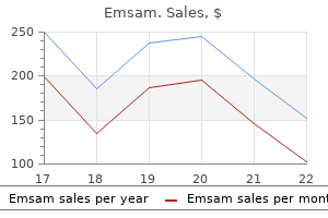
Emsam dosages: 5 mg
Emsam packs: 30 pills, 60 pills, 90 pills, 120 pills, 180 pills, 270 pills, 360 pills

Emsam 5 mg order mastercard
Laugier-Hunziker syndrome presents with mucosal pigmentation and pigmented nail streaks anxiety symptoms in 9 year old boy emsam 5 mg effective. A proteinlosing enteropathy might develop and is related to the degree of intestinal polyposis anxiety symptoms last for days emsam 5 mg discount visa. Onset is usually after age 30 on this sporadically occurring, generally benign condition. Hypogeusia (reduced taste) is the dominant initial symptom in Cronkhite-Canada, adopted by diarrhea and ectodermal changes. Hepatic dysfunction happens concurrently with the appearance of an eruption of discrete red-brown, erythematous papules within the intertriginous areas (areas of friction) of the decrease abdomen, buttocks, and lateral higher chest. Periungual pigmentation occurs in as much as half of acutely poisoned patients at three months. These varied types of arsenic could also be deposited into water, soil, and vegetation, producing serious well being dangers. Certain regions of Pakistan, India (West Bengal and Eastern India), Mongolia, China, Cambodia, and Vietnam have excessive levels of arsenic of their ingesting water, exposing millions of people to ranges of arsenic that lead to well being penalties. Evidence indicates that polymorphisms in arsenic-metabolizing (methylation) pathways, specifically changing monomethylarsonic acid to dimethylarsinic acid, may portend higher danger. Arsenic publicity can additionally be related to a major reduction in circulating helper T cells, perhaps contributing to increased most cancers threat. Skin lesions normally occur only when arsenic concentrations in ingesting water are 50 �g/L or extra. Patchy hyperpigmentation could also be accentuated within the inguinal folds, on the areolae, and on palmar creases. Areas of hypopigmentation may be scattered in the hyperpigmented areas, giving a "raindrop" appearance. Blackfoot disease-arsenic-induced peripheral vascular illness that may lead to vasospasm and peripheral gangrene-and a extreme peripheral neuropathy also can occur with chronic arsenic ingestion. Histologically, the arsenical keratosis on the palms and soles exhibits hyperkeratosis, parakeratosis, acanthosis, and papillomatosis. Approximately 6%�7% of hyperkeratotic pores and skin lesions will reveal basilar atypia, and about 1% will show cancer. Bowen illness represents nearly all of arsenic-induced skin cancers and should seem on sun-exposed or sun-protected pores and skin. Basal cell carcinomas are frequent, are normally a quantity of, are most common on the trunk, and can be in sun-protected sites. Two autosomal recessive types of juvenile hereditary hemochromatosis are described, caused by mutations within the Hemojuvelin and the Hepcidin gene. Mutations within the transferrin receptor 2 gene lead to a form of autosomal recessive adult-onset hemochromatosis. Ferroportin mutation leads to an adult-onset form of autosomal dominant hemochromatosis. Only a minority of individuals with the most typical genetic defects inflicting hemochromatosis will develop illness. With widespread genetic testing, the age of analysis has been decreased, and the number of asymptomatic affected females has dramatically elevated. The attribute cutaneous manifestation is grey to brown generalized mucocutaneous hyperpigmentation. Discolorations from medications containing silver and gold are mentioned n Chapter 6. The percentage of affected males with pigmentation is about 30%, and in ladies, fewer than 10% of recognized patients have pores and skin modifications. Biopsy of affected hyperpigmented skin reveals dermal iron deposition, however the visible pigmentation is definitely increased epidermal melanin within the basal cell layer. Porphyria cutanea tarda occurs more commonly in sufferers with hemochromatosis due to inhibition of uroporphyrinogen decarboxylase in the liver by iron overload. In sufferers with continual venous insufficiency, the risk of lower leg ulceration is increased sixfold in these additionally carrying the C282Y mutation, main some to counsel that this test ought to be ordered in at-risk patients on the preliminary stages of venous insufficiency. Consuming alcohol and smoking, in addition to coexistent hepatitis C virus an infection, make it extra doubtless that individuals with genetic predisposition will develop clinical disease. A rating of forty five or much less is regular, except in premenopausal girls, in whom higher than 35 could also be considered abnormal. All types of hemochromatosis are reated with phlebotomy till passable iron ranges are attained. Xia Y, et al: Well water arsenic exposure, arsenic-induced pores and skin lesions and self-reported morbidity in Inner Mongolia. Zamboni P, et al: Hemochromatosis C282Y gene mutation increases the danger of venous leg ulceration. Titanium screws used for orthopedic procedures, if near the pores and skin, may cause cutaneous blue-black hyperpigmentation. In circumstances of degeneration of artificial knee joints, periprosthetic black pigment can be seen, ensuing from titanium deposition. Rarely, this pigment might migrate to the pores and skin, leading to dermal blue-gray patches over the shin Titanium pigment was just lately reported due to intralesional and topical utility of triamcinolone in a affected person with alopecia areata. Melanin stains are constructive, but polarizing overseas material can be seen, and x-ray spectro photometry reveals titanium within the tissue. Fa mi Z, et al: Burden of pores and skin lesions of arsenicosis at higher exposure via groundwater of Taluka Gambat district Khairpur, Pakistan. It normally happens after age forty, and its prevalence will increase with age, exceeding 90% in Koreans over age 60. Arbache S, et al: Activation of melanocytes in idiopathic guttate hypomelanosis after 5-fluorouracil infusion utilizing a tattoo machine. Friedland R, et al: Idiopathic guttate hypomelanosis�like lesions in sufferers with mycosis fungoides J Eur Acad Dermatol Venereol 2010; 24: 1026. Rerknimitr P, et al: Topical tacrolimus considerably promotes repigmentation in idiopathic guttate hypomelanosis. Shilpashree P, et al: Therapeutic wounding: 88% phenol in idiopathic guttate hypomelanosis. Shin J, et al: the effect of fractional carbon dioxide lasers on idiopathic guttate hypomelanosis. The lesions are irregularly formed and sharply outlined, just like depigmented ephelides. Topical calcineurin inhibitors, by their stimulation of melanocyte migration and exercise, may be therapeutic. Recent stories have investigated the usage of tattoo needles to ship 5-fluorouracil, which can assist melanocyte migration by decreasing fibrosis. Vitiligo has developed in recipients of bone marrow transplant or lymphocyte infusions from sufferers with vitiligo. There could also be intermediate tan zones or lesions midway between the normal pores and skin colour and depigmentation, so-called trichrome vitiligo, especially in early illness. Blue-gray hyperpigmented macules representing melanin incontinence could also be present focally.
5 mg emsam purchase with mastercard
Markeeva E anxiety symptoms in men cheap emsam 5 mg line, et al: Combined surgical therapy of a pincer nail with chemical matricectomy anxiety 5 4 3-2-1 buy 5 mg emsam free shipping, median nail incision, and splinting. It might less often occur within the fingernails and, surprisingly, is often asymptomatic. Infrequently, ache, recurrent or continual infections, or even underlying osteomyelitis may complicate this situation. Some treatment success has been obtained utilizing commercial plastic braces after flattening of the nail. Urea ointment underneath occlusion, various surgical approaches, and chemical matricectomy with phenol and surgical nail bed repair have also been reported to be efficient. An incomplete shedding of the nail plate ends in the new, rising nail pushing the old, partially detached nail plate up and backward into the proximal nailfold. Longitudinal white strains are seen in Hailey-Hailey disease and with oncychopapillomas. Partial leukonychia might occur with tuberculosis, nephritis, selenium deficiency, advanced regional pain syndrome, Hodgkin illness, chilblains, metastatic carcinoma, or Hansen illness, or it might be idiopathic. Leukonychia could end result from irregular keratinization, with persistence of Longitudinal red bands within the nail plate that start within the matrix and prolong to the point of separation of the nail plate and nail mattress could occur on a quantity of nails with inflammatory situations, such as lichen planus, graft-versus-host disease, amyloidosis, acrokeratosis verruciformis of Hopf, or Dar er illness, or as an isolated discovering. When solely a localized single or bifid streak is present on a single digit, this may sign a benign or malignant tumor of the matrix. The fingernails of middle-aged persons are most frequently affected, with the thumbnail usually concerned. In solitary monodactylous, single or bifid bands in men over 50 a biopsy ought to be significantly thought of. If remark is the choice, as in longitudinal melanonychia, if the band broadens over time, excisional biopsy is indicated, as a end result of this could be secondary to an amelanotic melanoma or squamous cell carcinoma. Gerard E, et al: Risk elements, medical variants and therapeutic consequence of retronychia. A syndrome comprising leukonychia totalis, multiple sebaceous cysts, and renal calculi in a quantity of generations has been reported. As an alternative, the nailfold could be left in place and injected with a corticosteroid to scale back the following irritation. Liquid nitrogen spray to the area of tissue and nail involved for a freeze time of 20�30 seconds has been profitable in some patients. Melanonychia Black or brown pigmentation of the normal nail plate is termed melanonychia the whole nail may be concerned or a number of longitudinal or transverse bands on several nails may occur. The nail pigmen may be current as a traditional discovering on many digits in black sufferers. Longitudinal black or brown banding of the nails has been reported to occur in 77%�96% of black individuals and 11% of Asians. Other causes include trauma, systemic disease, or medicine; or as a postinflammatory occasion from such localized events as lichen planus or mounted drug response. Drugs may induce both transverse and longitudinal bands, with anticancer therapies inflicting most transverse bands. Friction might trigger longitudinal pigmented bands within the toenails, and subungual hemorrhage or black nail caused by Proteus mirabilis or quite a lot of fungi may enter into the differential diagnosis of a dark nail. The location within the matrix can be inferred from the situation of the pigment in the nail plate when considered end-on. A nevus, lentigo, onychopapilloma, onychomaticoma or squamous cell carcinoma may trigger melanonychia striata along with melanoma. Jin H, et al: Diagnostic standards for and medical evaluation of melanonychia in Korean patients. Although options beneath are helpful clinically, a biopsy in any grownup with the appearance of a longitudinal band of pigment in only one nail without a clear relation to a particular trigger should be significantly considered. Reasons to biopsy include a band that has a triangular shape (wider at proximal than distal part), a blurred lateral border of the band, a scarcity of homogeneity of the pigmentations (bands or strains of different color), a band over 6 mm in width, or pigmentation of the periungual pores and skin (Hutchinson sign). Finally, dermoscopic options that recommend melanoma are a brown coloration of the background and the presence of irregular coloration, spacing, or thickness of longitudinal strains or disruption of their parallelism Retracting the proximal nailfold to expose the origin of the streak on the matrix allows choice of the most effective biopsy site. The beneficial biopsy includes the entire lesion; this could be achieved by the tangential matrix excision, which can go away minimal scarring in some patients, or more actually, that is accomplished by longitudinal excision. Longitudinal melanonychia that appears in youngsters is usually benign in nature, and it is recommended that, as a result of an ungual melanocytic band can appear at an age when different nevi appear, the majority may be adopted. If the lesion is alarming in its look, however, especially if widening or darkening, sampling the whole lesion by tangential matrix or longitudinal excision is critical. A helpful diagnostic maneuver to distinguish nail plate staining from exogenous sources and nail plate pigmentation from melanin or endogenous chemical substances is to scrape the surface of the nail plate several occasions firmly with a glass slide or scalpel blade. Exogenous stains incessantly scrape off completely if the agent has not penetrated the complete nail plate. It is commonly related to systemic illness, most frequently lymphedema and compromised respiration. The nails are usually overcurved each transversely and longitudinally, grow very slowly (<0. Other less frequently related circumstances include autoimmune problems, immunodeficiency states, use of gold or d-penicillamine, and malignancies. In the latter instances, remedy of the underlying lymphoma or solid tissue tumor has resulted in enchancment of the nail findings. Although 30%�50% of patients experience spontaneous improvement within the situation of their nails, fluconazole taken in combination with vitamin E cured or improved all thirteen sufferers treated by Baran et al. Baran R, et al: Combination of fluconazole and alpha-tocopherol within the therapy of yellow nail syndrome. Kim Y, et al: A case of generalized argyria after ingestion of colloidal silver solution. Richert B, et al: Tangential excision of pigmented nail matrix lesions responsible for longitudinal melanonychia. Sawada M, et al: Proposed classification of longitudinal melanonychia based on medical and dermoscopic criteria. Causes of toe involvement embody physical strain on the toes, similar to that seen in surfboarding caused by a windsurfer attempting to maintain stability, or exogenous stress exerted from poorly becoming footwear. The blue colour in the latter might be associated to the changes in copper metabolism by the patient. Blue discoloration may end result from subungual hematoma, blue nevi, and melanotic whitlow. Signs heralding such neoplasms are paronychia, ingrown nail, onycholysis, pyogenic granuloma, nail plate dystrophy, longitudinal leukonychia or erythronychia, bleeding, and discolorations. Benign tumors of the nail unit embrace verruca, pyogenic granuloma, fibromas corresponding to superficial acral fibromyxomas and angiofibromas (Koenen tumors), nevus cell nevi, onychopapillomas, onychomatircomas, onychocytic matricomas, glomangiomas, and epidermoid and onycholemmal cysts. Pyogenic granuloma�like lesions might occur throughout treatment with isotretinoin, lamivudine, indinavir, the epidermal progress factor receptor inhibitor household of medication, or other focused most cancers therapies. Onychopapillomas are benign tumors of the nail mattress that often present as longitudinal erythronychia.
Emsam 5 mg on line
The physician must also consider the usage of electrocautery (heat solely anxiety symptoms joins bones emsam 5 mg buy without prescription, no electrical transmission) or a bipolar system (current transmitted between two tips) when treating these patients anxiety symptoms perimenopause emsam 5 mg generic line. Because contraction occurs in wound healing, wounds adjoining to a free margin could result in a pull and distortion. In some areas and conditions, nevertheless, permitting a wound to heal by second intention is acceptable. Advantages of flaps include better approximation of pores and skin texture and color, hiding incision traces, redirecting pressure vectors, and covering exposed cartilage and bone. Flap survival relies on the preservation of the random blood supply along the pedicle. The flap has the benefit of good survival secondary to the big pedicle and the power to recruit skin from a fantastic distance. A again minimize can be used to reduce pivotal restraint and provide greater tissue motion, but this will compromise the vascular pedicle. This is particularly helpful for defects which are adjacent to anatomic free margins. The key suture closes the secondary defect, and the flap is then lifted and transposed into place over the first defect. Choice of a particular type of flap involves a quantity of factors, including location of defect, availability of tissue movement, surrounding buildings, results of tissue motion, and blood supply. Full discussion of flaps is past the scope of this chapter and is on the market in a quantity of referenced texts. The profitable design and execution of flap repairs can be complicated and requires acceptable and extensive coaching. When used correctly, the Z-plasty alters the path of the scar, lengthens the contracted scar, and modifications a linear scar into geometric, damaged lines. Z-plasty can thus redirect a scar into the relaxed skin tension strains or can scale back anatomic distortion resulting from scar contracture by lengthening the scar. In the classic Z-plasty, the scar is the central diagonal, and two symmetric limbs are taken from the top of the scar, resulting in a "Z" appearance. The length and angle of these peripheral arms determine the degree of wound lengthening and redirection. Once the limbs are incised, the two triangular flaps are "flip-flopped" into place and sutured into place. One drawback of Z-plasty is the increase in incisions, which regularly are more difficult to camouflage. By definition, a graft is totally excised from the donor site and is devitalized. Success relies on the reattachment of vascular supply to the graft from the defect. However, the dearth of shade and texture match ensuing from the distant donor location of grafts is a possible disadvantage. Grafts can be categorized as full, cut up, or composite; when to use every is dependent upon the depth of the defect, vascular supply, and concern about pores and skin most cancers recurrence. Full-thickness skin grafts have a full dermis and are the most common grafts used in dermatologic surgery. The graft is defatted, trimmed to match the defect, anchored in place with peripheral and basting sutures, and secured with a tie-over dressing. Common donor websites embody the preauricular cheek, postauricular crease, conchal bowl, higher eyelid, upper inner arm, and clavicle. However, the elevated skin thickness ends in an elevated metabolic demand and the next fee of necrosis and failure. The graft is sustained by passive diffusion of vitamins from the wound mattress during this stage. The graft becomes edematous, and the fibrin community attaches the graft to the mattress. Inosculation is the next stage of wound healing, with revascularization resulting from linkage of dermal vessels in the graft to the wound bed. Neovascularization occurs from capillary ingrowth to the graft from the recipient base and facet partitions. Full circulation could be restored in 7 days and is dependent upon graft thickness and vascularity of the wound bed. Split-thickness pores and skin grafts have only a partial dermis and are useful for overlaying massive areas or bettering surveillance in tumors with a excessive threat of recurrence (see Video 37. Grafts can be meshed, which ends up in an expanded size to present protection for bigger defects. However, split-thickness grafts have a better degree of contraction, lack pores and skin appendages, and supply a poorer cosmetic match than full-thickness grafts. The unique approach used zinc chloride paste to fix tissue in vivo, adopted by surgical excision. Ted Tromovitch and Sam Stegman in San Francisco modified Mohs method within the Nineteen Seventies to a contemporary frozen tissue variant that con inues to be used right now. The fundamental surgical rules in Mohs micrographic surgery are just like these used in normal excision, although distinctive challenges are encountered with Mohs surgery. A full understanding of pathology, anatomy, cutaneous oncology, superior surgical reconstruction, and management of surgical complications is important to a profitable affected person consequence. Any dermatologist performing Mohs micrographic surgical procedure should be well skilled on this method and all of the accompanying challenges of surgical and postoperative care. Mohs micrographic surgical excision is a tissue-sparing technique that employs frozen-section management of 100 percent of the surgical margin (see Video 37. Immunohistochemical stains can be used in specific instances to help identify residual tumor. In an effort to assist determine which patients and tumors are acceptable for therapy with Mohs surgery, a joint task drive has established pointers for appropriate-use standards. These pointers ought to be adopted to stop overuse of Mohs surgery for inappropriate clinical situations. A smartphone "app" is available for easy reference when evaluating sufferers within the clinic. Other tumors that can be successfully treated by Mohs surgical procedure embrace dermatofibrosarcoma protuberans, atypical fibroxanthoma, and microcystic adnexal carcinoma. Several research have demonstrated comparable local recurrence, metastasis, and disease-specific survival charges in head and neck melanomas treated with Mohs micrographic surgery compared with commonplace excision. Dermatol Surg 2011; 37: 1069 Chin-Lenn L, et al: Comparison of outcomes for malignant melanoma of the face treated using Mohs micrographic surgery and extensive native excision. Foroozan M, et al: Efficacy of Mohs micrographic surgery for the treatment of dermatofibrosarcoma protuberans. Leibovitch I, et al: Cutaneous squamous cell carcinoma handled with Mohs micrographic surgery in Australia. Dermatol Surg 2017; forty three: 98 Nosrati A, et al: Outcomes of melanoma in situ treated with Mohs micrographic surgery in contrast with wide native excision. Pugliano-Mauro M, Goldman G: Mohs surgery is efficient for high-risk cutaneous squamous cell carcinoma. Topical agents offer the benefit of limiting photosensitivity to the appliance website and have become broadly used in dermatology.

5 mg emsam order with amex
These are likely to anxiety home remedies discount emsam 5 mg on-line be chronic situations anxiety 5 see 4 feel cheap emsam 5 mg on line, until the underlying disease course of is treated. Abnormal serum proteins behaving as cryoglobulins and cryofibrinogens may be IgG, IgM, or each Type I cryoglobulinemia results from monoclonal immunoglobulins, normally IgM and less frequently IgG, IgA, or light chains attributable to an underlying lymphoproliferative disorder, normally a quantity of myeloma or macroglobulinemia. This may happen in the summertime in addition to the winter, presumably because of indoor air-con, and has been reported within the intensive care unit in acutely febrile patients treated with cooling packs and ice. Marked brown hyperpigmentation of the dorsal feet, at times in a livedoid pattern, might recommend this diagnosis. An unusual medical presentation of sort I cryoglobulinemia in association with multiple myeloma is follicular hyperkeratosis of the central face, especially the nostril. Systemic problems in kind I cryoglobulinemia relate primarily to hyperviscosity and thrombosis. In monoclonal disease, the biopsy reveals amorphous, jelly-like, eosinophilic materials within the vessel lumen. Colantuono S, et al: Efficacy and safety of long-term treatment with low-dose rituximab for relapsing combined cryoglobulinemia vasculitis. Da Silva Fucuta Pereira P, et al: Long-term efficacy of rituximab in hepatitis C virus�associated cryoglobulinemia. Most sufferers should be suggested to maintain body heat, both with layers over the core and with acceptable gear to protect the extremities. The general treatment method varies considerably based mostly on the underlying illness. Rituximab is more and more reported as beneficial, even in cases of infection-associated cryoglobulinemia, and could additionally be essential to control cryoglobulinemia-related manifestations before or concurrent with antiviral therapy. Simple plasma exchange could be useful, however cryofiltration apheresis is the most effective method to remove cryoproteins within the remedy of cryoprecipitate-induced diseases. Often plasmapheresis or trade is mixed with one other therapy to forestall speedy reappearance of the cryoglobulin. Compared with cryoglobulinemia, cryofibrinogenemia is less often symptomatic and customarily extra readily treatable. Patients most frequently present with purpura, pores and skin necrosis, and arthralgias; ulceration and gangrene may result. Associated collagen vascular issues, infections, and malignancies are considerably more common in patients with each cryofibrinogens and cryoglobulins than in those with isolated cryofibrinogenemia. Cryofibrinogen has been related to calciphylaxis in the setting of renal disease and livedoid vasculopathy when accompanied by different prothrombotic risks. Familial primary cryofibrinogenemia manifests as painful purpura, with slow-healing ulcerations and edema of each toes through the winter months. Therapy beyond chilly avoidance is with aspirin, corticosteroids, or stanazol for average disease. A diffuse "peppery" distribution is often noted, resembling Schamberg disease. The petechiae could additionally be induced or aggravated by prolonged standing or strolling or by carrying constrictive garters or stockings. Serum protein electrophoresis demonstrates a broad-based peak (polyclonal hypergammaglobulinemia). The bulk of the protein oo Waldenstr�m Hyperglobulinemic Purpura (Purpura Hyperglobulinemica) sf ks ks ee Michaud M Pourrat J: Cryofibrinogenemia. Molinero C, et al: Cryoglobulinemia precipi ated by targeted temperature management. Roccatello D, et al: Improved (4 plus 2) rituximab protocol for extreme cases of combined cryoglobulinemia. Sidana S, et al: Clinical presentation and outcomes of patients with kind 1 monoclonal cryoglobulinemia. Visentini M, et al: Efficacy of low-dose rituximab for the remedy of blended cryoglobulinemia vasculitis. Disease manifestations relate to hyperviscosity and vascular complications resulting from the circulating paraprotein, in addition to from neoplastic lymphoplasmacytic infiltration of essential constructions such because the bone marrow, lymph nodes, spleen, and other organs. Immunoglobulin M is responsible for some of the pores and skin manifestations of the dysfunction. Nonspecific manifestations are associated to the hyperviscosity syndrome created by the circulating IgM and include purpura of the skin and mucous membranes. The IgM could behave as a cryoglobulin, leading to purpura, livedo, cutaneous ulcerations, and vasculitis. Urticaria (some sufferers satisfy the diagnostic criteria for Schnitzler syndrome or progress from that disorder), disseminated xanthoma, and amyloid deposition may be seen. The specific IgM deposits present clinically as subepidermal blisters (clinically and histologically resembling bullous amyloidosis) or translucent 1�3 mm papules. Histologically, the papules are composed of dermal nodular, homogeneous, and fissured pink deposits that are inclined to contain newly fashioned vessels. Acquired disorders usually end result from multiple coagulation factor deficiencies, as in liver illness, biliary tract obstruction, malabsorption, or drug ingestion. Hemorrhagic manifestations are common and could also be severe, especially in hereditary forms. Hyperglobulinemic purpura happens most incessantly in girls between ages 18 and 40 and is commonly seen with Sj�gren syndrome and rheumatoid arthritis. Hyperglobulinemic purpura could also be a manifestation or harbinger of connective tissue or hematopoietic diseases, and infrequently, development to myeloma has been reported. Small, red-brown to violaceous macules, papules, nodules, or plaques may be present, usually on the face. Widespread pores and skin involvement with a "deck-chair signal" (sparing the belly skinfolds) has been reported. Treatment is directed at reducing the amount of neoplastic cells and must be managed by an oncologist. Most asymptomatic patients are adopted or treated only when scientific illness happens. Chlorambucil, cyclophosphamide, fludarabine, systemic corticosteroids, and rituximab or bortezomib, used alone or together, are initial therapeutic choices. Rituximab, in combination with cyclophosphamide and dexamethasone, has been used as nicely. Plasmapheresis can be effective in controlling acute symptoms of hyperviscosity syndrome. With the emergence of monoclonal antibodies, proteasome inhibitors, and tyrosine kinase inhibitors, a quantity of focused therapeutic options exist. Oberschmid B, et al: M protein deposition within the pores and skin: a uncommon manifestation of Waldenstr�m macroglobulinemia. Paludo J, et al: Dexamethasone, rituximab, and cyclophosphamide for relapsed and/or refractory and therapy na�ve patients with Waldenstrom macroglobulinemia.
Emsam 5 mg quality
It is related classically with a food regimen nearly completely composed of corn anxiety box generic emsam 5 mg overnight delivery, millet anxiety free stress release formula 5 mg emsam generic visa, or sorghum. Malnutrition or other vitamin deficiencies especially pyridoxine, which interfere with the conversion of tryptophan to niacin, often coexist. Singer R, et al: High prevalence of ascorbate deficiency in an Australian peritoneal dialysis population. Subungual, subconjunctival, intramuscular, periosteal, and intraarticular hemorrhage may happen. The referring prognosis is often vasculitis because of the perifollicular hemorrhage with bone ache a distinguished function in superior disease; Henoch-Sch�nlein purpura can easily be confused with scurvy. Another attribute finding is keratotic plugging of the hair follicles, chiefly on the anterior forearms, stomach, and posterior thighs. The hair shafts are curled in follicles capped by keratotic plugs, a distinctive discovering called "corkscrew hairs. Gingival hyperplasia and bleeding may be absent or may be the sole sign of scurvy. Frequently, anemia is present and may be the outcomes of blood loss or related deficiencies of different vitamins, corresponding to folate. The analysis of scurvy is often made on clinical grounds and confirmed by a constructive response to vitamin C supplementation or a low vitamin C stage. Because vitamin C ranges normalize in a short time, if wanted, the blood test should be checked before any food regimen change. A biopsy will exclude vasculitis and show follicular hyperkeratosis, coiled hairs, and perifollicular hemorrhage within the absence of irritation. Treatment is with ascorbic acid, one thousand mg/ day for a couple of days to 1 week, and a upkeep dose of 100 mg/ day must be thought-about. The most characteristic cutaneous finding is the photosensitive eruption, which worsens within the spring and summer time. Compared with normal sunburn, the pellagrous skin takes about 4 occasions longer to get well from the acute phototoxic damage. After a number of phototoxic events, thickening, scaling, and hyperpigmentation of the affected skin happen. In protracted cases, the skin finally turns into dry, smooth, paper-thin, and glassy with a parchment-like consistency. The anticonvulsants, including hydantoins, phenobarbital, and carbamazepine, could rarely produce pellagra in a dose-dependent manner. Within 24 hours of niacin remedy initiation, the pores and skin lesions begin to resolve, confirming the prognosis. Alcoholism must be treated if present, and the elements which will have led to pellagra have to be corrected. Ladoyanni E, et al: Pellagra occurring in a patien with atopic dermatitis and food allergy. Yamaguchi S, et al: Depletion of epidermal Langerhans cells within the pores and skin lesions of pellagra patients. The most attribute discovering is pallor and vacuolar adjustments of the keratinocytes in a band in the upper layers of the stratum malpighii, slightly below the granular cell layer, which can be attenuated. If marked, a cleft may type within the upper epidermis, correlating with the blistering seen in moist pellagra. There is dull erythema of the bridge of the nose, with nice, yellow, powdery scales over the follicular orifices (sulfur flakes). Plugs of inspissated sebum could project from dilated orifices on the nose, giving it a tough appearance. At the onset, the patient has weakness, loss of urge for food, stomach ache, diarrhea, psychological depression, and photosensitivity. Skin lesions will be the earliest sign, with phototoxicity the presenting symptom in some circumstances. Apathy, despair, muscle weak spot, paresthesias, complications, and assaults of dizziness or falling are typical findings. Hallucinations, psychosis, seizures, dementia, neurologic degeneration, and coma might develop. Therefore deficiency is uncommon however can occur in sufferers with a short gut or malabsorption. Sometimes biotin deficiency happens in patients taking antibiotics or receiving parenteral nutrition. The three autosomal recessive syndromes of holocarboxylase synthetase deficiency (multiple carboxylase deficiency), biotinidase deficiency, and the rare syndrome of inability to transport biotin into cells all have related scientific options, referred to as "a number of carboxylase deficiency. Clinical presentation is variable, with some patients manifesting only certain features. Biotin deficiency may increase the inflammatory response from dendritic cells resulting in extra irritation. Alopecia, in some instances whole, together with lack of the eyebrows and eyelashes, can occur. Neurologic findings are outstanding; in adults these embrace melancholy, lethargy, hallucinations, and limb paresthesias, and in infants, hypotonia, lethargy, a withdrawn behavior, ataxia, seizures, deafness, and developmental delay. The prognosis of the inherited types is made by detecting organic aminoaciduria of 3-hydroxyisovaleric acid Measurement of serum biotinidase can distinguish biotinidase deficiency from holocarboxylase deficiency. Treatment consists of 10 mg/day of biotin, however depending on the severity of the enzyme mutation, higher doses could additionally be required. Skin lesions resolve rapidly, but the neurologic damage may be everlasting; thus early analysis is necessary. Valproic acid treatment in kids, could result in partial biotinidase deficiency, and that the pores and skin lesions (seborrheic dermatitis�like rash and alopecia) improved with biotin supplementation. Premature infants are at specific risk because of insufficient body zinc shops, suboptimal absorption, and high zinc necessities. Normally, human breast milk has sufficient zinc, and weaning classically precipitates clinical zinc deficiency in untimely infants and in infants with acrodermatitis enteropathica. However, medical zinc deficiency could happen in full-term and untimely infants still breastfeeding. This results from either low maternal breast milk zinc ranges or the next zinc requirement by the infant than the breast milk can present (even though zinc level in breast milk is normal). A rare syndrome of congenital myopathy, recurrent diarrhea, microcephaly, and deafness has been associated with a neonatal bullous eruption characteristic of nutritional deficiency. Infantile periorificial and intertriginous dermatitis preceding sepsis-like respiratory failure. Patients with severe erosive oral illness, similar to pemphigus or graft-versushost disease, may develop zinc deficiency from malnutrition.
Order emsam 5 mg amex
A molecule or ion will diffuse through the cell membrane whether it is lipid soluble anxiety quotes funny emsam 5 mg with amex, assisted by a provider molecule anxiety 60 mg cymbalta 90 mg prozac emsam 5 mg overnight delivery, or sufficiently small to move by way of membrane channels. Simple diffusion is further outlined as unassisted diffusion of very small or lipid-soluble particles. Oxygen constantly diffuses from the blood into the cells as a result of its concentration is always higher within the blood than in tissue cells. Oppositely, as a end result of carbon dioxide is in greater concentration inside the cells, it diffuses from tissue cells into the blood. It is similar to simple diffusion because it only moves molecules from areas of upper concentration towards areas of lower concentration. Substances that require facilitated diffusion embrace sure amino acids, ions, and molecules such as glucose and different sugars. Transported substances both bind to protein carriers in the membrane (and then move throughout it) or move by way of water-filled protein channels. Therefore, the two forms of facilitated diffusion are called carrier mediated and channel mediated. Movements Through Cell Membranes 69 Therefore, the provider alters its shape to envelop and later release the transported substance. Basically, these carrier changes move the binding site from one location on the membrane to the opposite. Just as in simple diffusion, substances transported by carrier-mediated facilitated diffusion transfer down their concentration gradients. For instance, glucose is normally in higher concentrations in the blood than in the cells. When all glucose carriers are engaged (saturated), glucose transport occurs at its fastest price. Channels are transmembrane proteins that move ions, water, and other substances by way of aqueous channels from one facet of a membrane to the other. Because of pore size and amino acid costs within the lining of the channel, they act selectively. Similar to carriers, channels can additionally be inhibited by some molecules, be specific, and show saturation. However, the facilitated diffusion rate is controllable, because membrane permeability may be altered by regulating the quantity or exercise of particular person channels (or carriers). Plasma membranes have selective permeability because of the dimensions, molecular shape, electrical cost, or lipid solubility of supplies in addition to different factors. Osmosis Osmosis is a particular sort of diffusion that happens when water molecules diffuse from an space of upper water concentration to an space of decrease water focus. Solutions containing larger concentrations of solutes have lower concentrations of water and vice versa. The ability of osmosis to create enough stress to raise a volume of water known as osmotic stress. Via osmosis, water equilibrates all through the body, so the focus of water and solutes in each intracellular and extracellular fluids is nearly the identical. Surprisingly, though highly polar, water passes via osmosis through the lipid bilayer. This might happen because of random actions of membrane lipids, which open small gaps in the membrane that permit water to transfer through. Osmosis is essential within the determination of water distribution in the cells, blood, and different fluid-containing body compartments. It principally continues until osmotic and hydrostatic pressures appearing upon the membrane are equal. Aquaporins are transmembrane proteins that construct water-specific channels that enable water to move freely and reversibly and water molecules to be subtle in a single file method. Whenever water focus differs on reverse sides of a membrane, osmosis happens. If the solute focus differs on either side of a membrane, water concentration also differs. The number (not the type) of solute particles determines the extent to which solutes decrease water focus. This is as a outcome of one water molecule is (basically) displaced by one molecule or one ion of solute. Osmolarity is defined as the entire concentration of all solute particles in an answer. When the water and solute concentration on both sides of the membrane is identical, equilibrium is reached. If a membrane is impermeable to solute particles, water diffuses shortly from the left to the best compartment. In this instance, equilibrium outcomes only from the movement of water as a result of the solutes are prevented from transferring. The movement of water causes dramatic modifications within the volumes of both compartments. In basic, the higher the amount of nonpenetrating (nondiffusible) solutes in a cell, the higher the osmotic strain. Also, which means a larger hydrostatic stress should happen to resist further internet water entry. The hydrostatic strain pushes water out, whereas the osmotic stress pulls water in. When an osmotic imbalance causes an animal cell to swell or shrink, one of two things happens: either the solute concentration would be the identical on both sides of the plasma membrane or the membrane will stretch till it breaks. This is predicated on two factors: the solute concentration and the solute permeability of the plasma membrane. The concentrations of nonpenetrating solutes are the identical as these found within the cells (5% glucose or zero. When a cell is exposed to an isotonic answer, it retains its regular shape with no internet gain or loss of water. Any resolution with a higher osmotic stress than physique fluids is recognized as hypertonic. This sort of resolution has a higher focus of nonpenetrating solutes than in the cells. A hypotonic solution is more dilute with a lower focus of nonpenetrating solutes than cells. The most extreme example of a hypotonic answer is distilled water, which contains zero solutes. These solutions trigger the excess water to be drawn out of the extracellular area. When dehydration is mild, the affected person is usually given hypotonic fluids similar to apple juice or a "sports activities drink," and rehydration often outcomes. Filtration Filtration is a passive cell mechanism that forces molecules through membranes. It is further outlined as any mechanical, bodily or organic operation that separates solids from fluids. Active Cell Mechanisms Particles generally move from a region of lower concentration to a area of higher concentration.
Emsam 5 mg cheap visa
Most of those sufferers are older Half the instances occur in lymphoma/leukemia sufferers and half in those with strong tumors anxiety symptoms palpitations buy emsam 5 mg free shipping. In some cases anxiety symptoms 6 months emsam 5 mg buy line, lesions are described as "atypical" in that they might be painful (see earlier), or current with less widespread clinical morphologic variants, corresponding to palmar lesions. Males and females present with the sudden onset of painful lesions on the arms and toes and a scattering of lesions at different sites. The lateral, dorsal, and marginal hands and, to much less extent, the feet are affected. Lesions are tender to palpation and, when present on the palms, are dusky and may vaguely resemble erythema multiforme. Patients may have associated arthralgias and diarrhea, they usually feel feverish, features of a "cytokine storm. The uveitis may be unilateral or bilateral, may be mild and may respond to topical therapy, or could also be aggressive, leading to visual impairment. Patients with granulomatous skin lesions and uveitis may also have sarcoidosis, and careful review of the histology and consideration for additional screening may be warranted. Lesions are most often located in the higher and middle reticular dermis but may contain the deep dermis or subcutaneous tissue. Immunoglobulin M (IgM) and C3 in the blood vessels of the pores and skin lesions are found in about half of sufferers. A patchy dermal infiltrate of histiocytes and other mononuclear cells with occasional neutrophils is interspersed between collagen bundles. The patchy distribution throughout the dermis is finest appreciated at scanning magnification. Despite laboratory proof of infection, therapy of the patient with acceptable antibiotics could not result in resolution of the skin lesions. Because the lesions are sometimes asymptomatic and spontaneous involution happens, no therapy is required in many delicate circumstances. For localized instances, the intralesional injection of triamcinolone suspension is effective and is an inexpensive preliminary remedy. Superpotent topical corticosteroids or topical calcineurin inhibitors, or imiquimod, may sometimes be efficient in some sufferers, especially these with extra macular lesions. Excimer laser, fractional photothermolysis, or photodynamic remedy can be efficacious for localized disease. For any remedy, 3�6 months of remedy seems essential for efficacy or failure to be demonstrated. The two therapeutic modalities more widely reported in the literature are phototherapy and antimalarial brokers. Antimalarials have been used for decades, with initial reports describing enchancment with chloroquine, and more recent information demonstrating good response charges to hydroxychloroquine, although chloroquine has successfully been used in patients who failed hydroxychloroquine. Although systemic corticosteroids may be very efficient, the high doses required and the standard quick relapse as the steroids are tapered make this strategy untenable in most situations. In addition, as a outcome of dyslipidemia or metabolic syndrome may be current, systemic corticosteroids may be comparatively contraindicated. Many systemic agents have been reported as effective, however few have been examined in giant numbers of patients or in blinded or managed trials. Antibiotics such as doxycycline; the mixture of rifampin, ofloxacin, and minocycline, once month-to-month; pentoxiphylline, four hundred mg thrice day by day; or excessive dose nicotinamide, potassium iodide, oral calcitriol, or dapsone, one hundred mg/day, may be efficient. Badavanis G, et al: Successful remedy of granuloma annulare with imiquimod cream 5%. Barzilai A, et al: Pseudorheumatoid nodules in adults: a juxtaarticular type of nodular granuloma annulare. Garg S, Baveja S: Generalized granuloma annulare treated with month-to-month rifampicin, ofloxacin and minocycline combination therapy. Garg S, Baveja S: Monthly rifampicin, ofloxacin, and minocycline remedy for generalized and localized granuloma annulare. Gualco F, et al: Interstitial granuloma annulare and borreliosis J Eur Acad Dermatol Venereol 2007; 21: 1117. Karsai S, et al: Fractional photothermolysis for the remedy of granuloma annulare. Klein A, et al: Off-label use of fumarate therapy for granulomatous and inflammatory pores and skin ailments apart from psoriasis vulgaris. Simpson B, et al: Triple antibiotic mixture therapy may improve but not resolve granuloma annulare. Wollina U Langner D: Treatment of disseminated granuloma annulare recalcitrant to topical therapy. The first is a single, asymptomatic, atrophic-appearing, yellow, skinny plaque on the forehead (Meischer granuloma). The second variant consists of multiple extensor upper extremity and sometimes trunk lesions, happens extra frequently in women, and favors sun-exposed areas. Although the vast majority of circumstances happen in adults, kids and even an infant have been affected. Most sufferers are in any other case well, though stories exist of rare associations with temporal arteritis. Lesions are regularly quite a few and may coalesce to cowl a lot of the exposed pores and skin. A history of onset after important sun exposure and the distribution on bodily examination ought to lead to suspicion of the diagnosis. Actinic granuloma may be related to transepidermal elimination of damaged connective tissue or lack of elastic tissue surrounding the follicular ostia, resulting in a Favre-Racouchot�like appearance. Repigmentation of gray hairs and enchancment in solar injury within the heart of lesions, have been described. This condition affects older adults (usually over age 50) and may be intensely pruritic. It is speculated that the vasculitis is also attributable to actinic harm to the connective tissue surrounding the temporal artery. The dermal infiltrate of macrophages is basically interstitial, and well-formed palisaded granulomas are unusual Multinucleated big cells, usually fairly massive, are numerous. Mucin is scant or lacking the macrophages characteristically comprise fragments of actinically broken elastic tissue (elastophagocytosis). When this typical histology is seen in concert with the traditional clinical features beforehand famous, it could be reasonable to make these particular diagnoses. Aggressive sun safety should be encouraged for patients with lesions primarily on sunexposed skin Topical and intralesional corticosteroids and topical calcineurin inhibitors can be utilized for particular person lesions. Many patients reply to systemic corticosteroids, bu relapse instantly when the steroids are tapered or discontinued.
5 mg emsam for sale
Periorbital swelling anxiety problems emsam 5 mg discount visa, edema of the face anxiety xanax benzodiazepines emsam 5 mg overnight delivery, neck, and ft, and sclerodermatous changes might occur. Disseminated deep dermal and subcutaneous metastatic nodules from a main bronchial carcinoid tumor have been documented. The scientific options of the carcinoid syndrome turn into evident only after hepatic metastases have occurred, or when the primary tumor is a bronchial carcinoid, or if the carcinoid arises in an ovarian teratoma, where the venous drainage bypasses the hepatic circulation. If that is impossible, long-acting somatostatin analogs provide good long-term symptomatic management of the flushing and diarrhea. Vitamin supplementation with niacin and avoidance of recognized set off components to flushing are recommended. Restriction of tryptophan-containing meals for brief durations may restrict serotonin manufacturing. The term epidermal nevus consists of several entities, together with keratinocytic epidermal nevi, nevus sebaceus, and nevus comedonicus, relying on which epidermal cell or structure comprises the lesion. Epidermal nevi of all types are considered an expression of cutaneous mosaicism with genetic mutation within the affected skin. Lesions follow the lines of Blaschko, suggesting that they symbolize postzygotic mutations. In basic, bigger lesions, more widespread lesions, and lesions of the pinnacle and neck are more likely to have related inside issues. The combination of an epidermal nevus and an related internal downside is termed epidermal nevus syndrome. For every histologic sort, the frequency and nature of related systemic problems may be attribute. If the lesion is widespread on half the physique, the term nevus unius lateris has been used. The most typical sample of keratinocytic epidermal nevus is linear epidermal nevus. Interspersed within the localized patch could additionally be attractive excrescences and infrequently comedones. The age of onset of epidermal nevi is generally at start but they may also develop within the first 10 years of life. The histologic adjustments in the dermis are hyperplastic and affect mainly the stratum corneum and stratum malpighii. Up to 62% of biopsies of epidermal nevi have this sample, so-called nonepidermolytic epidermal nevi. At times, other histologic patterns could also be discovered, including a psoriatic type, an acrokeratosis verruciformis�like type, and a Darier disease�like sort. It is assumed that every of these sorts can be related to a selected mutation in the affected pores and skin that if widespread, would give rise to the cutaneous disorder with the identical histology. For instance, epidermal nevi that present epidermolytic hyperkeratosis would have the identical gene mutation because the dysfunction of cornification, bullous congenital ichthyosiform erythroderma. These identical gene mutations are present in sporadic seborrheic keratoses, which, not surprisingly, have the identical histology. Large keratinocytic epidermal nevi of the trunk and extremities are extra frequently related to skeletal abnormalities. Because both nevus sebaceus and keratinocytic epidermal nevi were included n the unique and large stories of epidermal nevus syndrome, the exact characterization of the "keratinocytic epidermal nevus syndrome" remains to be defined. Epidermal hyperplasia, with acanthosis, papillomatosis, parakeratosis, and hyperkeratosis, is also current (the features of a keratinocytic epidermal nevus). In rare cases, instead of half the body being affec ed, massive quadrants of the physique, favoring folds, are the sites of the epidermal growths (ptychotropism). Any newly showing lesion inside a stable epidermal nevus should be biopsied to exclude this possibility. Management of keratinocytic epidermal nevi is tough as a outcome of, except the treatment extends into the dermis and thus could trigger scarring), the lesion recurs. Corticosteroids or a combination of a topical corticosteroid and calcipotriene could also be useful. Yarak S, et al: Squamous cell carcinoma arising in a a number of verrucous epidermal nevus. Cysts, abscesses, fistulas, and scars develop in about half the cases, which have been described as "inflammatory" nevus comedonicus. As with other epidermal nevi, lesions could also be localized to a small area or could have an in depth distribution. The lesions may develop any time from start to age 15 however are normally current by age 10. Follicular tumors, including trichofolliculoma and pilar sheath acanthoma, can seem inside the lesion. Histologic examination reveals massive dilated follicles filled with orthokeratotic attractive materials and lined by atrophic squamous epithelium. The interfollicular epidermis is papillomatous, as seen in typical epidermal nevi. Treatment of lesions not complicated by inflammatory cysts and nodules is primarily cosmetic. Pore-removing cosmetic strips and comedone expression might enhance the cosmetic appearance. Each variant has attribute cutaneous findings and at times comparatively specific inside findings. There are a minimum of 5 variants of organoid epidermal nevus syndrome: o oo s Epidermal Nevus Syndrome ee Chiriac A, et al: Extensive unilateral nevus comedonicus with out genetic abnormality. Polat M, et al: Bilateral nevus comedonicus of the eyelids associated with bladder cancer and successful therapy with topical tretinoin. Zhu C, et al: Ultrapulse carbon dioxide laser remedy for bilateral facial nevus comedonicus. The nevus sebaceus could have a flat, erythematous central area with an elevated margin displaying options of a nonorganoid epidermal nevus. A linear soft, thick, papillomatous keratinocytic nevus in the absence of cerebriform hyperplasia of the palms and soles, however with segmental glomerulosclerosis. Fibroblast progress factor receptor three epidermal nevus syndrome (Garcia-Hafner-Happle syndrome). A velvety-type nonepidermolytic epidermal nevus and cerebral defects establish this syndrome 6. Multiple, broadly separated areas may be affected, often on just one facet of the body; this will also be bilateral, analogous to different epidermal nevi. Nevus comedonicus with ipsilateral ocular, skeletal, or neurologic defects defines this syndrome. A linear epidermal nevus is covered with long, white hair rising from dilated follicular pores. Sebaceous nevus coexists with aplasia cutis, usually in close proximity to each other. Bafverstedt syndrome: horny excrescences in a linear pattern with psychological retardation and seizures. Diffuse ichthyosis-like hyperkeratosis covers the complete physique, including the palms and soles.