

Desyrel dosages: 100 mg
Desyrel packs: 30 pills, 60 pills, 90 pills, 120 pills, 180 pills, 270 pills, 360 pills

100 mg desyrel cheap with mastercard
It has been used off label for the treatment of postural orthostatic tachycardia syndrome anxiety symptoms 5 yr old 100 mg desyrel generic with mastercard. It has been utilized in combination with midodrine and fludrocortisone to treat patients with orthostatic hypotension and postural orthostatic tachycardia syndrome anxiety symptoms losing weight desyrel 100 mg purchase with visa. Caution is advised for delicate renal insufficiency, and use of the drug should be avoided in patients with average renal insufficiency. Sotalol ought to be averted in sufferers with acute hemodynamic decompensation or impaired baseline hemodynamic state. Sotalol can have a potent impact on the sinus node and can exacerbate sinus node dysfunction. Indications Prophylaxis and remedy of thromboembolic problems and issues. Warfarin is typically held 4 to 5 days before the procedure with want for bridging dependent upon the chance of thromboembolism versus bleeding. Chapter 12 Treatment of Arrhythmias 379 Comments Warfarin has no direct effect on present thrombus but can stop propagation and embolism of clot. Warfarin stays the anticoagulant of choice for sufferers with a mechanical valve prosthesis or end-stage kidney illness (CrCl <15 mL/minute). The four agents currently approved in the United States have each been directly compared to adjusted-dose warfarin in randomized controlled trials (Table 12. There has also been concern about possible elevated danger of thromboembolism when these agents are held, based on information throughout transitions again to warfarin from rivaroxaban and apixaban, though there was no improve with a transition protocol used for edoxaban. Selection of antithrombotic therapy for risk management of thromboembolism in sufferers with atrial fibrillation. Both free and clot-bound thrombin and thrombin-induced platelet aggregation are inhibited. Longer instances should be thought-about for major surgical procedures or spinal or epidural procedures. Comments Use with warning and cut back dosage in extreme renal impairment (CrCl 15 to 30 mL/minute); not recommended with CrCl lower than 15 mL/minute due to inadequate evidence to assist use. Dabigatran could be dialyzed with removing of roughly 60% of the drug over 2 to three hours. Patients should also open just one bottle at a time, keep the bottle tightly closed, and use the provision inside four months of opening. Idarucizumab is a humanized monoclonal antibody fragment (Fab) indicated when reversal of dabigatran is required for emergency surgery/urgent procedures or life-threatening or uncontrolled bleeding. Oral bioavailability is bigger than 80% when taken with meals (66% without food) with maximal anticoagulant effect at 2 to 4 hours. For warfarin: discontinue rivaroxaban and provoke warfarin and a parenteral anticoagulant when the subsequent dose of rivaroxaban would have been taken and continue until therapeutic warfarin is achieved. For parenteral anticoagulants: discontinue rivaroxaban and begin the parenteral anticoagulant on the time when the following dose of rivaroxaban would have been taken. Contraindications Active pathologic bleeding or historical past of extreme hypersensitivity to rivaroxaban or any component of the formulation. Apixaban inhibits free and clot-bound factor Xa, blocking the conversion of prothrombin to thrombin, reducing thrombus growth. For warfarin: discontinue apixaban and provoke warfarin and a parenteral anticoagulant when the following dose of apixaban would have been taken and proceed till therapeutic warfarin is achieved. Apixaban ought to be discontinued at least forty eight hours previous to procedures with a moderate-to-high danger of unacceptable or significant bleeding and a minimal of 24 hours previous to procedures with a low risk of bleeding and the place bleeding Chapter 12 Treatment of Arrhythmias 391 might be simply managed and never in a critical location. Contraindications Active pathologic bleeding or historical past of extreme hypersensitivity to apixaban or any part of the formulation. Dosage Oral bioavailability is 62% with most blood concentration achieved at 1. Renal clearance accounts for about 50% of edoxaban clearance and metabolism and biliary/intestinal excretion for the remaining. There may be an increased risk of bleeding with spinal/epidural anesthesia, puncture, or procedures. When edoxaban is discontinued for a purpose aside from pathological bleeding or completion of a course of therapy, coverage with another anticoagulant as described within the transition steerage is really helpful. The electrical shock is delivered through patches or paddles, which should be placed correctly. One is placed over the apex of the heart and the opposite in the higher proper scapular area or anterior chest, usually below the proper clavicle. If this is ineffective, an increase to 360 J could additionally be required or repositioning of the paddles or patches. Alternatively, repeating cardioversion after a specific antiarrhythmic drug is given could also be required. Most ablations use radiofrequency vitality and heating of the catheter tip; cryo or freezing ablation has been used for some rhythm disturbances. Radiofrequency ablation is generally protected and effective but requires invasive intracardiac testing. During radiofrequency ablation, alternating current between 370 and 50 kHz is used to achieve the tissue heating temperature of between 45�C and 100�C. The lesion diameter created by the catheter is 5 to 6 mm, and the depth is 2 to three mm. The ablation website is then focused and the affected person examined by attempting to reinduce the arrhythmia after the ablation process to decide whether or not there has been complete remedy of the arrhythmia. Ablation has changed antiarrhythmic drug remedy for therapy of many arrhythmias as a result of it may be healing. Each arrhythmia has its personal success price and ascribed complication price; this is, partially, associated to the experience and expertise of the operator. Complications of radiofrequency ablation are uncommon but embody death in roughly zero. Chapter thirteen Management of Arrhythmias in Pregnancy During pregnancy, preexisting arrhythmias could or could not enhance. Hemodynamic changes, particularly in the course of the third trimester, may predispose to some arrhythmias. Palpitations due to larger plasma volumes or ectopy are widespread and benign; noninvasive ambulatory monitoring may be thought-about if symptoms recommend sustained or symptomatic tachyarrhythmias. Management contains nonpharmacologic measures, corresponding to sustaining good hydration, assist stockings, avoidance of supine positioning, and presumably higher salt consumption. Dosing could additionally be complicated by the upper volume status, enhance in renal and hepatic blood circulate, decrease plasma protein focus, and hormonal modifications that may affect drug ranges and efficacy. Positive evidence of danger: Positive evidence of human fetal danger primarily based on adverse reaction knowledge from investigational or advertising experience or studies in people, but potential benefits could outweigh potential risks.
Desyrel 100 mg without prescription
The inside carotid artery was historically divided into four major segments: the cervical anxiety symptoms in children checklist 100 mg desyrel purchase overnight delivery, the petrous (horizontal) anxiety symptoms journal buy 100 mg desyrel, the cavernous (juxtasellar), and the intracranial (supraclinoid) parts. Today, there are seven recognized segments (C1 to C7): the cervical, petrous, lacerum, cavernous, clinoid, ophthalmic, and speaking (terminal) segments. At its origin, the inner carotid artery is somewhat dilated, forming the carotid bulb. The petrous phase, C2, of the inner carotid artery has three sections: the ascending (vertical), the genu (bend), and the horizontal portions. The clinoid phase, C5, could be very short, and begins after the artery exits from the cavernous sinus. C7 is that section of the artery extending from the origin of the posterior speaking artery to the carotid terminus, where the vessel divides into the anterior and middle cerebral arteries. There are additionally a quantity of inside carotid�vertebral artery anastomoses, which symbolize persistent embryonic circulatory patterns. One of these is seen not uncommonly, as a standard variant, and is the persistent trigeminal artery. Pial�leptomeningeal anastomoses are also present, and are an important potential supply of collateral blood flow in occlusive vascular illness. The proximal basilar artery is small, and terminates in its mid-section (small arrow). The distal basilar artery is provided from the right internal carotid artery, through a persistent embryonic connection (large arrow). The latter is then joined by the inferior sagittal sinus, which lies alongside the free edge of the falx, to form the straight sinus, which drains to the confluence of the sinuses (torcular herophili). Superficial cerebral veins over the convexity be a part of to kind the superior sagittal sinus, which lies alongside the midline, which then drains to the confluence of the sinuses. Flow continues via the transverse sinuses, which are often uneven (with the proper often dominant), to the sigmoid sinus, the jugular bulb, after which the interior jugular vein. There is the superficial center cerebral vein, which lies within the Sylvian fissure and drains into the cavernous or sphenoparietal sinus. The vein of Trolard joins the superior sagittal sinus and the superficial middle cerebral vein. The vein of Labbe joins the transverse sinus and the superficial center cerebral vein. Normal Myelination Myelination begins in the brainstem, progresses to the cerebellum and cerebrum, with the order of myelination from central to peripheral, inferior to superior, and posterior to anterior. T1-weighted photographs are particularly helpful to assess myelination within the first 9 months of life. With regular myelination, on T1-weighted photographs, white matter becomes larger in sign intensity. On T2-weighted images, with regular myelination, white matter becomes lower in sign intensity. Myelination is quite particular for age from the newborn to 2 years of life; nevertheless, for simplification, the traditional appearance is mentioned at solely 4 time frames. The dorsal pons, superior and inferior cerebellar peduncles, posterior limb of the internal capsule, and ventral lateral thalamus will demonstrate partial myelination, greatest seen on T1-weighted scans, in the new child. At 6 months of age, on T1-weighted scans, the white matter of the cerebellum, the anterior limb and genu of the internal capsule, the white matter of the occipital lobe, and the posterior centrum semiovale will all appear normally myelinated with excessive signal intensity. The corpus callosum at this age might be partially myelinated, but may also still seem to be skinny. At 6 months of age, on T2-weighted scans, only the posterior limb of the internal. Gray matter is predominantly of slightly larger sign depth when in comparison with white matter on a T1-weighted scan within the newborn, with a principal indicator of early myelination being excessive signal intensity within the posterior limb of the inner capsule (black arrow). At 12 months of age, on a T1-weighted scan, there might be a close to adult sample of myelination, specifically seen each within the deep and peripheral white matter. On a T2-weighted scan, the deep white matter, specifically the interior capsule, corpus callosum, and corona radiata, will appear mature, with low sign depth. The deep white matter of the parietal lobes, surrounding the ventricular trigones, is the last area to completely myelinate (the so-called zone of terminal myelination). Mild hyperintensity on T2-weighted scans on this area could persist up to 10 years of age. A cavum septum pellucidi is a common variant by which the two leaves are separated. This is a normal embryologic area, and is seen in all fetuses and untimely infants. The separation of leaves can persist into maturity and as such is taken into account a standard variant. A cavum septum vergae can additionally be a normal embryologic house, like a cavum septum pellucidi however less widespread. It is seen as a midline cavity posterior to the columns of the fornix, which ends at the splenium of the corpus callosum. The most common presentation is along with a cavum septum pellucidi, and on this occasion the time period cavum septum pellucidi et vergae. A cavum velum interpositum is a a lot much less widespread variant, describing a cyst between the fornices superiorly and the roof of the third ventricle inferiorly. An absent septum pellucidum is rare, and virtually at all times signifies main neurologic disease. It is associated with many congenital malformations, including septooptic dysplasia. An absent septum pellucidum can be an acquired abnormality, because of persistent hydrocephalus. Physiological Calcification the glomus portion of the choroid plexus, contained in the atria of the lateral ventricles, is the most frequent portion of the choroid plexus to calcify. Calcification and iron deposition are each dystrophic processes, and may happen together. Incidental Cystic Lesions A pineal cyst is a standard regular variant, and virtually at all times asymptomatic. These are ovoid in form, easily marginated, with a very thin wall, and rarely larger than 15 mm in diameter. Some giant, however still asymptomatic, pineal cysts appear to have slight mass effect upon the adjacent colliculi. Choroidal fissure cysts happen in the medial temporal lobe near the choroidal fissure. Choroidal fissure cysts have a attribute spindle form when seen in the sagittal airplane. Dilated Perivascular Spaces the terms Virchow-Robin area and perivascular space are used interchangeably.
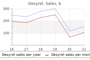
Desyrel 100 mg buy cheap on line
The bellows in a double-circuit design ventilator takes the place of the respiratory bag in the anesthesia circuit anxiety ocd desyrel 100 mg buy on-line. Pressurized oxygen or air from the ventilator energy outlet (45�50 psig) is routed to the area between the inside wall of the plastic enclosure and the skin wall of the bellows anxiety symptoms women desyrel 100 mg order free shipping. In contrast, throughout exhalation, the bellows ascend as stress inside the plastic enclosure drops and the bellows replenish with the exhaled fuel. A ventilator move management valve regulates drive gas circulate into the pressurizing chamber. Ventilators with microprocessors additionally make the most of feedback from move and stress sensors. If oxygen is used for pneumatic power it is going to be consumed at a price no much less than equal to minute air flow. Thus, if oxygen contemporary gasoline flow is 2 L/min and a ventilator is delivering 6 L/min to the circuit, a total of at least eight L/min of oxygen is being consumed. Some anesthesia machines scale back oxygen consumption by incorporating a Venturi gadget that attracts in room air to provide air/oxygen pneumatic power. This may be indicated by a better than anticipated rise in impressed oxygen concentration (if oxygen is the only pressurizing gas). Some machine ventilators have a built-in drive gasoline regulator that reduces the drive stress (eg, to 25 psig) for added security. Double-circuit design ventilators also incorporate a free respiratory valve that permits outdoors air to enter the rigid drive chamber and the bellows to collapse if the affected person generates adverse pressure by taking spontaneous breaths throughout mechanical air flow. Introduction of a negative-pressure reduction valve to the respiratory circuit could introduce the chance of air entrainment and the potential for dilution of oxygen and unstable anesthetic concentrations if the affected person breathes throughout mechanical ventilation and low recent gasoline flows. The major benefit of a piston ventilator is its capacity to ship correct tidal volumes to sufferers with very poor lung compliance and to very small patients. During volume-controlled ventilation the piston strikes at a continuing velocity whereas throughout pressurecontrolled ventilation the piston strikes with C. When the switch is turned to "bag" the ventilator is excluded and spontaneous/manual (bag) ventilation is possible. During exhalation, the pressurizing fuel is vented out and the ventilator spill valve is not closed. The ventilator bellows or piston refill throughout expiration; when the bellows is completely crammed, the rise in circle system stress causes the surplus fuel to be directed to the scavenging system through the spill valve. Sticking of this valve may end up in abnormally elevated airway strain throughout exhalation. Pressure & Volume Monitoring Peak inspiratory strain is the highest circuit pressure generated throughout an inspiratory cycle, and supplies an indication of dynamic compliance. Plateau stress is the strain measured during an inspiratory pause (a time of no gas flow), and mirrors static compliance. An enhance in each peak inspiratory strain and plateau strain implies an increase in tidal volume or a lower in pulmonary compliance. An increase in peak inspiratory stress with none change in plateau pressure signals an increase in airway resistance or inspiratory gas move fee (Table 4�3). Thus, the shape of the breathing-circuit stress waveform can provide essential airway information. Airway secretions or kinking of the tracheal tube could be easily dominated out with the usage of a suction catheter. Ventilator Alarms Alarms are an integral a half of all fashionable anesthe9 sia ventilators. To keep away from issues with ventilator�fresh gas move coupling, airway stress and exhaled tidal volume have to be monitored closely and extreme contemporary gas flows have to be averted. The first is at all times built into the ventilator whereas the latter two could additionally be in separate modules. A small leak or partial breathing-circuit disconnection may be detected by subtle decreases in peak inspiratory strain, exhaled volume, or end-tidal carbon dioxide earlier than alarm thresholds are reached. Most modern anesthesia ventilators even have integrated spirometers and oxygen analyzers that provide additional alarms. Excessive Positive Pressure Intermittent or sustained excessive inspiratory pressures (>30 mm Hg) during positive-pressure ventilation enhance the risk of pulmonary barotrauma (eg, pneumothorax) or hemodynamic compromise, or each, during anesthesia. Excessively excessive pressures could arise from incorrect settings on the ventilator, ventilator malfunction, recent gas circulate coupling (above), or activation of the oxygen flush through the eleven inspiratory phase of the ventilator. The mechanism of strain limiting could also be so simple as a threshold valve that opens at a certain pressure or digital sensing that abruptly terminates the ventilator inspiratory phase. Tidal Volume Discrepancies 12 Large discrepancies between the set and precise tidal volume that the patient receives are sometimes observed in the operating room during volume control air flow. A: In normal individuals, the peak inspiratory pressure is equal to or slightly greater than the plateau stress. B: An enhance in peak inspiratory stress and plateau stress (the difference between the 2 stays nearly constant) may be due to a rise in tidal quantity or a lower in pulmonary compliance. C: An enhance in peak inspiratory stress with little change in plateau pressure alerts an increase in inspiratory flow rate or a rise in airway resistance. For this reason respiration circuits for pediatric patients are designed to be much stiffer, with compliances as small as 1. Compression losses, usually about 3%, are as a result of gas compression inside the ventilator bellows and could additionally be dependent on breathing-circuit volume. Gas sampling for capnography and anesthetic fuel measurements characterize additional losses within the form of gas leaks until the sampled fuel is returned to the respiration circuit, as occurs in some machines. Accurate detection of tidal quantity discrepancies is dependent on where the spirometer is placed. Several mechanisms have been built into newer anesthesia machines to scale back tidal quantity discrepancies. During the initial digital self-checkout, some machines measure whole system compliance and subsequently use this measurement to modify the tour of the ventilator bellows or piston; leaks may also be measured however are normally not compensated. The actual technique of tidal volume compensation or modulation varies in accordance with producer and model. In one design a move sensor measures the tidal quantity delivered on the inspiratory valve for the first few breaths and adjusts subsequent metered drive gas flow volumes to compensate for tidal quantity losses (feedback adjustment). Another design frequently measures fresh gas and vaporizer circulate and subtracts this amount from the metered drive gas flow (preemptive adjustment). Alternately, machines that use digital control of fuel move can decouple recent gasoline flow from the tidal quantity by delivery of fresh gas move only throughout exhalation. Lastly, the inspiratory phase of the ventilator�fresh gasoline flow may be diverted by way of a decoupling valve into the breathing bag, which is excluded from the circle system throughout ventilation. During exhalation the decoupling valve opens, permitting the fresh fuel that was quickly stored within the bag to enter the breathing circuit. Pollution of the working room surroundings with anesthetic gases may pose a well being hazard to surgical personnel. Reduction to these trace levels is possible only with correctly functioning waste-gas scavenging methods.

100 mg desyrel discount fast delivery
The fusion fascia of the left colon (1) fixes the meso of the descending colon to the posterior primitive parietal peritoneum anxiety symptoms 8 weeks generic desyrel 100 mg mastercard. The superior restrict anxiety 10 months postpartum trusted 100 mg desyrel, which covers a half of the retroperitonealized pancreatic physique and tail, is the line connecting the origin of the superior mesenteric artery to the left angle of the transverse mesocolon. The inferior restrict begins a little left from the midline, in front of the promontory, and descends along the internal border of the psoas muscle, on the upper root of the sigmoid mesocolon. The retroduodenopancreatic fusion fascia of the duodenal loop (2) fixes the mesoduodenum and pancreatic head to the posterior primitive parietal peritoneum and to the fusion fascia of the left mesocolon, respectively, proper and left from the midline. The fusion fascia of the proper colon (4), positioned between cecum and transverse mesocolon, fixes the meso of the ascending colon to the posterior primitive parietal peritoneum and the duodenum and its fused meso, containing the caudal part of the pancreatic head. The left colonic compartment is demarcated from the primitive retroperitoneum by loose areolar tissue representing the left retromesenteric aircraft (black arrows). Anteriorly, the transverse mesocolon (black asterisks) attaches to the pancreatic neck, posterior to the stomach, and anterior to the duodenojejunal junction (white asterisk) in the left paraduodenal fossa. The proper pancreaticoduodenal compartment is demarcated posteriorly by the loose areolar tissue of the retropancreaticoduodenal fusion fascia (arrowheads), also referred to as fascia of Treitz, and anteriorly by the free areolar tissue of the cranial extension of the best retromesenteric airplane, also referred to as right fascia of Toldt (arrows). Note the continuity of the transverse mesocolon (asterisks) with the right colonic compartment, located anterior to the best perirenal house. The retropancreaticoduodenal fusion fascia (black arrowheads) is positioned posterior to the duodenum and anterior to the primitive retroperitoneum, aorta, and inferior caval vein. Anatomic landmarks of the completely different elements of the anterior pararenal house in a affected person with pancreatitis. The pancreatic head (P) is located posterior to the proper colonic compartment and transverse mesocolon (asterisk). White arrow � inferior mesenteric vein; white arrowheads � left retromesenteric aircraft. The left colic vessels (black arrowheads), within the left colonic compartment, are steady with the branches from the inferior mesenteric vein (black-and-white arrowhead) inside the cranial extension of the mesosigmoid. Extensive spread of pancreatic fluid to retromesenteric and retrorenal areas, with caudal extent. The pancreatic physique and tail are somewhat swollen (a, b), and posterior to it, extending caudally (c) into the left retromesenteric airplane (asterisks), fluid is present indicating significant trauma to the pancreatic parenchyma. Selective opacification of this compartment within the cadaver permits identification of the preferential pathway of unfold and the characteristic localizing features. This outline is the hallmark of perirenal collections, and its identification, due to this fact, on plain movies as properly as on other studies. Perforation of the renal capsule then results in contamination of the perirenal house. It is secondary to Escherichia coli, Aerobacter aerogenes, or, rarely, Clostridium and develops particularly in diabetics. In youngsters, hematogenous unfold sometimes occurs to the perirenal fat from distant sites of an infection, corresponding to furunculosis, wound infection, or upper respiratory disease. Its recognition is expounded directly to an understanding of the characteristic look of the acutely distended cone of renal fascia and the preferential spread by way of the rich perirenal fat dorsal to the kidney. The cone of renal fascia (arrows) envelops the adrenal gland, kidney (K), and perirenal fat. Note the conventional proper posterior renal fascia and open right perirenal house inferiorly (curved arrow). After the introduction of 450 mL of contrast medium, the distended cone of renal fascia is vertical and presents an inferiorly convex border overlying the iliac crest (arrows). Associated exudate distends the cone of renal fascia so that its lower border may be identified as an inferiorly convex shadow overlying the iliac crest. The inferiorly convex border of the distended perirenal area is a highly reliable localizing sign. Fulminating an infection might disrupt the perirenal fascial boundaries, allowing the gasoline to escape to different compartments. Bilateral perirenal gas-producing infections are unusual but their contours are once more distinctive. Perirenal Abscess Initially, fluid launched into the perirenal space is evenly dispersed throughout the perirenal fat. A convex decrease border (arrows) on the degree of the iliac crest characterizes the distended cone of renal fascia. Arteriography has been of particular value up to now in instances the place the standard radiographic findings are unsure or the place major renal infection is suspected to lengthen via the capsule. The hematoma is predominantly within the posterior renal area, displacing the left kidney anteriorly. To point out its pathogenesis and characteristic morphology, the most accurate designation for this situation ought to be uriniferous perirenal pseudocyst. Note fistulous tract between the left renal pelvis and posterior pararenal area (arrowhead). This occurred as a consequence of pyelosinus backflow secondary to partial distal ureteral obstruction from a left ovarian mass. Urinary extravasation into the perirenal fats results in speedy lipolysis, and a definite fibrous sac (false capsule or pseudocyst) is shaped within 12 days. Indeed, the tissue response itself ends in a seamless element of obstruction establishing a vicious cycle. Since perirenal effusions localize in accordance with the effect of gravity and planes of least resistance, extravasated urine seeks out the portion of the cone of renal fascia caudad to the kidney. Basic to an appreciation of the attribute complex of radiographic abnormalities is the reality that the pseudocyst usually conforms to the axis and dimensions of the cone of renal fascia. Surgical specimen of uriniferous perirenal pseudocyst and nonfunctioning hydronephrotic kidney. In addition, extravasation into the pseudocyst might confirm the actual point of leakage or point out gross communication with the collecting system. Its contours may be further outlined on plain movies by the distinction of other extraperitoneal fat (specifically within the posterior pararenal compartment) into which the pressure of the pseudocyst bulges. With big collections, the cone of renal fascia could turn into so distended that its axis appears more vertical. The pseudocyst could be identified as a soft-tissue density or as a lucent defect in the course of the part of whole physique opacification. Needle opacification of the pseudocyst could define precisely its contour, dimension, and attribute axis. The fat immediately around the kidney and upper third of the psoas muscle can be visualized intact, but the lower margin of the psoas muscle is obscured by the. Its contours are additional highlighted by the distinction offered by posterior pararenal fat into which it bulges posteriorly. The proximal ureter is displaced medially and is dilated, related to caliectasis and a mild obstructive nephrogram.
Desyrel 100 mg purchase amex
The authors undertook a cross-sectional and a longitudinal cognitive efficiency research anxiety x rays cheap 100 mg desyrel with visa. The cross-sectional work revealed that patients had gentle to average impairment on anterograde memory as defined by a bunch common Z score of -1 anxiety pictures purchase desyrel 100 mg mastercard. A delicate impairment in executive capabilities and language have been discovered with normal visuospatial function. This examine advised that the long-term sequelae might embody dysexecutive problems. Category fluency requires a search via conceptual data retailer for semantic extensions derived from a goal word. These longitudinal research are sometimes restricted by the comparatively small variety of neuropsychological exams administered and by their lack of integration with localized mind atrophy. Irani and colleagues imaged eight sufferers at convalescence who had regular medial temporal lobe imaging on scientific scans (except for one affected person with putaminal high signal). Patients have been found to have significantly smaller mixed hippocampal/total intracranial volumes and brain/total intracranial quantity ratios than controls. Regression evaluation found a significant adverse affiliation between brain/total intracranial volume and increasing age for each sufferers and controls, however no affiliation between either hippocampal/total intracranial volume or brain/total intracranial quantity with both cognitive impairment or the dosage of corticosteroids acquired. A second study86 quantified the longitudinal structural changes in volumes of each the hippocampus and amygdalae following autoimmune encephalitis using a totally automated software bundle. The disease is related to benign ovarian teratomas and is especially seen in females between the ages of 12 and forty years. Binding deficits in reminiscence following medial temporal lobe injury in sufferers with voltage-gated potassium channel complex antibody-associated limbic encephalitis, pp. In a number of patients, notably males, the presenting symptom may be seizures. Subsequently, and usually with the lag of 10 to 20 days, patients develop a motion disorder, dysautonomia, and, sometimes, central hypoventilation. When hyperkinetic, this movement dysfunction often reveals distinguished orofacial dyskinesias, particularly centred around the lips, and stereotyped, antigravity actions of legs and arms. The central hypoventilation is a much less widespread function however may necessitate intensive care unit admission which is itself a poor prognostic issue. Alongside this, the dichotomy of the timings of lymphocytosis and oligoclonal band appearances lend assist to two major phases to the disease course of. More recently, patients have been described with relatively limited presentations. While the antibodies could have effector functions in vitro, clear proof supporting an immunotherapy response is lacking. Inclusion of studies reporting imaging from two or more sufferers Reference Dalmau et al. In the first,109 nine sufferers were tested at a median time of 43 months (range: 23�69), with five receiving immunomodulatory therapy within three months of symptom onset, three receiving immunomodulation late within the disease, and one patient receiving no treatment. This examine discovered that spotlight was impaired in four patients, working memory was impaired in 4 patients, verbal reminiscence in two sufferers, non-verbal memory in one affected person, and executive function in 5 sufferers. Five sufferers have been impaired in as a lot as four exams, primarily affecting consideration or working reminiscence processes, but two sufferers had intensive neuropsychological impairments across a number of neuropsychological domains (attention, working memory, reminiscence and government function). A second study112 found evidence of government dysfunction (as measured by the digit span backwards, Stroop inhibition, and word fluency) but in addition anterograde memory impairment (as measured by Iizuka et al. They additionally found evidence of bilaterally decreased hippocampal connectivity in the anterior default mode community but no changes within the sensorimotor, major visual, or auditory resting state networks. The group who have been provided further immunosuppressive brokers had a better consequence (p = 0. Furthermore, relapse rates had been lowered in patients supplied extra immunotherapies. This describes the variable presence of cognitive disturbance with few seizures, but extra particularly oculomotor difficulties, ataxia, and a stiff-person-like phenotype (often startle, spasms, and ridgity). Pathophysiological hypotheses Our understanding of the pathophysiology of these diseases relies upon scientific and serological observations, in addition to recent in vitro and in vivo studies. The cells, and limited quantities of antibodies, then migrate throughout the blood�brain barrier. In this illness, the receptors are internalized by the divalent antibodies and this seems to be the main pathogenic mechanism. However, many questions related to pathophysiology stay unanswered, together with why the antibodies modulate hippocampal regions particularly given the widespread antigen distribution,127 why these ailments differ from those associated with genetic or pharmacological manipulations of the antigenic goal,17 how neuronal plasticity might account for patient recovery from the illness, and the relevance of IgM and IgA subclasses of autoantibodies. Atypical antipsychotics, particularly olanzapine, quetiapine, and benzodiazepines are used to sedate agitated, psychotic patients. Haloperidol must be prevented if attainable as it could worsen parkinsonism and will precipitate oculogyric crises. A thiopental coma may be induced and empirical therapies for management of blood pressure, bradycardia/asystole, and dyskinesias are sometimes administered. This disease is thought to have a pure history showing a continual course, usually over several years with relapses. Given the retained phylogeny of the mammalian hippocampus throughout species130 there are several mnemonic71,130�132 and computational accounts133 of the hippocampus which have but to be studied in humans. The questions requested had been extremely episodic in nature and so reveal the requirement of episodic neural apparatus for semantic recall, especially if relations between object and time are wanted. This is probably partially as a result of discrepancies in assay methodologies, as talked about earlier. Cell-surface central nervous system autoantibodies: scientific relevance and emerging paradigms. Potassium channel antibodyassociated encephalopathy: a potentially immunotherapy-responsive type of limbic encephalitis. Paraneoplastic limbic encephalitis: neurological signs, immunological findings and tumour affiliation in 50 patients. Clinical and experimental research of probably pathogenic brain-directed autoantibodies: current knowledge and future directions. Spectrum of neurological syndromes associated with glutamic acid decarboxylase antibodies: diagnostic clues for this affiliation. Clinical relevance of serum antibodies to extracellular N-methyl-D-aspartate receptor epitopes. Antibodies to Kv1 potassium channel-complex proteins leucine-rich, glioma inactivated 1 protein Conclusion the autoimmune encephalopathies related to antibodies directed towards cell surface neuronal proteins are an expanding group of immunotherapy-responsive situations with distinctive medical options and major potential implications for neurobiology studies. Future research will offer insights as to how specific prolonged receptor dysfunction is reversible, the mechanisms of plasticity, and the neuronal networks engaged within the illness process and restoration. Furthermore, the differing focality of these illnesses may offer novel insights into useful localizations. Novel antibody targets are prone to emerge in diseases with cognitive and psychiatric overlaps. Knowledge of their biology will feed into the ailments with recognized antibody targets and further inform a selection of brain neurotransmitter methods and neuronal populations. N-methyl-D-aspartate antibody encephalitis: temporal development of scientific and paraclinical observations in a predominantly non-paraneoplastic disorder of each sexes. Potentially reversible autoimmune limbic encephalitis with neuronal potassium channel antibody.
100 mg desyrel purchase amex
The alternate route follows the external pudendal artery to the nodes on the saphenofemoral junction anxiety symptoms preschooler cheap desyrel 100 mg overnight delivery, that are their sentinel nodes anxiety symptoms in men 100 mg desyrel safe. It is assessed primarily based on examination of pathologic specimen that retrieves 12 or more nodes. The lateral node (arrow) is present along the left middle rectal vessels outside the mesorectal fascia. Metastatic carcinoma of the anal canal to the node on the saphenofemoral junction and the deep inguinal node. The section of the small intestine anterior to the basis of the mesentery is the ileum (I) with its mesenteric vessels (curved arrow). Recurrent carcinoma on the root of the sigmoid mesocolon after segmental resection of the sigmoid colon. Patterns of Disease Spread Because of the non-specificity on anatomic imaging, additional imaging research and aspiration biopsy are regularly used to establish the diagnosis of metastatic illness earlier than therapy choice. Venous invasion with extramural extension from the primary could additionally be manifested as a tubular look accompanied by the artery extending into the mesocolon or mesorectum of the concerned phase. This statement can be more convincing if the thrombotic vein could be tracked and related with the traditional, bigger vein downstream within the mesocolon. On imaging studies, perineural infiltration might present as gentle tissue infiltration extending from the primary along the artery and nerve. However, these adjustments may be tough to distinguish from desmoplastic inflammatory reaction or venous invasion. The nerves round and throughout the mesorectum illustrated in a affected person with neurofibromatosis. Locally superior carcinoma of the rectosigmoid junction invading the bladder and increasing along the mesorectal fascia to involve the S2 sacral nerve. The hypointense tumor (arrowhead) extends posteriorly alongside the left mesorectal fascia (arrows) toward the retrorectal space. Adenocarcinoma of the ascending colon with tumor thrombus within the branches of the proper colic vein. Adenocarcinoma of the descending colon with tumor thrombus in the left colic vein. Kanamoto T, Matsuki M, Okuda J et al: Preoperative analysis of local invasion and metastatic lymph nodes of colorectal cancer and mesenteric vascular variations using multidetector-row computed tomography before laparoscopic surgery. Patterns of Spread of Renal, Upper Urothelial, and Adrenal Pathology 13 Introduction the kidneys and adrenal glands reside within the perirenal house, a subdivision of the extraperitoneum formed by the anterior and posterior renal fascia. The complicated designation of those vessels as ``capsular' is derived from the old nomenclature of the perirenal fats because the ``adipose capsule of the kidney. The proper adrenal gland lies posterior to the inferior vena cava, above the right kidney, lateral to the best diaphragmatic crus and medial to the bare space of the liver. The left adrenal gland is posterior to the splenic vessels, pancreas, and abdomen, lateral to the left diaphragmatic crus and left celiac ganglion, and superior to the left kidney. Patterns of Spread of Renal, Upper Urothelial, and Adrenal Pathology ureteral lymphatics to the exterior and inner iliac nodes. All the iliac nodes drain to the paraaortic nodes, cisterna chyli, and predominantly the left supraclavicular nodes by way of the thoracic duct. The proper renal artery courses in the perirenal area posterior to the inferior vena cava and right renal vein. During their course, the renal arteries cut up into dorsal and ventral branches earlier than getting into the kidney. Approximately 35% of people have accent renal arteries, which usually provide the decrease pole of the kidney. Blood supply to the ureters is regional, with the proximal ureters equipped by the renal arteries, aorta, gonadal artery, and customary iliac artery; the middle and distal ureters are supplied by the interior iliac artery and vesical arteries. Three arteries provide the adrenal glands: the superior adrenal artery from the inferior phrenic artery; the middle adrenal artery from the aorta; and the inferior adrenal artery from the renal artery. There are single adrenal veins, with the right draining into the inferior vena cava and the left into the left renal vein. Significantly, the renal and adrenal vasculature coursing inside the perirenal area arising from the aorta and draining to the inferior vena cava function a scaffold for unfold of disease to and from the kidneys and adrenal glands. It is necessary to recognize that these avenues interconnect the perirenal house with the remainder of the extraperitoneal space in addition to with the ligaments and mesenteries of the abdomen, forming a continuum within the subperitoneal house. Spread of Disease Renal Tumors Renal Cell Carcinomas Renal tumors account for 3% of all cancer cases and deaths. In post-mortem sequence, metastatic tumor involving the kidney is 2 to 3 times more frequent than primary renal tumors. The extra frequent detection at a lower disease stage permits for curative tumor resection. The tumor might invade the renal calyces and pelvis, mimicking transitional cell carcinoma. However, the papillary and chromophobe subtypes could be instructed by their much less intense Lymphatic Anatomy Lymphatics draining the kidney are derived from three plexuses: one beneath the renal capsule, a second across the renal tubules, and the third in the perirenal fat. Renal cystic tumors are thought of extremely suspicious for malignancy if any solid enhancing element is recognized. The Robson classification was developed in the 1960s and was based mostly on tumor confinement and spread related to anatomical landmarks. Cross-sectional imaging and therapeutic advances have made the Robson classification insufficient. The tumor could grow throughout the lumen with attachment to the wall at the initial site with the rest projecting into the lumen or invading the wall. The extent of tumor thrombus in the inferior vena cava is important in staging and remedy issues. Usually, thrombus in the inferior vena cava above the confluence with the renal veins is tumor thrombus and beneath the confluence bland thrombus. These include nodes alongside the renal arteries from the renal hilum to the paraaortic nodes at this stage. There is involvement of the principle renal vein in 25�35% 15 and inferior vena cava in 5�10% of circumstances. These embody involvement of non-regional lymph nodes, direct extension past the renal fascia, and hematogenous dissemination. Within the anterior pararenal area, the duodenum, colon, and pancreas could also be involved. Spread to the posterior pararenal space can result in further extension involving the posterior and lateral abdominal muscles. Hematogenous spread is most typical to the lung and adrenal gland, followed by bone, pleura, mind, pancreas, and liver. Lung metastases embody nodules, lymphangitic unfold, peripheral arterial spread, and endobronchial lesions, as properly as pleural and mediastinal involvement. Renal Lymphoma Renal lymphoma is rare as a primary tumor; nevertheless, extranodal unfold of lymphoma usually involves the genitourinary system, with the kidneys most incessantly 318 a thirteen. These patterns are derived from two mechanisms of unfold: hematogenous dissemination and contiguous subperitoneal extension. Lymphoma seeds to the renal cortex and then grows alongside the framework of the nephrons, amassing ducts, and blood vessels leading to a number of expansile lots.
Buy 100 mg desyrel visa
Liquid oxygen should be stored nicely under its important temperature of �119�C as a end result of gases may be liquefied by pressure only if saved beneath their important temperature anxiety synonyms discount desyrel 100 mg. To guard against a hospital gas-system failure anxiety online test desyrel 100 mg buy cheap, the anesthesiologist must always have an emergency (E-cylinder) provide of oxygen out there throughout anesthesia. Oxygen cylinder strain must be monitored earlier than use and periodically during use. Anesthesia machines usually additionally accommodate E-cylinders for medical air and nitrous oxide, and may settle for cylinders of helium. Compressed medical gases make the most of a pin index safety system for these cylinders to prevent inadvertent crossover and connections for different gasoline types. This metallurgic alloy has a low melting point, which allows dissipation of stress that might otherwise warmth the bottle to the point of ballistic explosion. Nitrous Oxide Nitrous oxide is manufactured by heating ammonium nitrate (thermal decomposition). It is almost all the time stored by hospitals in large H-cylinders related by a manifold with an computerized crossover function. Bulk liquid storage of nitrous oxide is economical solely in very massive institutions. Although a disruption in supply is normally not catastrophic, most anesthesia machines have reserve nitrous oxide E-cylinders. A larger reading implies gauge malfunction, tank overfill (liquid fill), or a cylinder containing a gas apart from nitrous oxide. Because energy is consumed in the conversion of a liquid to a gasoline (the latent heat of vaporization), the liquid nitrous oxide cools. The drop in temperature results in a decrease vapor stress and decrease cylinder stress. Nitrogen provide systems both incorporate the use of H-cylinders connected by a manifold or a wall system provided by a compressor driven central supply. Vacuum A central hospital vacuum system usually consists of unbiased suction pumps, each able to handling peak requirements. Traps at each person location forestall contamination of the system with international matter. Carbon Dioxide Many surgical procedures are performed utilizing laparoscopic or robotic-assisted methods requiring insufflation of physique cavities with carbon dioxide, an odorless, colorless, nonflammable and slightly acidic gasoline. Medical Air the use of air is becoming extra frequent in anesthesiology as the recognition of nitrous oxide and unnecessarily high concentrations of oxygen has declined. Dehumidified but unsterile air is provided to the hospital pipeline system by compression pumps. The inlets of these pumps must be distant from vacuum exhaust vents and equipment to decrease contamination. Pipes are sized such that the pressure drop throughout the entire system by no means exceeds 5 psig. Gas pipes are often constructed of seamless copper tubing using a special welding method. One end of a color-coded hose connects to the hospital medical gas supply system by means of a quick-coupler mechanism. The other end connects to the anesthesia machine via the diameter index security system. Operating room tools, including the anesthesia machine, interfaces with these pipeline system outlets by color-coded hoses. Quickcoupler mechanisms, which vary in design with totally different producers, join one end of the hose to the appropriate gas outlet. The different finish connects to the anesthesia machine via a noninterchangeable diameter index safety system becoming that stops incorrect hose attachment. E-cylinders of oxygen, nitrous oxide, and air three attach on to the anesthesia machine. Multiple washers positioned between the cylinder and yoke, which prevent correct engagement of the pins and holes, have unintentionally defeated this technique. The pin index security system can be ineffective if yoke pins are broken or the cylinder is crammed with the incorrect gasoline. The functioning of medical gas supply sources and pipeline systems is continually monitored by central and area alarm systems. Modern anesthesia machines and anesthetic fuel analyzers constantly measure the fraction of inspired oxygen (FiO2). Due to gasoline trade, move rates, and shunting a marked difference can exist between the monitored FiO2 and oxygen concentration on the tissue stage. However, scrub nurses and surgeons stand in surgical garb for hours under hot working room lights. As a basic precept, the comfort of working room personnel should be reconciled with affected person care. Hypothermia has been related to an increased incidence of wound infection, higher intraoperative blood loss (impaired coagulation assessed by thromboelastography), and prolonged hospitalization (see Chapter 52). Optimally humidity ranges in the working room should be maintained between 50% and 55%. Below this vary the dry air facilitates airborne motility of particulate matter, which is often a vector for an infection. At excessive humidity, dampness can have an result on the integrity of barrier gadgets such as sterile cloth drapes and pan liners. Operating room noise has been measured at 70�80 decibels (dB) with frequent sound peaks exceeding 80 dB. As a reference, if your speaking voice has to be raised above conversational degree, then ambient noise is approximated at eighty dB. Orthopedic air chisels and neurosurgical drills can approach the noise ranges of 125 dB, the extent at which most human subjects start to experience ache. These flow rates, often achieved by blending as much as 80% recirculated air with contemporary air, are engineered in a manner to decrease turbulent circulate and be unidirectional. Therefore, a separate anesthetic gas scavenging system should at all times supplement working room air flow. The operating room ought to keep a slightly optimistic strain to drive away gases that escape scavenging and must be designed so contemporary air is launched via or close to the ceiling and air return is dealt with at or near ground level. Air quality must be maintained by adequate air filtration using a 90% filter, outlined simply as one which filters out 90% of particles presented. For the anesthesia supplier radiation is often a component of either diagnostic imaging or radiation remedy. Examples embody fluoroscopy, linear accelerators, computed tomography, directed beam remedy, proton remedy, and diagnostic radiographs. Radiation-sensitive organs such as eyes, thyroid, and gonads have to be protected, in addition to blood, bone marrow, and fetus.

100 mg desyrel buy with amex
For sufferers with regular hearts anxiety high blood pressure desyrel 100 mg order without prescription, flecainide anxiety depression symptoms buy desyrel 100 mg without a prescription, propafenone, sotalol, amiodarone, and procainamide (in that order) could probably be used should there be no evidence for structural heart illness. Exerciseinduced, nonsustained repetitive monomorphic tachycardia is mostly catecholamine-dependent. Multiple monomorphic morphologies may be current in a single affected person at totally different instances. Understanding the underlying mechanism(s) is essential when planning therapy, both acutely and for the long term. Although they may not convert the rhythm, these drugs could nonetheless nonetheless successfully suppress arrhythmias after sinus rhythm has been achieved by different means. Digoxin antibodies are indicated, in addition to correction of electrolyte disturbances, similar to low potassium and magnesium. Antiarrhythmic medication, corresponding to amiodarone and sotalol, could also be used as adjunctive remedy to stop a quantity of recurrent shocks. Death will happen except a perfusing rhythm is restored within seconds to minutes; 0. If the affected person is hypothermic or has drowned in cold water, extended resuscitation could also be wanted, because the "diving reflex" in cool water might improve long-term survival even with no cardiac output for many minutes. This rhythm strip reveals ventricular fibrillation that degenerates additional into fantastic ventricular fibrillation. Genetically and phenotypically heterogeneous, more than 20 associated genes have been reported. These mutations are characterised by a highly variable expression and clinical course. Sudden death can occur in asymptomatic young people, including athletes, even as the initial presentation. These findings in a younger affected person with syncope suggest the diagnosis of hypertrophic cardiomyopathy, which was confirmed by echocardiography. Desmosomes are specialized intercellular junctions that anchor intermediate filaments of the cytoskeleton to the cytoplasmic membrane in adjoining cells. These are most prevalent in tissues, such as myocardium and skin epithelium, which are uncovered to frictional and shear stress. Because excessive myocardial strain may promote progression, limitation of competitive athletics, similar to long-distance biking, operating, swimming, or weight training, is also commonly really helpful. The arrhythmia mechanism may result from triggered exercise or increased automaticity. There are greater than 10 clinical syndromes, and each has been associated with mutations in particular genes or loci; nonetheless, the precise mutation can vary tremendously even though it could contain a specific gene. The age of onset is normally in childhood, but late onset (fourth decade) has been reported. A household historical past of sudden dying in relations less than forty years old is current in about one-third of patients. There may be a task for flecainide in lowering ventricular arrhythmias and may be thought-about in sufferers who proceed to have incomplete control of their arrhythmias on -adrenergic blockers. This has led to some medication being faraway from the market due to the danger of TdP even when only some sufferers out of several million developed the arrhythmia; in part, that is related to TdP being probably life-threatening, and plenty of medicine that are associated with it are used for relatively innocuous non�life-threatening conditions. A potential approach to scale back danger is to determine which patients can profit from specific long-term therapeutic methods. Assessing which sufferers are at highest threat for sudden cardiac death (but not dying from different causes) can modify outcomes and enhance long-term prognosis. However, all clinical predictors endure from lack of sensitivity, specificity, and predictive accuracy. Many of these tests reveal markers and never predictors of occasion and are sometimes population and illness specific. Furthermore, such sufferers may be at greater threat for whole mortality unbiased of the chance for sudden cardiac dying. Because all people are at some threat for sudden cardiac dying even with none danger issue, identification of danger is a complex and troublesome concern. In these syndromes the chance of whole mortality aside from sudden cardiac death is low. The specific scientific syndromes associated with sudden cardiac demise are often in a population of structural heart illness. The affected person should also have their underlying medical and arrhythmia conditions stabilized. The second letter indicates the chamber during which sensing of the intracardiac electrical sign is going on (atrium, A; ventricle, V; or each, D). The third letter signifies the response of the device to a sensed signal (inhibition of pacing stimulus output, I; triggering [causing to occur] of stimulus output, T; or each, D). The most tracking rate is that price at which ventricular pacing will be triggered by native P waves in a 1:1 relationship (atrial based); the utmost sensor-based price is the best programmed rate dictated by sensor input to the pulse generator. Whereas these rates are sometimes programmed to be the identical, the sensor-based price could be programmed to exceed the tracking price in response to exercise, thereby avoiding speedy ventricular paced charges triggered by supraventricular tachycardias. It varies with the manufacturer; a number of manufacturers set a continuing magnet rate nicely above the expected spontaneous rate. Because magnet placement eliminates sensing, pacing output happens regardless of the existence of a spontaneous cardiac rhythm; repetitive atrial or ventricular beating is only very rarely a scientific consequence. This can happen because of too low a programmed voltage output, an increase in myocardial stimulation threshold (such as happens throughout hyperkalemia or flecainide treatment), pacing lead insulation break or fracture, lead dislodgement, or battery finish of life; failure to capture can also be "useful" because of refractoriness of the myocardial tissue. Pacing system interrogation by way of manufacturer-specific programmers is commonly essential to outline the nature of the issue. Rapid paced ventricular charges typically occur in response to supraventricular tachycardias. V1 rhythm strip illustrating myopotential oversensing, during which irregular and longer-than-programmed ventricular stimuli happen. Morphology in biventricular pacing is dependent on lead location and the programmed relative timing of right and left ventricular pacing impulses. High rates in either atrium or ventricle can be interrogated, and saved intracardiac electrograms may be seen for affirmation of the rhythm and acceptable management undertaken. Pacing devices additionally retailer other clinically related data, such as coronary heart fee histograms, percentage of atrial and ventricular pacing and sensing, and number of mode switches. If interrogation can be achieved, pulse turbines nearing end of life ("elective substitute time") will show a warning. Unlike regular ventricular pacing from the right ventricle, which displays a left bundle branch block sample, these ventricular paced beats show a right bundle department block sample. The purpose for that is that the ventricular lead is in the left ventricle (instead of the conventional location in the right ventricular apex). The ventricular pacing lead in this affected person was inadvertently handed through a patent foramen ovale into the left atrium, via the mitral valve, and into the left ventricle. This represents biventricular pacing from the right ventricular apex and an epicardial left cardiac vein. The implant process is much like that for a pacemaker, aside from the dimensions of the device and the necessity for particular leads that incorporate defibrillation coils alongside the physique of the leads.