Citalopram
Citalopram dosages: 40 mg, 20 mg
Citalopram packs: 30 pills, 60 pills, 90 pills, 120 pills, 180 pills, 270 pills, 360 pills
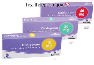
Citalopram 40 mg buy discount on line
However medicine bow wyoming buy discount citalopram 40 mg, lumbosacral transitional anomalies could be missed when solely sagittal and axial views are obtained medications without a script discount citalopram 20 mg visa. There are only only a few alterations that are uncommon in asymptomatic individuals younger than 50 years [272], i. She was admitted to an intensive rehab program with emphasis on stabilizing workouts which resolved her symptoms. Computed tomography Computed tomography is the imaging modality of choice for the assessment of spinal fusion. A detailed description of the strength and weaknesses of those diagnostic research is included in Chapter 10. Currently, discography predominantly serves as a pain provocation take a look at to differentiate symptomatic and asymptomatic disc degeneration. However, careful interpretation of the findings continues to be mandatory with reference to the clinical presentation [43]. We suggest utilizing distinction injection to doc the correct needle position and filling of the joint capsule (Case Study 1). It is due to this fact necessary to perform repetitive infiltrations to improve the diagnostic accuracy [239]. Stabilization of the putative irregular segments by an external transpedicular fixator has been instructed by a quantity of authors [74, 237, 254] with combined outcomes by method of end result prediction. Nonetheless, in the presence of selected factors (see Chapter 7), surgical procedure ought to at least be delayed till attempts have been made to modify danger factors which are amenable to change and all attainable conservative means of remedy are exhausted. The first essential side is a multidisciplinary functional restoration program and psychological interventions to affect affected person conduct (see Chapter 21). The longer ache and useful limitations persist, the much less likely is ache reduction, useful restoration and return to work (see Chapter 6). These sufferers should promptly be included in a multidisciplinary practical work conditioning program. Favorable indications for non-operative remedy embrace (Table 4): Cognitive behavioral interventions are necessary to address fears and misbeliefs Table four. Since its introduction in 1911 by Albee [3] and Hibbs [127], spinal fusion was initially only used to deal with spinal infections and high-grade spondylolisthesis. Today approximately seventy five % of the interventions are done for painful degenerative problems [66]. Despite its frequent use, spinal fusion for lumbar spondylosis remains to be not solidly based on scientific proof in phrases of its medical effectiveness [66, 102, 103, 264]. For a very long time it was hoped that consequence of spinal fusions could possibly be considerably improved when the fusion rates come near a hundred %. If a pathomorphological alteration in concordance with the scientific signs may be discovered, the affected person must be selected for potential surgical procedure. As outlined above, the duration of signs should be quick to keep away from the adverse effects of a continual pain syndrome. Biology of Spinal Fusion A primary understanding of the final ideas of bone improvement and bone therapeutic as nicely as the biologic requirements for spinal fusion within the lumbar spine are a prerequisite to selecting the optimum fusion technique [13]. Spinal arthrodesis may be generated by a fusion of:) adjacent laminae and spinous processes) aspect joints) transverse processes) intervertebral disc area An osseous fusion of the transverse processes is the commonest kind of fusion carried out in the lumbar backbone [16]. MacNab was one of the first to understand that the success of intertransverse fusion over posterior fusion. The prerequisite of profitable spine fusion is three distinct properties of the utilized graft materials, i. Particularly cancellous bone with its porous and highly interconnected trabecular architecture permits easy ingrowth of surrounding tissues. Osteoconduction can also be noticed the optimum graft material should be osteogenic, osteoconductive and osteoinductive Vascular provide to the fusion area is necessary 556 Section Degenerative Disorders in fabricated materials which have porosity much like that of bone construction. Osteoinduction indicates that primitive, undifferentiated and pluripotent cells are stimulated to turn into bone-forming cells [4]. Autologous bone for spinal fusion is harvested from the anterior or posterior iliac crest as cancellous bone, corticocancellous bone chips or tricortical bone blocks. The downside of autologous bone is expounded to the limited amount and potential donor website pain [63, eighty, 125]. These drawbacks have led to using allograft bone early in the evolution of spinal fusion. Fresh allografts elicit both native and systemic immune responses diminishing or destroying the osteoinductive and conductive properties. Gamma irradiation of a minimum of 34 kGy is recommended to considerably reduce the infectivity titer of enveloped and non-enveloped viruses [220]. Femoral ring allografts for anterior interbody fusions have gained increasing reputation because of their capability for an initial structural support [191]. Bone Graft Substitutes Bone graft substitutes are more and more getting used for spinal fusion because of the minimal however inherent danger of a transmission of infectious disease with allografts Degenerative Lumbar Spondylosis Chapter 20 557 [115]. These natural ceramics are derived from sea corals and are structurally similar to cancellous bone. These supplies are available in varied preparations including putty, granular material, powder, pellets or injectable calcium phosphate cement [20]. However, solely growing expertise and long run follow-up will show whether these new fusion strategies will surpass the level of security and scientific feasibility and could be established as a cheap remedy. So far, no evidence has been reported to demonstrate that these new strategies are superior to spinal fusion. The scientific literature reveals a plethora of articles masking the result of surgical therapy. These information greatly restrict treatment suggestions on degenerative lumbar spondylosis. The scientific evidence for spinal fusion in lumbar spondylosis is poor Degenerative Lumbar Spondylosis Chapter 20 559 Non-instrumented Spinal Fusion Lumbar arthrodesis could be achieved by three approaches. The so-called mixed or 360 degree fusion is the combination of each techniques. Posterolateral Fusion Posterolateral fusion was first described by Watkins in 1953 [270] and stays the gold standard for spinal fusion. This technique has been modified by Truchly and Thompson [255], who used multiple skinny iliac bone strips as graft materials instead of a single corticocancellous bone block because of frequent graft dislocation [255]. In 1972, Stauffer and Coventry [245] introduced the technique nonetheless used right now by most surgeons, which consisted of a single midline method. Furthermore, graft insertion necessitates a substantial retraction of the nerve roots which carries the danger of nerve root accidents and important postoperative scarring. Based on an evaluation of 1 372 cases reported in eight studies [53, 56, 130, 131, 165, 171, 194, 219], imply fusion price was 89 % (range, eighty two � 94 %) and the typical fee of passable end result was eighty two % (range, 78 � ninety eight %) [24]. Iliac tricortical bone autograft as well as femoral, tibia, or fibula diaphyseal allografts have been used for this technique. On the other hand, the belly access is related to specific approach related problems such as retrograde ejaculation in male patients (range, 0.
Buy citalopram 40 mg free shipping
Reproduced with permission from Underwood (2006) Portion Alcohol Beer 335 ml Sprits forty four ml Wine 118 ml Food: Meat Seafood (fish)* Purine rich vegetables Total day by day merchandise Low-fat day by day products High-fat daily merchandise 1 medications questions 40 mg citalopram discount visa. Relationship between gout and hyperuricaemia Hyperuricaemia is necessary for the event of gout medications in mothers milk effective 20 mg citalopram. Serum urate can fall during an acute Gout, Hyperuricaemia and Crystal Arthritis 61 8 Annual instance of gout (%) Table 10. Reproduced with permission from Underwood (2006) attack, and patients on urate-lowering medication can still be affected until crystal deposits have cleared from the joints. Investigations if pseudogout suspected Calcium, magnesium, ferritin, thyroid operate Hyperuricaemia and cardiovascular disease There is a well-recognized association between hyperuricaemia and cardiovascular disease. However, assessing cardiovascular threat in folks presenting with gout is price it. Untreated, acute gout usually resolves spontaneously inside 7�10 days but can on occasion final a quantity of weeks. The other mostly affected areas are the opposite joints within the foot, ankle, knee, wrist, finger and elbow. The affected joint is heat, tender and swollen, and generally, the overlying pores and skin is erythematous. The predilection for peripheral joints might be because crystals usually have a tendency to type in cooler joints. However, few generalists (or their patients) will relish aspirating an acutely inflamed first metatarsophalangeal joint. Pyrophosphate crystals, that are weakly positively birefringent underneath polarized mild, are present in pseudogout. Both types of crystals coexist in about 10% of crystal-associated synovial effusions. Bursitis of the first metatarsophalangeal joint can mimic podagra and is commonly mislabelled and mistreated as gout, particularly in younger girls. Investigations-All sufferers with a suspected first episode of gout should be investigated to get hold of some confirmatory proof to assist the analysis, to look for underlying causes and to establish associated co-morbidities (Table 10. Chronic gout Chronic, poly- or oligoarticular gout could cause inflammatory arthritis in older people, particularly those on diuretics. Tophi are chalky deposits of urate embedded in a matrix of lipid, protein and calcific debris. They are often subcutaneous, but might happen in bone and different organs, together with heart valves and the attention. These can be seen radiographically as soft-tissue swellings (occasionally with related calcification) and there are characteristic X-ray modifications of subcortical cysts with out erosions and geodes (punched-out type erosions with sclerotic margins and overhanging edges). Uricosuric drugs should be avoided in patients with history of urate containing renal stones. Patients with ileostomies are susceptible to urate stones as a consequence of producing concentrated acidic urine. Adults with new onset gout could additionally be heterozygous for one of these circumstances, but investigations should be restricted to these with indicative household histories. Recommendations for the remedy are largely based mostly on clinical experience quite than randomized controlled trial proof. There are actually some suggested quality standards for the management of gout/hyperuricaemia that can be utilized to audit follow (Box 10. Acute gout the selection of drug therapy is dependent on the balance of dangers and benefits. Our view, backed by some empirical data, is that for lots of patients with acute gout a short course of oral steroids often supplies one of the best balance of advantages and dangers. Decades of scientific expertise attest to the efficacy of those drugs for acute gout. Colchicine-There is one small managed trial of colchicine in comparability with placebo for acute gout; everyone taking colchicine developed diarrhoea and vomiting, regularly before ache reduction. Severe diarrhoea when immobilized with acute gout could be an disagreeable expertise. Traditionally excessive doses of colchicine are really helpful; nonetheless, a decrease dose of zero. One trial, in a Hong Kong emergency division, in contrast indometacin a hundred and fifty mg/day with prednisolone 30 mg per day for five days. Among the sufferers that received indometacin, 5% had a gastrointestinal haemorrhage and 11% had been admitted to hospital for treatment of a severe opposed impact. A second trial, in Dutch main care, in contrast naproxen 1 g/day with prednisolone 35 mg per day and located effectiveness to be equal and an analogous incidence of antagonistic results. Clinical expertise helps the usage of intra-articular steroids, however septic arthritis must be positively excluded. Intra-articular injections in acute gout could be difficult and really painful, particularly in smaller joints. Other treatments-Experience and some controlled trial evidence recommend that some non-drug pain-relief modalities such as the utilization of ice packs could give additional pain aid. Desensitizing regimens of allopurinol may be tried in milder circumstances of hypersensitivity. Uricosuric drugs-Uricosuric drugs lower serum urate by inhibiting its tubular reabsorption. Uricase drugs-Uricase drugs work by oxidizing uric acid to the more soluble allantoin. Other drugs-Several other drugs have, coincidentally, been found to have urate-lowering results. Intercritical and chronic gout the mainstay of treatment for prevention of recurrent acute gout and chronic gout is lowering serum urate enough to permit crystals to clear. Patients with two or more assaults of gout per year must be provided urate-lowering medication. Starting urate-lowering drugs throughout an assault might delay resolution and should be avoided as decreasing serum urate can set off acute gout. Medication review-Consider stopping diuretics or some other drugs identified to increase serum urate. Lifestyle interventions-Patients must be suggested to: shed weight; reduce the quantity of meat and fish eaten; cut back alcohol intake and keep away from beer; increase consumption of low-fat dairy products. Xanthine oxidase inhibitors-These prevent the purine breakdown merchandise xanthine and hypoxanthine being converted into urate. Allopurinol-Allopurinol reduces serum urate, however its effect on recurrent gout is unclear. Only a minority of patients taking the everyday dose of 300 mg/day obtain a target urate of 0. Allopurinol hypersensitivity might happen in as a lot as 2% of sufferers; this Pseudogout Pseudogout, which may be simply confused with gout, is caused by deposition of calcium pyrophosphate crystals.
Generic 40 mg citalopram with visa
The commonest acute injuries contain ligaments (sprain) or muscle tissue (strain) and differ enormously in severity by way of the extent of injury (from a simple sprain/strain to a Box 22 medications beta blockers buy citalopram 40 mg with mastercard. Although most stress accidents are fatigue-related cancer treatment 60 minutes discount citalopram 20 mg with amex, the risk of insufficiency fractures, particularly in lightweight athletes, must always be thought of. Most stress fractures will respond to relative relaxation, assist and correction of the underlying cause (training, biomechanics, gear errors). However, certain stress fractures are related to an elevated danger of poor healing/ completion, together with the superior floor of the femoral neck, anterior tibial cortex and navicular. These are areas that are underneath rigidity (rather than compression) and/or have poor vascular supply. Common sports activities injuries-the chronic/overuse harm Whereas acute injuries are more common while working in a aggressive sporting surroundings, the damage that presents most commonly to a sports drugs clinic is the overuse damage. Overuse injuries are defined by the inability of a standard structure to cope with an extreme load, versus an insufficiency damage, in which a pathologically weak construction is unable to address a normal load. Exercise prescription the advantages of exercise in the prevention and administration of illness are well established. Many sufferers with rheumatological ailments ought to be given an exercise prescription, as many are at elevated risk of medical problems such as osteoporosis and cardiovascular events. Current recommendations are that adults aged 18 to 65 years need moderate-intensity cardio physical activity for no less than half-hour on 5 days each week or vigorousintensity cardio physical activity for a minimum of 20 minutes on 3 days every week and strengthening train two to three times weekly. This might must be modified for those with illnesses such as rheumatoid arthritis, however provides a target. Patients in moderate- to severe-cardiac-risk teams should be assessed with an exercise check before commencing a programme. Compliance is enhanced by education and counselling, cautious prescription in alternative of activities, written details about the programme, objective setting and frequent follow up, which may be carried out by telephone. It is subsequently essential that rheumatologists are assured within the evaluation and rehabilitation of the exercising particular person. Summary the evidence supporting the benefits of exercise/an lively lifestyle each in preserving and restoring health is irrefutable. Medium-vessel vasculitis Classical polyarteritis nodosa A multi-system vasculitis characterized by formation of microaneurysms in medium-sized arteries. Systemic necrotizing vasculitis could be rapidly life-threatening, so early correct prognosis and remedy is vital. The severity of vasculitis is expounded to the scale and website of the vessels affected. Classification is based on vessel measurement and determines the remedy strategy (Table 23. Kawasaki disease (mucocutaneous lymph node syndrome) An acute vasculitis that primarily affects infants and younger children. Coronary arteries turn out to be affected in as a lot as one-quarter of untreated sufferers; this will lead to myocardial ischaemia and infarction. Microscopic polyangiitis this is characterized by a vasculitis that commonly impacts the kidneys. Lung involvement usually presents with haemoptysis attributable to pulmonary capillaritis and haemorrhage (pulmonaryrenal syndrome). Biopsy of the kidney exhibits a focal segmental necrotizing glomerulonephritis with few immune deposits (sometimes called pauci-immune vasculitis). Churg�Strauss syndrome this syndrome is characterized by atopy (especially late-onset asthma), pulmonary involvement (75% of sufferers have radiographic evidence of infiltration) and eosinophilia in the tissues and peripheral blood (>1 � 109/l). Cardiac involvement is a particular function of Churg�Strauss syndrome and determines prognosis. These may occur at any age, with the height incidence at 60�70 years and are barely extra frequent in men. The symptoms depend on the scale and site of the vessel affected and on the individual analysis. Small-vessel vasculitis Small-vessel vasculitis (leucocytoclastic or hypersensitivity) is often confined to the skin, however it could be a part of a systemic illness. Biopsy exhibits a mobile infiltrate of small vessels typically with leucocytoclasis (fragmented polymorphonuclear cells and nuclear dust). Small-vessel vasculitis has a variety of causes, of which medicine and infection are the commonest. Symptoms in the ear, nostril and throat (such as epistaxis, crusting and deafness) are notably related to this condition, and they want to be sought in all sufferers with suspected vasculitis. A sturdy hyperlink exists between an infection with hepatitis C virus and essential combined cryoglobulinaemia: 80�90% of such sufferers are constructive for antihepatitis C virus antibodies. Ocular involvement occurs early in the disease course and impacts 50% of sufferers. The pathergy phenomenon is characteristic and is a non-specific hyperreactivity in response to minor trauma. Henoch�Sch�nlein purpura this can be a type of small-vessel vasculitis that happens primarily in kids and younger adults. Deposits of immunoglobulin A could be detected histologically in the pores and skin and renal mesangium. Urine analysis this is crucial investigation, as a outcome of the severity of renal involvement is amongst the key determinants of prognosis. Detection of proteinuria or haematuria in a affected person with systemic sickness needs immediate additional investigation, and the patient is a medical emergency. Leucopaenia is related to vasculitis secondary to a connective tissue disease (typically systemic lupus erythematosus). Liver perform checks Abnormal results counsel viral an infection (hepatitis A, B or C) or may be non-specific. Rheumatoid factors and antinuclear antibodies might indicate vasculitis associated with connective tissue disease. Immunological checks � Antineutrophil cytoplasmic antibodies (including proteinase three and myeloperoxidase antibodies) � Other autoantibodies (rheumatoid issue, antinuclear antibodies, anticardiolipin antibodies) � Complement � Cryoglobulins Differential analysis � Blood cultures � Viral serology � Echocardiography Biopsy Tissue biopsy is necessary to affirm the analysis before therapy with potentially toxic immunosuppressive drugs. Blood cultures, viral serology and echocardiography are important to exclude an infection and other circumstances that may present as systemic multi-system disease and mimic vasculitis (Boxes 23. Bacterial infections Direct bacterial an infection of small arteries and arterioles causes a necrotizing vasculitis or thrombosis. Infective endocarditis Several organisms-streptococci, staphylococci, Gram-negative bacilli and Coxiella-can cause endocarditis. Typical cutaneous manifestations are ischaemia of the digits, significantly the toes, from stomach atheroma, emboli and livedo reticularis. Digital ischaemia often presents as sudden onset of a small, cool, cyanotic and painful space of the foot (usually the toe). The lesions are tender to touch and will progress to ulceration, digital infarction and gangrene; this mimics systemic vasculitis. Constitutional symptoms and systemic embolization might lead to a mistaken prognosis of vasculitis. Antiphospholipid antibody syndrome Antiphospholipid antibody syndrome may present as catastrophic widespread thrombosis, and this will mimic systemic vasculitis. Livedo reticularis is the commonest cutaneous lesion, and it occurs in association with thrombosis and recurrent fetal loss.
Order citalopram 40 mg otc
Remind your self of the major divisions of the nervous system (the central and peripheral nervous systems) and their elements which have been launched in Lesson 4 treatment menopause order citalopram 40 mg otc. The name of each spinal cord region corresponds to the extent at which spinal nerves move by way of the intervertebral foramina medicine joint pain cheap 20 mg citalopram amex. Immediately adjacent to the brain stem is the cervical area, adopted by the thoracic, then the lumbar, and finally the sacral area. The spinal twine has two areas where the diameter of the spinal twine is enlarged because of elevated neural buildings associated with the appendages. The cervical enlargement is caused by nerves moving to and from the arms and is positioned from approximately C3 by way of T2. Some of the largest neurons of the spinal cord lengthen from the cauda equina including the motor neuron that causes contraction of the big toe which is situated within the sacral area of the spinal cord. The neuronal cell physique that maintains that long fiber can additionally be necessarily quite massive, presumably a quantity of hundred micrometers in diameter, making it one of many largest cells within the body. Immediately superior to the cauda equina, the spinal wire terminates at the medullary cone (also known as the conus medullaris) at approximately vertebra L1. Beyond the medullary cone, the meninges that cowl the spinal wire (discussed below) continue as a thin, delicate strand of tissue known as the terminal filum, which anchors the spinal cord to the coccyx. There are eight pairs of cervical nerves designated C1 to C8, twelve thoracic nerves designated T1 to T12, five pairs of lumbar nerves designated L1 to L5, five pairs of sacral nerves designated S1 to S5, and one pair of coccygeal nerves. The first nerve, C1, emerges between the first cervical vertebra and the occipital bone. The identical happens for C3 to C7, however C8 emerges between the seventh cervical vertebra and the first thoracic vertebra. For the thoracic and lumbar nerves, each one emerges between the vertebra that has the same designation and the next vertebra within the column. The sacral nerves emerge from the sacral foramina along the length of that unique vertebra. The Meninges the spinal twine and mind are lined by the meninges that are a steady, layered unit of tissues that present assist and protection to the fragile structures of the nervous system. The outermost layer, the dura mater, is anchored to the within of the vertebral cavity. The arachnoid mater is the thin center layer, connecting the dura mater to the pia mater. The arachnoid mater gets its name from its web-like look and is linked to the pia mater by way of tiny fibrous extensions that span the subarachnoid area between the 2 layers. It is thin and rich in blood vessels, although the pia mater is thicker and fewer vascular in the spinal wire than within the mind. One example of a disease generally identified via lumbar puncture is meningitis, which is an irritation of the meninges caused by both a viral or bacterial an infection. Symptoms include fever, chills, nausea, vomiting, sensitivity to gentle, soreness of the neck, and severe headache. More severe are the potential neurological symptoms, similar to modifications in psychological state including confusion, reminiscence deficits, different dementia-type signs, hearing loss, and even death as a result of the close proximity of the infection to nervous system constructions. Cross-sectional Anatomy Each part of the spinal cord has its associated spinal nerves forming two nerve routes that embody a combination of incoming sensory axons and outgoing motor axons. For instance, the radial nerve accommodates fibers of cutaneous sensation within the arm, as well as motor fibers that transfer muscle tissue in the arm. The cell our bodies of sensory neurons are grouped together on the posterior (dorsal) root ganglion, causing an enlargement of that portion of the spinal nerve. Inside the spinal twine, the anterior and posterior nerve roots type the grey matter of the spinal wire. The posterior horn receives data from the posterior nerve root and is due to this fact answerable for sensory processing, whereas the anterior horn sends out motor alerts to the anterior nerve root to move skeletal muscular tissues. The lateral horn, which is simply discovered in the thoracic, higher lumbar, and sacral areas, is a key part of the sympathetic division of the autonomic nervous system. The anterior median fissure marks the anterior midline and the posterior median sulcus marks the posterior midline. Each side of the gray matter is linked by the grey commissure and situated within the middle of the gray commissure is the central canal, which runs the size of the spinal wire. The central canal is steady with the ventricular system of the brain and transports nutrients to the spinal cord. Comparable to the gray matter being separated into horns, the white matter of the spinal cord is separated into columns. Ascending tracts of nervous system fibers in these columns carry sensory data from the periphery to the brain, whereas descending tracts carry motor commands from the mind to the periphery. Looking at the spinal twine longitudinally, the columns lengthen along its length as steady bands of white matter. In cross-section, the posterior columns may be seen between the 2 posterior horns of gray matter, whereas the anterior columns are bounded by the anterior horns. The white matter on either facet of the spinal twine, between the posterior horn and the anterior horn, are the lateral columns. The posterior columns are composed of axons of ascending tracts, whereas the anterior and lateral columns are composed of many alternative groups of axons of both ascending and descending tracts. Nerve Plexuses Spinal nerves extend outward from the vertebral column to innervate the periphery. This happens at four locations along the size of the vertebral column, each identified as a nerve plexus which have beforehand been described within the context of the peripheral nerves. The cervical plexus consists of axons from spinal nerves C1 via C5 and branches into nerves within the posterior neck and head, as nicely as the phrenic nerve, which connects to the diaphragm on the base of the thoracic cavity. Spinal nerves C4 through T1 reorganize by way of this plexus to give rise to the nerves of the arms (ex: radial nerve), as the name brachial suggests. The lumbar plexus arises from all the lumbar spinal nerves and provides rise to nerves innervating the pelvic region and the anterior leg (ex: femoral nerve). The sacral plexus comes from the lower lumbar nerves L4 and L5 and the sacral nerves S1 to S4. Apply learning outcomes 1 to describe signaling pathways by way of spinal nerves, together with sensory and motor data. Identify the following features on the spinal twine mannequin: o Posterior (dorsal) median sulcus o Anterior (ventral) median fissure o Posterior (dorsal) horn o Anterior (ventral) horn o Lateral horn o Gray commissure o Posterior (dorsal) root o Posterior (dorsal) root ganglion o Anterior (ventral) root o Posterior (dorsal) column o Anterior (ventral) column o Lateral column o Central canal o Pia mater o Arachnoid mater o Subarachnoid space o Dura mater o Spinal nerve Check Your Understanding 1. Fill in the following table: Example of a Muscle Nerve Innervated by Nerve Triceps brachii Flexor carpi radialis Deltoid Adductor Longus Ulnar Musculocutaneous Femoral Sacral Nerve Plexus Cervical three. Describe the composition of grey and white matter and supply examples of mind constructions made of every. Background Information Nervous System Review In Lesson four nervous tissue and the nervous system was introduced.
40 mg citalopram purchase with mastercard
In addition symptoms meaning citalopram 20 mg order without a prescription, extensor digitorum longus pulls the dorsal facet of the big toe toward the anterior leg symptoms xanax overdose citalopram 40 mg purchase on-line, thus extending the first digit. Notable Muscle Facts Extensor hallucis longus is essential in the course of the swing part of walking, as it helps to hold the foot lifted off of the floor. Likewise, this muscle helps to management the speed of descent of the foot because it comes to the ground just after heel stake. Palpation and Massage Extensor hallucis longus may be palpated and massaged deep within the anterior leg compartment. Location Tibialis anterior is the most important and most superficial muscle in the anterior leg compartment. The tendon of insertion of tibialis anterior crosses the anterior side of the ankle joint on its approach to the medial side of the foot. Because it inserts on the medial facet of the foot, it pulls the medial side of the foot superiorly, inflicting inversion. It is used concentrically once we pull the dorsal aspect of the foot closer to the anterior leg as we swing our leg with each step. Also, we use tibialis anterior eccentrically right after our heel strikes the bottom, to management the rate of descent of the foot to the bottom. We use tibialis anterior even more when going uphill and more eccentric contraction is required when going downhill. Base of 1st metatarsal and medial cuneiform Notable Muscle Facts Tibialis anterior is certainly one of the strongest muscle tissue within the physique (per unit of volume). Along with the plantarflexors, tibialis anterior helps us maintain stability as we shift our weight on our ft. A lengthened or weakened tibialis anterior causes the foot to slap or drop to the bottom, simply after heel strike when walking. Synergists Dorsiflexors: extensor digitorum longus, extensor hallucis longus, and peroneus tertius; inverter: tibialis posterior Antagonists Palpation and Massage Tibialis anterior is easy to palpate and massage in the anterior leg, between the tibia and fibula. Effleurage, friction, and direct strain are all effective strokes to apply to this muscle. Plantarflexors: gastrocnemius, soleus, plantaris, tibialis posterior, flexor hallucis longus, flexor digitorum longus, peroneus longus, and peroneus brevis; evertors of the foot: peroneus longus, peroneus brevis, and peroneus tertius Innervation and Arterial Supply How to Stretch this Muscle Plantarflex the ankle whereas everting the foot. They are a bunch of 4 interosseus muscles, every of which strikes a single digit in one path. Location these muscles are situated between the metatarsals on the dorsal facet of the foot. Origin and Insertion Origin of each dorsal interosseus: adjacent sides of the metatarsals it lies between Insertion of every dorsal interosseus: base of the proximal phalanx of both the second, third, or forth digit Dorsal interossei Adjacent metatarsals 1-5 Origin Actions the sum of the actions of dorsal interossei is claimed to be abduction of the toes, which is the movements of the digits away from the midline of the foot, outlined as the second digit. In reality, every interosseus muscle strikes a single digit both medially or laterally. One muscle strikes the fourth digit laterally, one strikes the third digit laterally, one strikes the second digit medially, and one strikes the second digit laterally. Palpation and Massage Palpating and frictioning deep between the metatarsals on the dorsal aspect of the foot will discover and handle dorsal interossei. Antagonists Plantar interossei (adducts the toes) Implications of Shortened and/or Lengthened/ Weak Muscle Shortened: Limited capability to adduct the toes is famous. Location the plantar interossei are positioned on the plantar side of the foot, deep between the metatarsals. Medial aspect of metatarsals three,four and 5 Plantar interossei Origin and Insertion Origin: metatarsals three, four, and 5 Insertion: plantar sides of the proximal phalanges of digits 3, 4, and 5 Origin Origin Insertion Insertion Actions As a group, the plantar interossei adduct the toes. Individually, every plantar interosseus moves both the third, fourth, or fifth digit towards the second digit, which is the midline of the foot. Medial facet of proximal phalanges 3,4 and 5 Explanation of Actions One plantar interosseus muscle originates on the medial side of the third metatarsal. This interosseus muscle inserts on the medial facet of the proximal phalanx of the third digit. When the muscle shortens, it pulls the proximal phalanx of the second digit medially. The plantar interosseus muscle that originates on the medial aspect of the fourth metatarsal inserts on the medial facet of the proximal phalanx of the fourth digit. Thus, when it shortens, it pulls the proximal phalanx of the fourth digit medially. The plantar interosseus muscle that originates on the medial side of the fifth metacarpal inserts on the medial aspect of the proximal phalanx of the fifth digit, and thus pulls the fifth digit medially when it shortens. The combined actions of the three interossei muscular tissues is to deliver digits three, four, and 5 closer to digit 2, which is identical as adducting the toes. Antagonists Dorsal interossei (abducts the toes) Implications of Shortened and/or Lengthened/ Weak Muscle Shortened: Limited capacity to abduct the toes is noted. The word brevis informs us that this muscle is shorter than flexor hallucis longus. Location Flexor hallucis brevis is a third-layer intrinsic foot muscle, located on the plantar floor of the foot and masking the primary metatarsal. Palpation and Massage this muscle could be palpated on the plantar aspect of the primary metatarsal. Extensor hallucis longus and brevis (extend the massive toe) Innervation and Arterial Supply Innervation: medial plantar nerve Arterial provide: medial plantar artery Notable Muscle Facts There are two tendons of insertion of flexor hallucis brevis, each of which accommodates a sesamoid bone. Notable Muscle Facts Adductor hallucis helps to help the transverse arch of the foot. Adductor hallucis is much like adductor pollicis in that each muscle tissue have a transverse head and an indirect head. Location Adductor hallucis is a third-layer intrinsic foot muscle, positioned on the plantar surface of the foot. Implications of Shortened and/or Lengthened/ Weak Muscle Shortened: Inability to abduct the nice toe is famous. Because the origin is proximal to the insertion, and the muscle crosses the plantar surface of the big toe, adductor hallucis additionally flexes the large toe. Brevis indicates that the digiti minimi of the foot is smaller than that of the hand. Location Flexor digiti minimi brevis is a third-layer intrinsic foot muscle, located on the plantar floor of the foot. Palpation and Massage Flexor digiti minimi brevis could be palpated and massaged by applying direct strain or friction to the muscle on the plantar surface of the fifth digit. Origin and Insertion Origin: base of the fifth metatarsal Insertion: base of the proximal phalanx of the fifth digit How to Stretch this Muscle Extend the fifth digit of the foot. Thus, the plantar floor of the proximal phalanx is pulled toward the fifth metatarsal.
40 mg citalopram cheap with mastercard
Myosin cross-bridges interact with actin receptor websites and skinny myofilaments are drawn in direction of the centre of every sarcomere medicine assistance programs citalopram 40 mg with visa. Calcium ions mix with troponin which pushes tropomycin away from motion receptor sites symptoms kidney disease citalopram 20 mg purchase with amex. Enzymic motion breaks the bond between myosin crossbridges and actin receptor sites. Myosin crossbridges rejoin other actin receptor sites, every rejoining drawing the skinny filaments nearer to the centre of the sarcomere. As each sarcomere shortens the whole muscle fibre contracts Calcium ion is reabsorbed by the sarcoplasmic reticulum. Troponin and tropomysin once more inhibit the interaction of myosin and actin myofilaments, and the muscle fibre relaxes. Anaerobic respiration is in the end limited by depletion of glucose and a build up of lactic acid inside the muscle fibre. Muscle pain that lasts for a couple of days following train, however, results from injury to connective tissue and muscle fibres inside the muscle. Instead, we cease because of psychological fatigue, the sensation that the muscle tissue have tired. A burst of activity in a tired athlete as a end result of encouragement from spectators is an instance of how psychological fatigue can be overcome. The increased amount of oxygen wanted in chemical reactions to convert lactic acid to glucose is the oxygen debt. Types of muscle contraction Muscle contractions are categorized as both isotonic or isometric. In isotonic contractions, the quantity of tension produced by the muscle is constant throughout contraction, however the size of the muscle adjustments; for instance, motion of the fingers to make fist. Clenching the fist 119 Human Anatomy and Physiology tougher and harder is an example. For instance, when shaking arms, the muscular tissues shorten a lengthy way (isotonic contractions) and the degree of rigidity increases (isometric contractions). Isometric contractions are additionally responsible for muscle tone, the fixed tension produced by muscular tissues of the physique for lengthy periods. Muscle tone is responsible for posture; for instance, keeping the again and legs straight, the head held in upright place, and the abdomen from bulging. Muscle attachments Most muscular tissues lengthen from one bone to another and cross a minimal of one movable joint. Muscle contraction causes most physique actions by pulling one of many bones in course of the opposite across the movable joint. For instance, some facial muscles attach to the skin, which moves because the muscle tissue contract. The origin is probably the most stationary end of the muscle and the insertion is the top of the muscle connected to the bone present process the best motion. Some muscles have multiple origin, but the principle is the same-the origin act to anchor or 121 Human Anatomy and Physiology maintain the muscle so that the drive of contraction causes the insertion to transfer. For instance, the biceps brachii causes the radius to move, resulting in flexion of the forearm. The triceps brachii muscle has three origins; two on the humerus and one on the scapula. The insertion of the triceps brachii is on the ulna and contraction leads to extension of the forearm. Of all the muscle tissue contracting concurrently, the one mainly responsible for producing a specific movement is called the prime mover for that motion. As prime movers and synergist muscle tissue at a joint contract, different muscle tissue known as antagonists, loosen up. When those antagonist muscle tissue contract, they produce a movement opposite to that of those prime movers and their synergist muscles. Naming skeletal muscular tissues Most of the skeletal muscular tissues are named based on one or more of the next basis: 1. Direction of muscle fibres relative to the midline of the body or longitudinal axis of a structure Rectus means the fibres run parallel to the midline of the physique or longitudinal axis of a construction. Example, rectus abdominis 122 Human Anatomy and Physiology Transverse means the fibres run perpendicular to the midline longitudinal axis of a structure. Example, transverse abdominis Oblique means the fibres run diagonally to the midline longitudinal axis of a construction. Location-structure to which a muscle is discovered carefully related Example: Frontal, a muscle close to the frontal bone Tibialis anterior, a muscle near the front of tibia three. Origin and insertion-sites where muscular tissues originates and inserts 123 Human Anatomy and Physiology Example, sternocleidomastoid-originates on sternum and clavicle and inserts on mastoid process of temporal bone. Example, depressor labii inferioris Supinator: turns the palm upward or anteriorly. Example, obturator externus Example, Example, 124 Human Anatomy and Physiology Principal skeletal muscles Although there are over seven-hundred individual skeletal muscle tissue within the human physique, an appreciation and understanding of skeletal muscle tissue can be accomplished by concentrating on the large superficial muscles and muscle teams. Table 6-1 via Table 6-4 summarizes the origin, insertion, and motion of those muscular tissues. Head and neck muscle tissue Muscle Origin Insertion Action Muscles of facial expression Occipitofrontalis orbicularis oculi Orbicularis oris Buccinator Zygomaticus muscular tissues Levator labii superioris Corrugator supercilli Depressor anguli oris Mandible Lower lip near nook of mouth Frontal bone Skin of eye forehead Lowers and draws together eye brows Depresses corner of the mouth Maxilla Upper lip Occipital bone Maxilla & frontal Maxilla & mandible Mandible & maxilla Zygomatic bone Corner of mouth Elevates corner of mouth Elevates upper lip Skin of eye brow Skin across the eye Skin across the lips Corner of mouth Flattens cheeks Closes lip Elevates eye brows Closes eye a hundred twenty five Human Anatomy and Physiology Muscles of mastication Temporalis Massetor Muscles rapezius Sternocleidomas toid that Temporal area on aspect of the cranium Zygomatich arch Occipital and vertebrae Sternum clavicle & bone Mandible Closes jaw Mandible Scapula and Clavicle Closes jaw Extends head and neck move the head Mastoid process of temporal bone Rotates head and flexes neck Table 6-2. Trunk muscle tissue Muscle Origin Insertion Action Muscles that transfer the vertebral column Erector spinae Deep muscles Rectus abdominis External belly indirect Internal abdominal indirect Transversus abdominis Ribcage, vertebrae and iliac crest Iliac crest and vertebrae Rib cage Pubis Xiphoid strategy of sternum & lower ribs Iliac crest & facia of rectus abdominis Lower facia of rectus abdominis Xiphoid strategy of sternum, facia of rectus, abdominis pubis and ribs and Flexes vertebral Flexes vertebral & & rotates column; rotates column; back Ilium, sacrum, Superior vertebrae Ribs Vertebrae Extend, abduct, and rotate vertebrae Extend, abduct, and rotate vertebrae Flexes vertebrae; compress abdomen vertebrae Vertebrae compress stomach compress stomach Compress stomach 126 Human Anatomy and Physiology Table 6-3. Upper limb muscular tissues Muscle Origin Insertion Action Muscles that transfer the scapula Trapezius Serratus anterior Occipital bone and vertebrae Ribs Scapula clavicle Medial border of the scapula holds scapula in place rotates scapula Rotates scapula and pulls anteriorly Muscles that move the arm Pectoralis main Lattismus dorsi Deltoid Sternum, and clavicle Vertebrae Scapula clavicle Scapula Scapula and ribs, Tubercle humerus Tubercle humerus Shaft humerus Tubercle humerus Tubercle humerus of Adducts arm and flexes of of Adducts and extends arm Abducts, flexes, and extends arm Adducts and extends arm Extends arm Teres main Infraspinalis of of Muscles that transfer the forearm Brachilis Shaft of humerus Coracoids process of ulna Radial tuberosity Flexes and forearm Biceps brachii Coracoids means of Supinates scapula Shaft of humerus and lateral border of scapula Triceps brachii Olecranon process of ulna Extends forearm Muscles that move the wrist and fingers 127 Human Anatomy and Physiology Anterior arm muscles Posterior forearm muscle tissue Intrinsic muscle tissue fore Medial epicondyle Lateral epicondyle Carpals,metacar pals, and phalanges Carpals,metacar buddies, and phalanges Phalanges Flex wrist, fingers and thumb; pronate forearm Extend wrist, fingers and thumb; supinate forearm Abduct, adduct, flex, and prolong fingers and thumb hand Carpals metacarpals Table 6-4. Define fascia, muscle bundle, muscle fibre, myofibril, myofilament, and sarcomere. Name and describe the main actions and innervations of the principal muscle tissue of the pinnacle and neck, upper extremities, trunk, and decrease extremities. List the components of a reflex arc List the divisions of the nervous system Identify the main anatomical components of the mind and spinal cord and briefly remark in the operate of each. Discuss spinal and cranial nerves 133 Human Anatomy and Physiology - Discuss the anatomical and functional traits of the two divisions of the autonomic nervous system Classify sense organs as particular or common and clarify the fundamental variations between the 2 teams. Selected Key Terms the following terms are defined within the glossary: Accommodation Acetylcholine Action potential Afferent Autonomic nervous system Axon Brain stem Cerebellum Midbrain Nerve Nerve impulse Neucleus Neuron Neurotransmitter Ossicle Plexus 134 Human Anatomy and Physiology Cerebral cortex Cerebrum Choroid Cochlea Conjunctiva Cornea Dendrite Diencephalons Effector Efferent Epinephrine Ganglion Gray matter Hypothalamus Lacrimal Medulla oblongata Meninges Pons Proprioceptor Receptor Reflex Refraction Retina Sclera Semicircular canal Spinal wire Stimulus Synapse Thalamus Tract Tympanic membrane Ventricle Vestibule White matter General Function None of the body system is able to functioning alone. All are interdependent and work together as one unit so that normal circumstances inside the body may prevail. Both techniques transmit info from 135 Human Anatomy and Physiology one part of the physique to one other, however they do it in numerous ways. The nervous system transmits information very quickly by nerve impulses performed from one body space to another.
Generic citalopram 20 mg visa
The capacity to mix this responsive and flexible approach to systematic enquiry is demanding in phrases of scientific reasoning skills and as quickly as once more is facilitated by a sound data base treatment hypothyroidism citalopram 40 mg generic line. There is a suspicion that named approaches such because the Bobath Concept characterize guru-led philosophies and the perpetuation of traditional beliefs associated to the nature and impression of presenting impairments on operate medications 1 buy citalopram 20 mg cheap, the precise effects of therapeutic intervention and the precise goals of the intervention process (Turner & Allan Whitfield 1999; Rothstein 2004). In addition to this, there are significant problems in using a positivist research methodology such as the randomised managed trial to take a look at the effectiveness of a theoretical framework for evaluation and remedy (Higgs et al. The necessary constraint of a managed trial in standardising intervention for a given homogenous group of topics is a direct contradiction of the application of a set of ideas to individual medical presentations and social and psychological circumstances. Attempts have been made to compare the effectiveness of the Bobath Concept with control interventions or other methodologies. As one might predict, these have essentially been inconclusive (Paci 2003; van Vliet et al. The Bobath Concept as currently practised is entirely supportive of the philosophy of evidence-based follow and fully embraces the use of scientific proof in the remedy and management of patients. It recognises, nevertheless, the constraints of current research and the need for the application of data from the essential sciences to individual scientific conditions. The elementary areas of information underpinning evaluation and decision-making utilizing the Bobath Concept are movement analyses, including kinetics, fifty two Assessment and Clinical Reasoning within the Bobath Concept kinematics and biomechanics, allied to an appreciation of related neuroscience within the areas of motor control, neuroplasticity and muscle and motor studying (Raine 2006, 2007). Key Learning Points the Bobath Concept fully embraces an evidence-based practice paradigm, recognising the need to underpin clinical selections with the best available proof. The Bobath Concept represents a framework for scientific reasoning that integrates knowledge gained from the fundamental sciences and medical analysis with the non-public and social context of the individual affected person to produce individually tailored evaluation and intervention. Illustrating clinical reasoning using the Bobath Concept this section will search to provide a brief instance of the clinical reasoning course of within an assessment situation to be able to demonstrate the greatest way by which underpinning data is used to direct the systematic enquiry and analysis of the clinical presentation. The medical reasoning process contains factors similar to: preliminary knowledge gathering primarily based on motion evaluation; initial hypothesis technology; refinement and testing of hypothesis with specific intervention; evaluation of end result and further speculation generation. He had no functional use of his left higher limb and some non-neural muscle adaptation within the elbow flexors limiting full extension. Associated lateral placement of the strolling stick to the best to increase biomechanical stability and supply postural support taken by way of the best higher limb. Left decrease limb being maintained in an alignment of knee hyperextension and relative inner rotation/flexion of the hip. Reduced extension and abduction at the left hip resulting in a lack of selective lateral pelvic tilt within stance phase. Analysis and initial speculation era A primary problem of postural hypotonia principally affecting the left decrease limb and trunk resulting in decreased postural stability over the left lower limb in stance is observed. During locomotion, this lack of stability is compensated for by lively limitation of the motion of the centre of gravity towards the left lower limb in stance and through the use of a strolling stick for a level of postural assist. This produces not only a level of mechanical assist but also a set alignment which severely limits postural changes and balance. The left lower limb alignment also negates the potential for ahead transition of physique weight over the left foot during stance phase. There is a subsequent posterior displacement of the centre of gravity in stance which produces each an related response of the left upper limb into flexion and a posture of flexion/inversion inside the left foot, leading to adaptive shortening of plantar constructions. The secondary adaptation throughout the left foot further interferes with the recovery of selective postural activity within the left decrease limb and trunk due to the shortage of lively interplay with the support surface in stance. The associated response to flexion throughout the left higher limb produces interference to gaining appropriate alignment and stability of the left scapula on the thorax which additional limits the development of environment friendly postural activity. The lack of selective extension (weakness) within the left upper limb and repeated motion into flexion has resulted in adaptive muscle shortening. The preliminary medical speculation, due to this fact, in respect of addressing the movement dysfunction would recommend the following: An improvement in distal mobility inside the foot and ankle allied to elevated left hip and core stability will present a better foundation for environment friendly weight bearing in the course of the left stance part of locomotion. This shall be facilitated by the potential for enhanced feed-forward postural control and improved stability in stance such that there may be extra environment friendly forward development of the centre of gravity over the left foot. This will end in less dependence upon the walking stick for postural assist and in a discount within the associated response within the left arm as an involuntary response to postural instability. Refinement and testing of hypothesis through particular intervention Assessment of specific movement parts with associated intervention enables additional refinement and testing of the scientific speculation. Evaluation of outcome and further hypothesis era Key adjustments in medical presentation and the next improvement of the clinical hypothesis is detailed below: Increased motion of the centre of gravity towards the left lower limb in stance. Improved left hip extension/abduction on the left hip with improved pelvic alignment. Further hypothesis generation may relate to the extent of left shoulder girdle instability and its potential interference to additional growth of left hip and decrease trunk stability. The enchancment in postural stability and weight bearing over the left lower limb positive aspects higher control over the related response within the left higher limb. This would allow more specific assessment and analysis of scapula stability and the potential for selective exercise throughout the left higher limb. Inversion on the left ankle/foot with nice toe extension and adduction resulting in poor foot contact to the plinth. This case presentation provides a brief example of the systematic decisionmaking process and the interaction between evaluation and therapy. This lively reasoning process might be additional illustrated in subsequent chapters in relation to key features of useful motion. Summary the Bobath Concept represents a holistic strategy to assessment recognising the interaction of physical, psychological and social factors. Undoubtedly, its primary 57 Bobath Concept: Theory and Clinical Practice in Neurological Rehabilitation (a) (b). Distal initiation of limb motion will facilitate anticipatory activation of belly and hip musculature (core stability). Selective movement of right lower limb is used as a facilitator of postural stability within left hip and decrease limb. Clinical reasoning is facilitated by the use of a scientific, flexible and responsive method to the evaluation process. The integration and interaction of specific features of intervention within the evaluation calls for an energetic reasoning course of in order to totally set up potential for enchancment. This is underpinned and enhanced by a sound data of motion science and relevant neuroscience. The Bobath Concept fully embraces an evidence-based apply paradigm recognising the necessity to underpin clinical selections with the most effective out there evidence. The Bobath Concept represents a framework for medical reasoning that integrates data gained from the fundamental sciences and medical analysis, with the personal and social context of the person patient to produce individually tailor-made assessment and intervention. Length maintained throughout the left foot for good heel contact and control of involuntary toe flexion. Control of related response within the left upper limb with delicate elbow flexion secondary to non-neural muscle tightness within the elbow flexors. Strong tactile and proprioceptive enter along with acceptable ground response forces selling anti-gravity exercise for stance on the left decrease limb. In: Science-Based Rehabilitation Theories into sixty one Bobath Concept: Theory and Clinical Practice in Neurological Rehabilitation Practice (eds K. International Bobath Instructors Association (2007) Theoretical assumptions and scientific practice. A comparison of two completely different approaches of physiotherapy in stroke rehabilitation: A randomised controlled research.

