Toradol
Toradol dosages: 10 mg
Toradol packs: 60 pills, 90 pills, 120 pills, 180 pills, 270 pills, 360 pills
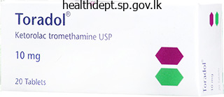
Toradol 10 mg cheap otc
Their colour is pale to deep brown neuropathic pain treatment guidelines iasp purchase toradol 10 mg line, relying on the pores and skin colour of the person pain medication for dogs cancer toradol 10 mg buy on line. Predisposing components Part 12: NeoPlasia There have been reports of lentigines creating after topical immunotherapy with tacrolimus, squaric acid dibutylester and diphencyprone [8,9]. Pathology There is a slight improve within the variety of melanocytes along the dermal�epidermal junction, with out nesting. The epidermal rete ridges are normally elongated and there might be a gentle inflammatory infiltrate within the upper dermis. Differential analysis the differential diagnosis of lentigines from freckles is made clinically by their comparatively darker color, more scattered distribution and by their unchanged standing in relation to daylight publicity. In distinction to freckles, lentigines present histologically with an elevated variety of melanocytes. The differentiation of lentigines from small junctional naevi is usually impossible on clinical grounds. On histology, naevi present Genetics Multiple lentigines arising early in life on both exposed and non uncovered areas are usually a manifestation of inherited syndromes Simple lentigo 132. However, there are circumstances of larger lesions that clinically appear as lentigines, however have small nests of naevus cells alongside the dermal�epidermal junction. These transitional lesions may be considered precursors of future melanocytic naevi [1]. This overlap is clinically insignificant, as each are markers of light pores and skin complexion, excessive sun exposure and a sure threat of pores and skin most cancers (see Chapter 143). As the bulk arise in sunexposed areas, photoprotection could lower the speed of new lesions developing. For beauty reasons, a wide range of depigmenting topical brokers and dermatological procedures corresponding to chemical peels, lasers and photodynamic remedy reduces their pigmentation (see Chapters 159 and 160) [8]. In patients with a number of lentigines arising early in life on nonsunexposed sites, the potential of a hereditary multisystem syndrome (Table 132. In the very uncommon circumstances of generalized lentiginoses, individuals could additionally be at elevated risk for melanoma, and thus must be educated on avoidance of sunburn and self skin examination. Environmental elements Solar lentigines are associated with both intermittent and chronic solar exposure [3]. Solar lentigines on the again have additionally been associated with a historical past of sunburns before the age of 20 years, whereas facial solar lentigines have been related to cutaneous indicators of photodamage [3]. They are macular, tan colored and may be very giant, with a putting irregular border. There is regularly a history of acute sunburn, followed by the sudden look of huge numbers of those irregular macular lesions [13,14]. Once again the lesions are large and macular, have an irregular edge and are often a uniform shade of brown. They are situated in an area of clinically evident sundamaged dermis and infrequently manifest a scientific and pathological overlap with flat seborrhoeic keratoses. Solar or actinic lentigo Definition and nomenclature A photo voltaic lentigo is a brown macule appearing after excessive sun exposure. Synonyms and inclusions � Senile freckle � Lentigo senilis � Old age spot Epidemiology Differential analysis Differential analysis consists of simple lentigo, seborrhoeic keratosis and, in some instances, lentigo maligna and melanoma in situ. Pathophysiology the pathogenetic mechanism resulting in the event of solar lentigines remains unclear. Disease course and prognosis It has been proposed that solar lentigo may generally evolve into seborrhoeic keratosis. Investigations Dermoscopy is similar as in easy lentigo, whereas adjacent pores and skin might show features of photodamage. Solar lentigo ought to be differentiated from seborrhoeic keratosis exhibiting milia like cysts, cerebriform patterns and sharply demarcated borders. In some instances, distinction between photo voltaic lentigo and lentigo maligna or melanoma in situ could be very difficult when traditional pathology is used. Part 12: NeoPlasia Predisposing elements A historical past of occupational radon publicity, in addition to leisure solar publicity, has been implicated in the improvement of multiple lentigines in a single case report [11]. Pathology the histological features of photo voltaic lentigo are the same as for easy lentigo. Solar elastosis of the dermis and photoactivation features of melanocytes are normally current. Management Patients usually request treatment for these pigmented lesions located in visible physique areas such because the face and back of hands. A consensus has supported the usage of cryotherapy as first line therapy, with topical remedy. There is, nonetheless, an intensive literature on the beauty treatment of lentigines utilizing a variety of other remedies corresponding to intense pulsed gentle and pigmentspecific (Qswitched) laser systems (see Chapter 160). Solar lentigines are benign lesions, however any pigmented lesion wants cautious analysis prior to laser remedy. There have been reviews of instances that have been referred for beauty laser therapy as photo voltaic lentigines and had been actually melanomas [17]. Furthermore, noninvasive confocal imaging has revealed remaining melanocytes on the site of photo voltaic lentigines of all topics after Qswitched ruby laser treatment, although there was hardly any observable skin pigmentation on clinical examination [18]. Investigations Dermoscopy helps to establish the prognosis, revealing similar findings to solar lentigines. There is a slight enhance of melanocytes between the epidermal basal cells, and melanophages in the papillary dermis could be seen. Synonyms and inclusions � Mucosal melanosis/genital lentiginosis (these terms have been used interchangeably) (a) Introduction and common description Some degree of macular pigmentation of the mucosa in the mouth or on the genitalia is regular. These macules outcome from native extreme pigment production with regular numbers of melanocytes [27]. Rarely, these melanotic macules might broaden to a number of centimetres in measurement and be patchy in distribution. They could develop irregular deep pigmentation, which resembles melanoma and causes concern. Since melanoma in the genital area could begin with a protracted in situ phase that clinically resembles a mucosal melanotic macule, a biopsy is usually carried out to exclude this possibility. Age on uncovered areas, such because the higher back, and although the identical patient may have a number of solar lentigines, inkspot lentigines are commonly solitary lesions. Differential diagnosis Due to their black color, differential diagnosis primarily includes lentigo maligna and melanoma in situ. Ethnicity Pigmented melanotic macules occur in all races, however are extra common in people with darker pores and skin complexion. Investigations Dermoscopical features include a outstanding black network with thin and/or thick traces. Associated ailments Pigmented macules in the female genitalia have been occasionally related to lichen sclerosus [30]. Histological examination of those lesions reveals features of atypical melanocytic naevi.
Cheap toradol 10 mg amex
Other issues in repositioning embrace: � Maintaining the head of the mattress at lower than 30 levels to minimize shearing blue ridge pain treatment center harrisonburg order 10 mg toradol. Air of gel stuffed systems Nonpowered Air fluidized Powered Alternating pressure Powered Part 11: ExtErnal agEnts 124 florida pain treatment center miami fl buy toradol 10 mg fast delivery. A patientcentred approach should tackle concerns similar to pain, mobility and private elements. Both preventative and remedy measures must happen simultaneously for optimum management of those complicated sufferers. Nonhealable wound Examples: � Untreated Illness � Adherence issues � Health care system issues (funding) Examples: � Inadequate blood supply � Untreatable illness � Lack of nutrition Riet 1995 [41]. Moderate proof was found that wound enchancment was superior with air fluidized beds compared to standard hospital beds [37,38]. In particular, the evaluation of powered versus non powered support surfaces show inconsistent results in treating stress ulcers [39]. This will give the patient, household and other well being care professionals sensible expectations. A wound is considered healable when the underlying points contributing to the wound could be corrected. A upkeep wound is a wound that could heal; nonetheless, there are well being care delivery points or patientrelated elements stopping the wound from therapeutic. Examples of this embrace the shortage of pressure offloading devices or anticipated patient difficulties in adhering to the therapy plan. In a stress ulcer this might be due to an irreversible vascular downside, an underlying malignancy or comorbidities. Intrinsic elements Mobility Perfusion Skin changes Other Extrinsic elements Pressure Shear Skin microclimate Patient centred Pain Mobility Social issues Nutrition There has been interest in the results of nutritional supplementation in the promotion of pressure ulcer healing. In 12 research, protein supplementation was found to be useful in the discount in ulcer measurement [37]. A research from 1974 confirmed that vitamin C 500 mg twice every day did cut back the scale of pressure ulcers compared to placebo [42]. There could also be a task for vitamin C supplementation, however at current unambiguous evidence for benefit is lacking [44]. Dressing kind Films Uses in stress ulcers � Protects skin in danger for friction � Autolytic debridement � Uninfected wounds that are granulating with minimal exudate � Autolytic debridement � Clean stage 2 ulcers � Low to moderately exudative wounds � Moderate to heavy exudating wounds � Useful for haemostasis � A randomized management trial found this to be superior to dextranomer paste [46] � Stage 2 or shallow stage 3 � May shield from shearing and friction � Moderately exudating wounds � For highly exudative wounds � Autolytic debridement � Moderately exudating wounds � Avoid in deep wounds � Antiinfective � Gauze not at all times optimal as a result of it might go away dressing residue within the wound � May use gauze strips to fill deep ulcers or sinuses � May be impregnated with polyhexamethylene biguanide Debridement Necrotic tissues (dead cells and debris) along with slough (fibrin, cells and proteinaceous material) can type on the surface of wounds, promoting the growth of bacteria and affecting the moisture steadiness. It may cause pain and bleeding; nevertheless, this methodology can encourage a stalled wound to begin healing as a response to the now acute wound. It includes applying a saline soaked gauze to the wound, permitting it to dry, and then eradicating it and pulling off the lifeless tissue. However, these methods can cause injury to the healthy granulation tissue and are sometimes painful for the patient. Hydrogels Hydrocolloids Calcium alginates Foams Hydrofibres Cadexomer iodine Gauze Dressings the dressing ought to be chosen to optimize moisture at the wound bed, to prevent injury to the encompassing skin and to treat superficial an infection when present. If packing is required, the dressing have to be of enough power to be eliminated intact, stopping strands being misplaced in the ulcer or parts of the dressing being retained within the ulcer [41]. There is some controversy relating to brokers corresponding to poviodineiodine; nevertheless, proof from non randomized managed trials has supported their use for these specific kinds of ulcers (Table 124. It is unknown if biological agents such as plateletderived progress factor are costeffective in comparison with standard wound care. The prognosis of an infection in a continual wound may be difficult, as the traditional indicators of an infection will not be current. Patients with diabetes have a 10fold elevated danger of being hospitalized for softtissue an infection in comparability with persons with out diabetes [47]. The reference standard for an infection using a deep tissue biopsy is a hundred and five microbes per gram of wound tissue or any stage of haemolytic Streptococcus [49]. In evaluating the signs often related to wound an infection, including odour, ache, pink granulation tissue, exudate, delayed therapeutic, heat, purulent discharge and pocketing; only ache was predictive of infection [51]. Other studies not using a reference standard and combining at least three of five indicators (non therapeutic, exudate, friable tissue, debris and smell) showed a sensitivity of 73% and specificity of 80% for superficial infection [50]. For deeper infection, combining three of seven signs (increasing Part 11: ExtErnal agEnts 124. The Infectious Disease Society of America defines contaminated diabetic foot ulcers as these producing purulent discharge or bearing two or more indicators of inflammation (pain, erythema, induration, heat or oedema) [51]. If pain is increased, it ought to alert the clinician to think about infection as the cause [51]. Diagnosis of infection therefore remains challenging and the subject of additional investigation. While the consensus is that stress ulcers are largely preventable, there could also be individuals for whom stress ulcers are unavoidable [59]. There are few highquality research that consider pressure ulcer development in sufferers with advanced disease [60]. The potential unavoidability of stress ulcers can solely be determined after preventative care has been totally applied. It can be clear that pores and skin failure in the palliative care setting is a separate entity from strain ulcers. This pear or butterfly formed ulcer normally at the sacrum develops shortly and incessantly indicates death is imminent [63]. With superior sickness, physiological modifications occur within the pores and skin as part of multiorgan failure. The goal for each unavoidable and endoflife pores and skin adjustments is to present these fragile patients comfort measures specific to their wants. Adjunctive therapies Negative pressure wound therapy involves the appliance of controlled suction to the wound mattress via a computerized unit hooked up to an opencell foam dressing positioned in the wound and held in place by an adhesive clear dressing [52]. In: International Review: pressure ulcer prevention; pressure, shear, friction and microclimate. Infectious Diseases Society of America clinical practice guideline for the prognosis and remedy of diabetic foot infections. Pressure ulcer remedy strategies: a systematic comparative effectiveness review. The two commonest reconstructive procedures are the musculocutaneous and the fasciocutaneous flap. There is a high recurrence fee with surgical procedure and sufferers should be fastidiously screened. Exposure to chilly causes constriction of the arterioles and veins by a direct mechanism mediated partially by endothelial synthesis of the vasoconstrictor peptide endothelin1 [1]. A reflex increase in sympathetic tone is triggered by chilly receptors in the skin and, if the blood temperature falls, by the hypothalamic heatregulating centre. Heat conservation is additional enhanced by a countercurrent trade system between the arteries and veins within the limbs. Coldinduced vasoconstriction causes shunting of blood to the deep venous system which allows warmth to be transferred from the arteries to veins. Consequently, arterial blood passing into the limbs is cooler, venous blood returning to the body is hotter and less heat is misplaced to the skin setting.
Buy toradol 10 mg fast delivery
However dfw pain treatment center buy generic toradol 10 mg line, in a small Australian study pain treatment center west hartford ct buy toradol 10 mg on-line, employees with epoxy resin allergy had a worse outlook than could be expected in spite of avoidance [9]. Greater age, associated atopy, and longer duration and severity of dermatitis at analysis correlated with poorer prognosis in this survey. It is obvious from a selection of different research that poor compliance and understanding results in a higher price of ongoing exposure to the causative allergen, and is associated with a worse prognosis [10�12]. As the pores and skin integrity is compromised, there are enhanced opportunities for new sensitivities to medicaments or other substances to develop during the course of dermatitis. During a long course of relapsing dermatitis, sensitivity to numerous allergens might accumulate, and this increases the danger of recurrence or persistence [13]. Contact dermatitis of the palms is often of combined origin, with alternating or simultaneous exposure to allergens and irritants. In one examine of the prevalence of dermatitis of the palms, half the patients had suffered from their dermatitis for more than 5 years. Follow up after 6�22 months revealed onequarter had healed completely, half had improved and onequarter had been unchanged or worse. The concept of persistent postoccupational dermatitis despite avoidance of the original cause(s) is now well established, and will occur following each irritant and allergic contact dermatitis [15]. The degree of sensitivity might decline until boosted by repeated exposure, but with a high initial stage of sensitivity it usually remains demonstrable even several years later [17]. Relapse or chronicity is due not only to unavoidable or unrecognized reexposure to allergens and irritants but also to other contributory mechanisms. Recovery is prevented by exposure to allergens or irritants in concentrations that might properly be tolerated by regular skin. The methods have developed into a usually standardized methodology worldwide, although there are some variations, significantly with regard to reading times and check units. Patch testing relies on the observation that primed antigenspecific T lymphocytes will be present all through the body, and therefore allergen within the patch check may be applied to normal skin, often on the upper again the place the checks are least likely to be disturbed. The take a look at depends on the allergen being absorbed in sufficient quantity to induce a reproducible irritation of the pores and skin at the web site of utility in sensitized topics. Indications It is nicely established that aimed patch testing with a few suspected allergens is suboptimal. The cause is that even experienced dermatologists are poor predictors of the finish result of patch checks; 17% of sufferers with allergies were missed on a prepatch test assessment in a single large clinic [1]. This parallels our own expertise, with 20% of allergic sufferers considered positively not allergic prior to patch exams and, conversely, 16% of patients thought to have contact allergy who have been negative when patch examined. An audit of patch testing has instructed that the investigation is underused, and consequently essential opportunities to improve or resolve doubtlessly disabling and wrongly categorized eczema/ dermatitis are lost [2]. The audit concluded that facilities should be obtainable to patch test no much less than 142 per a hundred 000 inhabitants annually and that patient with the indications listed in Box 128. Furthermore, the investigation has been shown to be costeffective and to scale back the worth of remedy in sufferers with severe allergic contact dermatitis [3,6]. The amount of allergen is outlined by its focus in the vehicle and the quantity applied. By testing the identical allergens in parallel, the approach has been confirmed to be typically reproducible [7,8]. The procedure ideally must be delayed till the take a look at website has been away from eczema for at least a fortnight. This information must be given to the patients before they book their appointments. Corticosteroids and other immunosuppressive medication must be stopped (if this is feasible) before patch testing as they might reduce or extinguish positive patch checks in sensitized topics. Young youngsters, even infants, can be patch tested when indicated, but the variety of allergens examined may should be lowered due to lack of area [10]. Test materials Allergens are obtainable from the next manufacturers or from their native distributors. The commonest system used to apply allergens is the Finn chamber (Epitest Ltd, Oy. The chambers are provided in strips of 5 or 10 Methods the premise of testing is to elicit an immune response by challenging already sensitized individuals to outlined quantities of allergen and Investigations 128. They are mounted on nonocclusive tape with an acrylic primarily based adhesive backing that has been chosen for its hypoallergenicity. Other methods consist of sq. plastic chambers (Van der Bend chambers), and oval plastic chambers (Epicheck). This system has been tested in parallel with the established Finn chamber system and there was close correlation of outcomes [11]. It is a consistent, handy, portable methodology for those wishing to check only a few allergens, however supplementary tests are necessary to achieve a comprehensive range of investigations [12]. It has been estimated that by utilizing preprepared checks alone between 60% and 70% of relevant allergic reactions could additionally be missed. In order to avoid an irritant effect, they must be blended or dissolved in a automobile to achieve a suitable check concentration. If a dispersion of allergen in petrolatum is used, contact with the pores and skin is determined by the scale of the particles and on their solubility or dispersion in petrolatum. Many substances may additionally be dissolved in water, alcohol, acetone, methylethylketone or olive oil, as appropriate. False constructive or false negative reactions might happen when inappropriate vehicles are used. Petrolatum is mostly extra reliable, and has the added benefit of being occlusive, which helps to stop oxidation and prolongs shelflife. In sizzling climates, petrolatum may not be perfect, as it melts too rapidly between preparation and application of the patch take a look at. A collection in modified Plastibase has been devised for the Indian Contact Dermatitis Group [14]. Patch test concentrations the choice of an acceptable focus is of basic importance. Excessive concentrations end in false positive reactions, due to their irritant effect, and will even sensitize patients; insufficient concentrations produce false adverse outcomes. The focus of allergen routinely employed for patch tests may, under some situations and in some people, give rise to false unfavorable or false positive reactions. The selection of focus is thus a compromise, but most have been chosen by long expertise with commonly used allergens. The concentrations used for patch testing are often a lot larger than these encountered during the improvement of dermatitis. To demonstrate the existence of nickel dermatitis produced by the minute quantities dissolved from nickelplated objects, a 5% focus of nickel sulphate in petrolatum is critical. Lists of suitable concentrations and automobiles are provided within the text by De Groot (see Resources list).
Buy generic toradol 10 mg online
This in turn might current the protein allergens to Thelper 2 cells pain medication for dogs dosage buy 10 mg toradol amex, inducing a delayedtype hypersensitivity response resulting in eczematous lesions pain treatment in dogs 10 mg toradol generic overnight delivery. Genetics There is a powerful affiliation of allergic contact urticaria with inherited atopic disease of all varieties. There can additionally be a reported association between the event of immediatetype hypersensitivity reactions, specifically to peanut, with filaggrin mutations in these people [8]. Environmental factors Contact sensitization develops following exposure, and changes in the publicity to an allergen in the setting will affect the event of the allergy. This has been demonstrated by the management of latex allergy by environmental measures and using lowproteinpowderfree gloves [9]. Pathophysiology the mechanisms underlying contact urticaria are divided into two main types � immunological and nonimmunological. Nonimmunological contact urticaria is the extra frequent type of the condition and is secondary to the discharge of vasogenic mediators without the involvement of immunological processes. The medical reaction is commonly erythema without oedema quite than a true weal and flare response. Substances capable of producing nonimmunological contact urticaria are often lowmolecular weight chemical compounds that simply cross the barrier of the pores and skin, corresponding to vegetation, animals or chemical substances. Many of these chemicals are used as flavourings, fragrances and preservatives within the cosmetic, pharmaceutical and meals industries [5]. The pathogenesis of immunological (allergic) contact urticaria entails the traditional kind 1 hypersensitivity response mediated by particular immunoglobulin E in a presensitized individual. IgE binding on mast cells, its subsequent degranulation and release of histamine and different vasoactive mediators is described in Chapter 8. Clinical options Presentation the signs often occur within 1 h and fade by three h. Local signs embody itching and burning, and the development of erythema and the attribute weal and flare response. Early symptoms are commonly missed by physicians though properly recognized by sufferers. Exposure to allergens in those who are highly sensitized, topically or through the oral or respiratory route, may lead to widespread urticaria and swelling of the mucous membranes, resulting in conjunctivitis, rhinitis, oropharyngeal swelling, bronchoconstriction and rarely anaphylaxis. The commonest causes of contact urticaria are foodstuffs, which can provoke oropharyngeal symptoms (the oral allergy syndrome) following ingestion, or cutaneous hand symptoms in food handlers. The oral allergy syndrome (pollen�fruit syndrome) results from consuming raw/uncooked/ unprocessed fruits, greens and nuts. Symptoms are normally confined to the localized mucosal surfaces with irritation or tingling. In the birchrich areas of northern and central Europe almost all birch pollenallergic sufferers are sensitized to Bet v1, the main allergen component of pollen from Betula verucosa. It has been reported that in patients with atopic eczema and raised IgE levels, the IgE might attach to the highaffinity IgE receptors on the surface of Langerhans cells or other antigenpresenting cells. In Scandinavia, the most common reason for occupational contact urticaria is from cow dander in farmers [14]. Of those staff uncovered to contact allergic urticants, bakers, butchers and agricultural staff seem to be most vulnerable to becoming sensitized, more so than health care staff. The allergens are present within the watersoluble protein moiety of the sap collected from the rubberbearing tree Hevea braziliensis, harvested mainly in Malayasia and SouthEast Asia. These gadgets are vulcanized at a lower temperature than stable rubber merchandise corresponding to tyres, seals and gaskets. During the manufacturing course of, the pure rubber latex was not left to stand in holding tanks as lengthy, the process was shortened by decrease vulcanization temperatures, and there was much less thorough washing of the final product [16]. All these measures led to an increase within the protein content of the gloves and this, coupled with their increasing use, resulted in a rise within the incidence of allergy to pure rubber latex. Many of the allergenic peptides in pure rubber latex crossreact with those found in other plants [17], such as banana, lychees, chestnuts and avocado, and sufferers allergic to latex could exhibit sensitivity to such meals. Due to several interventional measures together with an increased use of powderfree latex gloves, and nonlatex gloves and medical equipment, the frequency of pure rubber latex allergy in health care workers seems to have peaked [9]. Hjorth and RoedPeterson (1976) outlined this as an immediate dermatitis induced by contact with proteins. They described 33 caterers affected by itch within 10�30 min following contact with meat, fish and vegetables, which was adopted by erythema and vesicle formation. Application of the relevant meals to the affected pores and skin resulted in both urticaria or eczema [18]. Characteristically, the condition involves skin websites that have been affected beforehand by dermatitis. Damaged pores and skin in all probability facilitates penetration of the allergens, and inflammatory cells already current within the dermis may clarify the accelerated medical response. An affiliation between atopy and protein contact dermatitis occurs in approximately 50% circumstances. Other clinically important meals allergies generally associated with Bet v1 are reactions to the Apiaceae family. The Bet v1 homologous proteins in the Rosaceae household are very sensitive to warmth and proteases and the scientific reactions are often aborted when eating cooked or tinned types of the fruit. The most necessary molecular basis of the latex�fruit syndrome is the homology between the hevein (Hev b 6. This immunological phenomenon of crossreactivity has penalties for the diagnosis and remedy of sure meals allergies (Table 128. It is essential to decide the clinical relevance of these crossreactions, and whether or not they symbolize sensitization only or precise medical reactivity (allergy). The dermatologist may be called upon to decide the risk of reactions to related foods and a selection of other plantderived foods that will share proteins with pollens, latex and each other. Clinical evaluation requires a cautious historical past, skin prick exams and IgEmediated blood tests. The use of componentresolved diagnostics that detects IgE antibodies to single allergen parts can be used to assist in this course of. Investigations the prognosis of contact urticaria relies on a full medical history and pores and skin testing with suspected substances. In explicit, the examine of persistent hand eczema in skilled food handlers ought to include both immediate and delayed exams. A detailed historical past concerning the prevalence of instant signs � whether or not confined to the pores and skin or not � and their affiliation, notably with occupational publicity, should at all times be considered. With an unknown allergen, exposure should be graded with an initial application check (open and subsequently occluded) adopted by a prick check and, if acceptable, an intradermal check. Von Krogh and Maibach produced tips for evaluating immediate kinds of cutaneous response [22]. If that is negative, the test is then repeated on slightly affected pores and skin or beforehand affected skin. The use of alcohol vehicles or the addition of propylene glycol to the car enhances the sensitivity of the test [23]. A constructive end result occurs when oedema and/or erythema is observed, usually within 15 min.
Buy toradol 10 mg with amex
The faulty group of the dermal�epidermal junction or of the superficial dermis seen in the mechanobullous issues predisposes to blister formation with trivial trauma pain management for dogs with pancreatitis generic 10 mg toradol mastercard, and individuals with disorders of the connective tissue sciatica pain treatment youtube generic toradol 10 mg line, such as Ehlers�Danlos syndrome and Marfan syndrome, show irregular fragility to mechanical harm. Some drugs, notably corticosteroids, can modify the structural integrity of the skin. Occasionally, structural changes within the pores and skin defend patients from mechanical harm. In amyotrophic lateral sclerosis, pressure ulcers happen less than in comparably bedridden patients, in all probability because of extra dense packing of collagen fibrils [11]. Finally, there appear to be reproducible variations in response between people which might be poorly understood. The discussion of mechanical injury to the pores and skin on this chapter is limited to those results that will concern the dermatologist. Little is known concerning the pathogenesis in other conditions in which the Koebner phenomenon occurs. Isomorphic (Koebner) response Introduction and common description Koebner initially described the localization of psoriasis to pores and skin injured by a variety of stimuli [1] but the time period has been used for the same phenomenon in different illnesses [2]. The isomorphic response is the event of lesions in beforehand regular pores and skin that has been subjected to trauma [1,2]. The term Koebner response is greatest not used when a dermatosis results from the unfold of an infective agent. It differs from the isotopic response [3,4], by which a dermatosis happens on the website of a earlier healed and unrelated dermatosis. In psoriasis, the Koebner response happens in about 20% of patients, but reported series range widely [5]. The latency is about 10�14 days, and a Koebner response is extra more probably to happen when the illness is lively. As well as in psoriasis [9], the Koebner response is commonly seen in lichen planus [2] and vitiligo [10,11�13]. It has been nicely recorded in lots of other diseases, a few of that are proven in Table 123. Utilization of mechanical stimuli Selective use could additionally be made of mechanical stimuli to affirm the prognosis or to permit for the biopsy of early lesions in conditions by which dynamic adjustments and secondary effects happen quickly [1]. Simple frictional trauma, corresponding to that attributable to twisting a Biomechanical considerations desk 123. Disease Carcinomas Darier illness Erythema multiforme Granuloma annulare Hailey�Hailey illness Leukaemia Lichen planus Lichen sclerosus Lupus erythematosus Scleromyxoedema Multicentric reticulohistiocytosis Necrobiosis lipoidica Pemphigus foliaceus Perforating collagenosis and folliculitis Psoriasis Myxoedema, pretibial Vasculitis Vitiligo Xanthoma reference Fisher et al. The pustular reaction to pores and skin puncture (including venesection) is evidence of an energetic stage of Beh�et syndrome. Firm stroking of the pores and skin could elicit purpura in amyloidosis, is routinely used to diagnose dermographism, and has additionally been used to research the early lesions of vasculitis [7] and to verify a diagnosis of delayed stress urticaria [8]. The era of mechanical forces within the skin and subcutis has long been of interest to surgeons, with early contributions by Langer on the oval shapes produced when spherical punctures are made in the skin, and the popularity of relaxed pores and skin rigidity traces [6]. The mechanical qualities of skin, particularly creep, are critical to understanding expansion strategies utilized in dermatological surgery [7]. The effects of mechanical forces have additionally been studied extensively in relation to wound therapeutic and the consequences of extra fluid in tissues [8]. The utility of mechanical drive intermittently leads to extra cellular activity than a constant drive [12]. The importance of weight bearing on bones for their normal structural integrity was recognized long ago, and the stress imposed by gravitational forces can also be essential for the upkeep of dermal constituents [14]. A quantitative evaluation of the mechanical properties of skin and subjacent tissues must begin from engineering principles [1,15]. Most research involve the measurement over time of deformation produced by a given constant drive. Stresses perpendicular to the surface are termed normal, whereas those in different instructions are termed shearing stresses. Many organic materials mix the traits of elastic solids and viscous liquids and are termed viscoelastic. Skin, in common with different viscoelastic materials, has nonlinear stress�strain properties and has timedependent behaviour even with low masses [16]. These timedependent properties are thought in part to be a function of the bottom substance. The elastic element is broadly analogous to a linear spring and the viscous component to a dashpot shock absorber. Much of the sooner work on the mechanical properties of the pores and skin was carried out on tissues or their elements (collagen, elastin, and so on. Strengthrelated values corresponding to breaking strain, timedependent creep and nontimedependent parameters, similar to elasticity and viscosity, have been measured, but these research could be unhelpful in predicting the behaviour of complete pores and skin in vivo. External forces embrace friction, stretching, compression, vibration and penetration. The major mechanical properties of pores and skin are stiffness (resistance to change of shape), elasticity (ability to recuperate the preliminary shape after deformation) and viscoelasticity. Quantification of the behaviour of skin subjected to mechanical forces is difficult by many factors. The pores and skin consists of not one however multiple and interrelated functional parts, and its behaviour is topic to the confounding results of physiological phenomena, such because the earlier experience of the tissue, nutritional status, sweating and sebum excretion. Other variables of practical relevance [5] are associated to body site, age, sex and disease � not solely cutaneous illness but in addition systemic. Deformation Uv Uf Ue Ur Ua Time Mechanical properties of the pores and skin Methods of evaluation [1,2,3,4] Many strategies have been used to derive information about the mechanical properties of the pores and skin. Most strategies measure properties of the dermis, though some give data predominantly in regards to the stratum corneum [7,8], and all have limitations. Details of methodology, similar to the realm of skin subjected to suction, are of great importance in understanding which zone of the pores and skin is being evaluated. One of the few studies that immediately compared totally different methods concluded that the suction cup device mainly measures elasticity, whereas the ballistometer predominantly measures stiffness [44]. Application of drive Ue = quick distension Uv = delayed distension Ur = quick retraction Uf = ultimate deformation Ua = restoration after stress elimination Ur /Uf = biological elasticity Uv /Ue = viscoelasticity with respect to quick distension Ua/Uf = gross elasticity determine 123. Determinants of the mechanical properties of the pores and skin Stratum corneum the primary perform of the stratum corneum is to present a limited barrier throughout which exchanges occur with the setting. This pattern is anisotropic and associated to the anisotropy of the underlying dermis. These adjustments cause secondary alterations in the form of the cells in the Malpighian layer and the underlying papillary dermis [2]. The elastic modulus of the individual corneocyte is far greater than of the whole stratum corneum, suggesting that the biomechanical properties of the latter are largely a operate of the substances binding the cells to one another [3,4]. Direct measurements of the biomechanical properties of the stratum corneum using nanotechnology strategies have recently been revealed [5]. The extensibility of the stratum corneum is significantly influenced by the relative humidity and its state of hydration [4,6,7�9].
Discount toradol 10 mg line
Cabinet makers regularly develop a genital dermatitis from the buildup of sawdust on the clothes throughout sawing and planing homeopathic treatment for shingles pain generic toradol 10 mg visa, and by hand contact flourtown pain evaluation treatment center discount 10 mg toradol mastercard. Exotic woods usually tend to sensitize than fir or spruce, though the latter could cause dermatitis in patients delicate to colophony and turpentine. Dermatitis in woodworkers may also be brought on by liverworts and lichens on the bark of bushes. Colophony also can give an publicity sample of dermatitis from its presence in solder fluxes, paper dust, polish and linoleum flooring [3,4]. Resin techniques, notably epoxy resins, including the more unstable amine hardeners, might induce an airborne sample of allergy, especially in the occupational setting. Other causes of this pattern include perfumes, metals, many industrial and pharmaceutical chemical compounds, pesticides, fungicides, animal feed additives, textile dyes and matches [1]. Equivalent patterns of airborne dermatitis could also be seen with sort I allergens, similar to housedust mite antigens in atopics [5]. Photocontact allergy causes a similar distribution, and is mentioned in a separate part on this chapter. Similarly, prior exposure to nickel and chromate in orthodontic home equipment seems to scale back the danger of later sensitization [2,3]. Contact inflammation of the mucous membranes is rare, and is usually secondary to skin sensitization with the identical substances. The oral mucosa of sensitized animals has been shown to have an equivalent mobile phenotype and cytokine expression as the pores and skin when challenged by an allergen [4]. Allergic reactions in the mouth show erythema and swelling, but vesicles are not often seen, besides on the vermilion border. Eczematous reactions of the adjacent skin might happen, and these may be the only signs of an allergic contact dermatitis. The burning mouth syndrome is an entity during which psychological components are necessary (see also Chapter 84). Sometimes, allergens similar to metals, rubber, food components and flavourings have been recognized and the symptoms relieved by contact avoidance [6], however the return from investigations is commonly disappointing [7]. Orofacial granulomatosis has been related to contact allergy to meals components and a few of these individuals may get hold of a beneficial response, often solely partial, to dietary elimination of the identified allergens [8] (see additionally Chapter 110). Allergic reactions to denture materials have been present in some cases [9], due to traces of residual acrylic monomer following the carrying of latest dental home equipment or following their restore with coldcuring resins. Candidal infection can also play a role [10], and is usually current in angular cheilitis. Acrylate allergy has additionally been seen not often after dental restorative work [11], however may be seen secondary to major sensitization from their use in artificial nails. Mercury from amalgam fillings could cause native mucosal [12] or lichenoid [13,14] reactions; perioral dermatitis after dental filling may occur, as may generalized skin eruptions [15]. Contact reactions to different metals have additionally been reported, particularly gold used in dental restorative supplies, and nickel and palladium in orthodontic appliances [16,17]. Toothpaste flavours can cause stomatitis, glossitis, gingivitis, cheilitis and perioral eczema [19]. The nasal mucosa could react to drugs containing antibacterial agents and antihistamines. Corticosteroid allergy from nasal sprays has been reported, with one case of related perforation of the nasal septum [20]. In the conjunctivae, varied medicine are reported to have elicited allergic contact reactions, for instance blocking compounds for the remedy of glaucoma, antibiotics, and preservatives in each medicine and make contact with lens options [21]. Genital mucous membranes may be affected by allergens, notably medicaments, causing dermatitis on the encircling pores and skin [22]. In common with cobalt, however not like chromium, the steel itself sensitizes and is, in follow, essentially the most frequent supply of sensitization [2]. Nickel is probably the most frequent contact allergen, and sensitivity is extra common in girls than in males. The prevalence of nickel sensitivity recorded in a patch test clinic is between 15% and 30%, and is influenced by the relative number of females tested. All age teams are affected, but the prevalence of nickel allergy amongst females tends to rise from 10 years of age onwards [4]. The incidence of nickel dermatitis in Denmark diversified with the total imports of nickel throughout and after the Second World War [5], which means that the level of sensitivity is determined by the total nickel exposure in the environment. In Denmark in the early Nineties it was found that the prevalence of nickel dermatitis within the common inhabitants was eleven. A more modern research from northern Norway showed the prevalence of nickel allergy in ladies was 27. In Kuwait, nickel allergy was commoner in males [8], and in Nigeria [9] and Japan [10] the prevalence was related in each sexes in patchtested sufferers. The prevalence may be higher in some occupational teams, for instance hairdressers, in whom research have shown that 27�38% are nickel allergic [11,12]. Legislation was introduced in Denmark in 1990 with the intention of controlling using nickelreleasing objects in touch with the skin. These laws only relate to metallic items in prolonged and direct skin contact, for instance jewellery, clothes gadgets and spectacle frames. In a big panEuropean retrospective research of patch test data from 1985 to 2010, a statistically vital decrease of nickel allergy was observed in Danish, German and Italian girls beneath the age of 30 years. The reduction in the prevalence of nickel allergy in younger girls was contemporaneous with the introduction of the nickel laws. The prevalence of allergy to nickel in under18s attending for patch checks in Denmark has decreased significantly, from 24. Sensitization is chiefly the results of frequent skin contact with corroded objects containing nickel. A excessive price of corrosion has been documented from nickelplated items, nickeliron, German silver, coin and several other alloys [17]. Chromiumplated steel is usually first nickelplated, and after long use the nickel may reach the floor, for instance on water taps. Stainless steels contain nickel however most are incapable of releasing sufficient quantities to elicit allergic contact dermatitis. Quantitative research point out that repeated exposure to occluded metallic gadgets releasing nickel at a price higher than zero. Jewellery and metal elements of clothing are the standard sources of nickel in prolonged contact with the pores and skin. Transient but probably frequent and repeated publicity might occur from handling cash, keys, scissors, knitting needles, thimbles, scouring pads and different metallic instruments and utensils. Platers and a few steel machinists are essentially at threat of occupational nickel allergy. Other sources embrace pigments in glass, pottery and enamel, electrocautery plates [20], mobile phones [21], laptop computer systems [22], bindi [23], intravenous cannulae [24], tattoo pigment [25],orthodontic home equipment [26], metallic scouring pads and even soaps [27] and detergents [16].
10 mg toradol with mastercard
No treatment has but been demonstrated to be efficient in decreasing the dimensions of pores and skin lesions or in inducing remission [6] pain medication for dogs tramadol dosage toradol 10 mg buy with visa. Synonyms and inclusions � Disseminated xanthosiderohistiocytosis � Montgomery disease Pathology Histologically pain management treatment center wi 10 mg toradol order with amex, this can be a dermal illness with neither epidermal involvement nor epidermotropism. Early lesions present an accumulation of xanthomatized and scalloped histiocytes with some infiltrating lymphocytes. In older lesions, the histiocytes are spindle formed and arranged in a storiform pattern. Superficial papules are 2�10 mm in diameter and yelloworange, while deep subcutaneous nodules are 1�5 cm in diameter and could also be pores and skin coloured or reddish orange due to overlying telangiectasia [5]. Xanthoma disseminatum is characterized by proliferation of histiocytic cells in which lipid deposition is a secondary event, with involvement of the skin, mucous membranes of eyes, higher respiratory tract and meninges. Rarely, other organs could additionally be affected, together with the liver, spleen and bone marrow [1]. Epidemiology the illness predominantly impacts male children and younger adults, but can happen at any age and in either intercourse. Fifty per cent of lesions appear earlier than the age of 25 and 36% of sufferers are youngsters [2]. The lesional cell appears to be an inflammatory lipidladen macrophage with a attribute foamy look which may represent increased uptake, synthesis or decreased efflux of lipids [3]. In older lesions, extra foamy histiocytes are evident and Touton large cells may be observed. At the ultrastructural level, histiocytic cells contain myeloid bodies and membranebound fat droplets. Some described variations in morphology of the cells, similar to foamy xanthoma cells versus oncolytic or epitheliod cells, could also be a perform of when the biopsy was accomplished in the midst of evolution of the disease rather than a distinction in analysis [4]. In 39�60% of sufferers, the mucous membranes are affected, with particular involvement of the lips, pharynx, larynx, conjunctivae and bronchus. Meningeal involvement is frequent, with infiltration at the base of the brain leading to diabetes insipidus in as a lot as 40% of circumstances, which can, however, be transient. Other manifestations of meningeal involvement are seizures and progress retardation. Hepatic and bone involvement have been reported however symbolize uncommon complications of the illness. Diffuse aircraft xanthomatosis Definition and nomenclature In this disease, multiple flat xanthomatous macules and plaques develop in the skin in association with a monoclonal paraproteinaemia. Three scientific patterns have been identified: a uncommon selfhealing form, a persistent, often progressive form and a progressive multiorgan type, which can be fatal. Other types of surgical removing, excision, dermabrasion and electrocoagulation have been used with moderate impact [2,9]. Glucocorticoids, chlorambucil, azathioprine and cyclophosphamide have been efficient in some patients with cutaneous disease [3,10]. Interestingly, one patient had a partial response to a combination of three lipidlowering brokers, rosiglitazone, simvastin and the niacin analogue acipimox [3], and a second patient had a dramatic response after 5 months of simvastatin alone [12]. Epidemiology the disease typically happens in adults however rare paediatric circumstances have been reported [1]. Pathophysiology the condition arises on account of perivascular deposition of lipoprotein�immunoglobulin complexes. Although serum lipid ranges are often regular, they may be raised, probably due to lowered clearance of the lipoprotein�antibody complexes. Pathology the histological options embrace each xanthomatous and inflammatory components. Accumulations of foamy macrophages infiltrate the dermis, with a distinct perivascular accentuation, and are associated with a variable degree with a blended inflammatory cell response [2]. However, association with many different lymphoproliferative problems has been reported, including continual myeloid leukaemia, acute monoblastic leukaemia, persistent lymphatic leukaemia, chronic myelomonocytic leukaemia, lymphoma, Sezary syndrome and Castleman illness [3]. Clinical features Presentation the clinical presentation can vary from asymptomatic to fulminant organ failure. Diagnosis is made on the idea of radiological features of osteosclerosis along with the classic histology. Disorders during which skin may be concerned however systemic component predominates Erdheim�Chester illness Definition and nomenclature this disease is characterized by infiltration of viscera, bones, retroperitoneum and pores and skin. Pulmonary involvement is seen in 25�50% of patients [6], and although largely asymptomatic, the presence of cough and progressive dyspnoea carries a poor prognosis. However, pulmonary involvement was not found to be an impartial predictor of decreased survival in a single massive series [7]. Neurological imaging can reveal thickening of the dura or more rarely intracerebral lots that may resemble meningiomalike tumours [9]. This is predominantly a disease of adults, with a mean age of fifty three years (range 7�84 years) [2]. Disease course and prognosis Erdheim�Chester disease appears to have a significantly worse prognosis in contrast with different histiocytoses, with an general mortality of around 60% in one collection [2], primarily from cardiorespiratory failure from lung fibrosis or renal failure from retroperitoneal fibrosis. Histological examination reveals a xanthogranulomatous infiltration by lipidladen histiocytes inside a mesh and surrounded by fibrosis. These embrace surgical debulking, highdose corticosteroids, ciclosporin, interferon, systemic chemotherapy and radiation therapy [2,11]. The commonest chemotherapy drugs utilized have been vinca alkaloids, anthracyclines and cyclophosphamide [12]. Interferon has been recently recommended as first line therapy due to the sturdy response seen in three patients with advanced illness [13,14]. Although the exact role of imatinib within the treatment paradigm remains undefined, it provides an alternative therapeutic choice for sufferers who fail to respond or are intolerant to interferon. Cells might have a lacuna spacelike clearing at the periphery and scalloped cytoplasm. Followup in 12 patients, with a median followup of thirteen years, showed no recurrence following major excision. Familial seablue histiocytosis Introduction and basic description this could be a rare inherited abnormality of lipid metabolism in which attribute histiocytic cells are found within the bone marrow and different tissues. Seablue histiocytes have been described in a wide selection of problems of lipid metabolism such as Niemann�Pick illness, in patients with prolonged use of intravenous fats emulsions, and in cases of partial sphingomyelinase deficiency and persistent myelogenous leukaemia [1]. Synonyms and inclusions � Solitary epithelioid histiocytoma � Solitary histiocytoma Epidemiology It normally presents in younger adulthood with hepatosplenomegaly and thrombocytopenia, though the age at presentation ranges from 1 to eighty three years [1]. Pathophysiology the organic abnormality is poorly understood, however the situation in all probability represents a storage disease during which glycolipid, phospholipid or each accumulate in histiocytic cells in numerous organs together with the bone marrow, liver and spleen. Introduction and basic description Reticulohistiocytoma is the localized variant of multicentric reticulohistiocytosis.
10 mg toradol fast delivery
There may be coexisting signs of publicity previous to anterior knee pain treatment exercises 10 mg toradol purchase overnight delivery or along with neuropathic pain treatment guidelines 2010 buy 10 mg toradol amex evidence of pores and skin cancer. These may embrace oil folliculitis and hyperkeratoses, described in folks working with mineral oil, and pitch or tar warts. Oil hyperkeratoses were described as being flat, white, round, hyperkeratotic easy plaques, small in diameter and infrequently clustered. Introduction and common description the pathogenesis of vibration white finger is poorly understood. Chronic vibration publicity may harm endothelial vasoregulatory mechanisms by disturbing the endothelialderived stress-free factormediated vasodilatory perform [3]. Differential analysis Skin cancer totally coincidental with the present occupation. Sirtuin 1 single nucleotide polymorphism may be a diagnostic marker for this disease [5]. Pathophysiology Management Prevention of the event of skin cancers is most necessary. In the workplace, it is essential to think about substitution of carcinogens where attainable; an instance is the declining exposure to polycyclic hydrocarbons in latest decades. Protection of the pores and skin, with both protecting clothes or with engineering control corresponding to machine guarding, is important. Since many of the pores and skin cancers are associated with a very lengthy latency interval, you will want to have continued surveillance of older or retired employees. Clinical options Operatives using vibrating instruments, corresponding to lumberjacks, coal miners and street and building workers, are vulnerable to developing vibration white finger. Affected people develop symptoms of Raynaud phenomenon on publicity to chilly or vibration, normally after a few years of working with vibrating tools [2]. Episodic numbness, tingling, colour change and loss of handbook dexterity are typical. Management Widespread data of its causes, controls over period of use of relevant equipment and improved private protecting gear have led to a reduction in the incidence of vibration white finger [2]. It is generally believed that signs of vibration white finger regress a while after cessation of exposure [2]. Impact of preventative methods on pattern of occupational pores and skin disease in hairdressers: inhabitants primarily based register examine. Skin irritation thresholds in hairdressers: implications for the event of hand dermatitis. Allergic contact dermatitis: immunological elements and customary occupational causes. Occupational relevance of optimistic standard patchtest results in employed persons with an initial report of an occupational skin illness. Evidence based mostly pointers for the prevention, identification and management of occupational contact dermatitis and urticaria. Chloracne: a crucial evaluation including a comparability of two series of circumstances of pimples from chlornaphthalene and pitch fumes. Occupational publicity to ultraviolet radiation and danger of nonmelanoma skin cancer in multinational European examine. Treatment ladder First line � Primary prevention Second line � Redeployment Third line � Standard remedies for Raynaud phenomenon such as keeping heat, stopping smoking, nifedipine, etc. Clinical course of occupational irritant contact dermatitis of the palms in relation to filaggrin genotype status and atopy. Cnidarians have tentacles bearing batteries of stinging cells (nematocysts) which are used for defense and capturing prey. Within every nematocyst is a spirally coiled thread that can be everted, uncoiled and forcibly ejected. In contact with prey, or with human skin, the nematocysts are discharged and the threads inject venom. Many species inflict a minimal of some discomfort on humans, and some are probably dangerous [3�7]. This class contains the fire corals and freefloating members of the subclass Siphonophora. The Siphonophora are colonial organisms in which a quantity of individuals, specialised for various functions, are structurally related. Pathology Histopathology of the acute eruption from a cnidarian sting demonstrates intracellular oedema of the keratinocytes, lots of which have pyknotic nuclei, and a lymphocytic infiltrate in an oedematous superficial dermis [8]. Nematocysts had been visible penetrating the dermis in a 5yearold baby who suffered deadly envenomation from Chironex fleckeri [9]. Histology of recurrent reactions reveals a spongiotic vesicular dermatitis with a dense, perivascular, lymphohistiocytic infiltrate, usually containing massive numbers of eosinophils [10,11]. The most infamous is Chironex fleckeri, which has been answerable for a number of deaths in Australian waters. This class contains a quantity of thousand species, including the sea anemones, the delicate corals and the stony or true corals. The reefforming corals could cause injury to the skin with their nematocysts, or with their calcareous outer skeletons [17]. Clinical options Contact with Physalia tentacles often results in a linear erythematous eruption accompanied by severe local ache [3,18�20]. In humans, pain and skin lesions are normally the boundaries of toxicity, but often extra extreme reactions happen [21]. Haemolysis and acute renal failure in a 4yearold girl [22], and fatalities, have been reported [23,24]. The native effects of field jellyfish tentacle contact are immediate, severe ache and linear weals with a white, ischaemic centre. Box jellyfish could also be responsible not only for local lesions but also for severe systemic results, which can lead to dying [3,four,eight,25�28]. In addition to acute skin lesions, that are regarded as poisonous in nature, there may be delayed, persistent or recurrent eruptions on the original sites of cnidarian envenomation [10,29�35]. Recurrent episodes may be single or multiple, and will take the form of erythema, urticarial lesions, papules or plaques. A delayed hypersensitivity response to jellyfish antigens has been demonstrated by a positive patch test response to a nematocyst preparation from Olindias sambaquiensis [36]. Other reported sequelae of jellyfish stings embody erythema nodosum [37], cold urticaria [38] and Mondor disease [39]. Envenomation by fireplace corals normally produces instant burning or stinging pain, followed by urticarial lesions on the site of contact. These could in turn be followed by a localized vesiculobullous eruption, and subsequently continual granulomatous and lichenoid lesions [40�42].

