

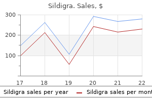
Sildigra dosages: 120 mg, 100 mg, 50 mg, 25 mg
Sildigra packs: 30 pills, 60 pills, 90 pills, 120 pills, 180 pills, 270 pills

Discount 50 mg sildigra
Vitamin D deficiency results in decreased intestinal absorption of calcium and phosphate erectile dysfunction diabetes type 2 treatment buy sildigra 50 mg on line. There is increased osteoblastic activity (A) in rickets laying down an increased quantity of osteoid in an effort to compensate for weak bones impotence effect on relationship sildigra 25 mg generic overnight delivery. Distal interphalangeal joints and large weight-bearing joints are most commonly concerned. Cartilage is weak to damage primarily because cells of the immune system have limited entry to it, mostly due to its avascular nature. Clear fluid with increased inflammatory cells (A, B) is usually found in rheumatoid arthritis. Purulent fluid with elevated inflammatory cells (D, E) is commonly seen in septic arthritis. Multiple pathological fractures (E) may be commonly encountered in multiple myeloma and osteogenesis imperfecta. Correct: Hypertrophic zone of chondrocytes (C) Rickets related to vitamin D deficiency is characterised by growth of the hypertrophic layer. Cells in this layer are metabolically energetic that divide, produce matrix, and initiate vascular invasion. The expansion of the rachitic growth plate is a consequence of impaired apoptosis of the late hypertrophic chondrocytes, an occasion that precedes alternative of those cells by osteoblasts throughout endochondral bone formation. The reserve zone (A, D) normally exhibits no cellular proliferation or matrix manufacturing. The proliferating zone (B, E) contain cells that divide and kind matrix (by collagen synthesis, etc. Aggrecan, with facet chains of chondroitin sulfate and keratan sulfate, is essentially the most abundant proteoglycan of hyaline cartilage. Also, these proteoglycans are answerable for the basophilia of the cartilage matrix. Cutaneous erythema 41 5 Cartilage and Bones Histology and heat and bone tenderness are found over affected areas of the skeleton, reflecting tremendously increased blood flow via the bone. Initial lesions are sometimes osteolytic, with focal radiolucencies; means of accelerated bone reworking leaves cortical thickening and coarse trabeculation, and alternating lytic and sclerotic lesions. Osteoporosis (A) is a metabolic skeletal illness defined as a reduction of bone mass that presents as homogeneously osteolytic lesions. Primary osteoporosis occurs in postmenopausal women or in older men and women as a end result of age-related factors. Secondary osteoporosis results from specific medical problems, such as endocrinopathies, trauma, and inflammation. Osteomalacia (B) is attributable to vitamin D deficiency leading to abnormal mineralization of osseous tissue that happens after the epiphyseal plates have closed. This ends in broad osteoid seams, and a large fraction of bone is covered by non-mineralized osteoid. Multiple myeloma (C) is the most typical major malignant bone neoplasm in adults (> 70% of cases are recognized between 50 and 70 years of age) arising from red marrow because of monoclonal proliferation of the plasma cells. The destruction of bone manifests as osteolytic lesions, bone ache, and pathologic fractures. Rise in the parathyroid hormone results in mobilization of skeletal calcium via rapid osteoclastic turnover of bone to keep regular serum calcium ranges. Radiographic options embody well-defined, purely lytic lesions that provoke little reactive bone. Calcium and phosphate levels, subsequently, are often maintained despite enormously elevated skeletal turnover charges. Correct: C (C) Appositional development types new cartilage on the floor of existing cartilage and includes progenitor cells in the perichondrium (C) differentiating into chondroblasts. Correct: D (D) Interstitial growth forms new cartilage within present cartilage and involves division of lacunar chondrocytes (D). In practically all sufferers, the wrist, metacarpophalangeal joints, and proximal interphalangeal joints are affected, whereas the distal interphalangeal joints are spared. The typical X-ray discovering in rheumatoid arthritis, periarticular bone erosion (loss of joint space), could not develop till later within the illness process. Distal interphalangeal joints and enormous weightbearing joints are mostly concerned in osteoarthritis. Lupus (D), whereas it could have similar medical options, usually presents with asymmetric polyarthritis. Osteoporosis (E) is a noninflam- the image represents hyaline cartilage, recognized by isogenous groups of chondrocytes scattered in a glassy matrix. Because this kind of cartilage lacks particular perichondrium (B), interstitial but not appositional is the first mode of cartilage progress. Cells in fibrocartilage largely happen in isolation, somewhat than in isogenous groups (A). Subperiosteal bone resorption is a classic radiographic discovering for renal osteodystrophy. Osteomalacia (C) could have similar clinical features, but the affected person may have hypocalcemia and hypophosphatemia (since without renal failure). No robust recommendations about morning stiffness, absence of redness or swelling of the affected joints, and sparing of the hand practically precludes the diagnosis. In contrast to other types of osteolytic bone illness, serum alkaline phosphatase exercise is decreased or inside the normal range. Rheumatoid arthritis (A) is an autoimmune inflammatory disease that originally impacts the hands and wrists in a characteristic symmetric, proximal distribution. Primary osteoporosis (C) occurs in postmenopausal women or in older women and men due to age-related elements. Correct: Scanty bony trabeculae (B) Decreased bone density in osteoporosis may be appreciated radiographically by decreased cortical thickness and loss of bony trabeculae. Decreased mineral-to-osteoid ratio (A) is frequent in defective mineralization of bone (rickets, for example). Numbers of each osteoblasts (E) and osteoclasts (D) are unaltered in osteoporosis. Describe neural responses to harm inside the central and peripheral nervous system. A biopsy obtained from the lower brainstem of a 26-year-old male is examined underneath the microscope. A 26-year-old sexually active girl presents with vesicular lesions on the cervix and bilateral painful vesicles on the exterior genitalia. She was treated with oral antivirals and was counselled fastidiously for prevention of recurrence. Virions travel from the preliminary web site of an infection on the mucosa to the dorsal root ganglion, the place latency is established.
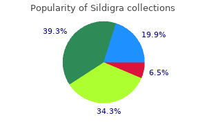
Cheap sildigra 100 mg on line
Her serum -fetoprotein level is raised erectile dysfunction pills canada sildigra 25 mg purchase on line, and an ultrasound reveals a defect within the anterior belly wall of the fetus erectile dysfunction age at onset sildigra 25 mg generic fast delivery. A gastroenterology resident was explaining to his intern how the enteric nervous system includes a number of neural circuits that management motor functions, local blood circulate, and mucosal transport and secretions and modulates immune and endocrine functions. An 18-month-old boy is dropped at the clinic with the chief criticism of passing giant quantities of darkish pink blood from his rectum, and black jellylike stools of two days length. A Tc-99m pertechnetate scintigraphy scan (readily taken up by parietal cells) demonstrates immediate tracer localization within the stomach and in the right decrease abdominal quadrant. A 75-year-old man with a medical history of recurrent pancreatitis was hospitalized for epigastric pain and vomiting. Laboratory reports got here back with elevated levels of serum amylase and serum lipase. Drainage of most of the pancreas through the dorsal pancreatic duct, which opens into the most important duodenal papilla F. Drainage of many of the pancreas by way of the ventral pancreatic duct, which opens into the major duodenal papilla 17. A rotational defect causes the cranial limb of the first intestinal (midgut) loop to endure ischemic necrosis throughout physiologic herniation. A newborn girl presents on her first day of life for evaluation of a perineal mass. The child had regular external feminine genitalia and a sacral dimple, however the resident and the attending have been unable to locate the anal opening. On further inspection, a small opening was famous in the posterior facet of the vaginal vestibule. Failure of rupture of the cloacal membrane and defective growth of the urorectal septum E. Failure of rupture of the cloacal membrane and failure of migration of neural crest cells to the gut wall muscularis externa, the internal and exterior anal sphincters, and serosa/adventitia are derived from mesoderm. The simple columnar absorptive cells lining hindgut derivatives and the upper anal canal, goblet cells, and enteroendocrine cells comprising the intestinal glands are derived from endoderm (A). Extrinsic innervation of the gastrointestinal tract develops from neuroectoderm (C). Correct: Elevated -fetoprotein degree in the amniotic fluid (E) the fetus is affected by gastroschisis, during which intestinal loops herniate into the amniotic cavity via a lateral defect, usually to the proper of the umbilicus. A second- or third-trimester ultrasound and an elevated -fetoprotein (in maternal serum and/or amniotic fluid) are prenatal diagnostic indicators. In distinction to omphalocele, gastroschisis is normally unrelated to chromosomal abnormalities (A) or different severe defects (B, C). Correct: Peritoneal fold that connects the abdomen to the liver (D) the ventral mesentery connecting the abdomen (lesser curvature) to the ventral physique wall is referred to because the ventral mesogastrium. The liver grows in it and divides it into lesser omentum (connects the liver to the stomach) and falciform ligament (connects the liver to the ventral body wall). Peritoneal folds that join the sigmoid colon to the body wall (A, sigmoid mesocolon), jejunum to the physique wall (B, the mesentery), stomach to the transverse colon (C, larger omentum), and stomach to the spleen (E, gastrosplenic ligament) are derived from the dorsal mesentery. Esophageal atresia leads to the lack to drink the amniotic fluid for the fetus, thus the pregnancy is sophisticated by polyhydramnios (not oligohydramnios, A). Chest X-ray following a failed attempt to insert a nasogastric tube (C) confirms the prognosis of esophageal atresia. Dorsal deviation of the tracheoesophageal septum or failed recanalization of the intestine tube ends in esophageal atresia. Neither faulty lateral folding of the embryo (D) nor faulty migration of neural crest cells (E) causes the defect. Correct: Mesoderm (E) In the gastrointestinal tract, the lamina propria, muscularis mucosae, submucosa, inside circular and outer longitudinal (tenia coli) clean muscle of the the neonate has annular pancreas, which happens when pancreatic tissue surrounds and constricts the second part of the duodenum. The affected person often presents with options of duodenal obstruction (abdominal ache, bilious vomiting, nonperistaltic phase distal to and hyperperistaltic segment proximal to obstruction, double-bubble sign, electrolyte imbalance, and so forth. The double-bubble signal is seen in infants and represents dilatation of the proximal duodenum and abdomen. Also, the most common presentation within the toddler 187 22 Digestive System Embryology with ileal diverticulum is painless hematochezia, not intestinal obstruction. Absence of fundic fuel shadow (C) is an indication of obstruction proximal to the stomach. Correct: Failure of the omphalomesenteric duct to involute (C) the omphalomesenteric or vitelline duct is the connection between the embryonic midgut and the yolk sac. This duct provides nutrition to the growing embryo until the placenta is established. If the duct is patent by way of its whole course from the small bowel to the umbilicus, fecal umbilical drainage happens. The streaming of the contrast from the umbilicus through the barium enema confirms an enterocutaneous fistula. Failure of the midgut to recanalize (B) will present as gut atresia leading to intestinal obstruction. Failure of the urorectal septum to develop normally (E) in a male infant would lead to recto vesical or recto urethral fistula. Meconium-stained urine dribbles during micturition (recto vesical) or continuously (recto urethral) with urinary fistulas. Correct: Splenic vein (D) the proximal hindgut is drained by the inferior mesenteric vein, which drains into the splenic vein. The splenic vein then joins the superior mesenteric vein (C) to kind the hepatic portal vein. Ileal (A, ileum) and proper colic (B, ascending colon) veins drain portions of the midgut. The most distal part of the hindgut (lower rectum) drains into the inferior vena cava (E), by way of the interior pudendal and center rectal veins. Correct: Absence/agenesis of the frequent bile duct (C) Elevated conjugated (direct) bilirubin and alkaline phosphatase, dark urine, and pale stool recommend obstructive jaundice. From the listing, only agenesis of the widespread bile duct could cause a posthepatic (obstructive) jaundice where conjugated bilirubin would fail to attain the intestine. The infant is affected by extrahepatic biliary atresia, the most typical form of infantile pathological jaundice, which happens as a result of failure of recanalization of the extrahepatic bile ducts. Typical signs embrace variable levels of jaundice, darkish urine, and light stools (develop over the first few weeks of life). Agenesis of the liver (A) is incompatible with life, and if something, will produce unconjugated hyperbilirubinemia. Correct: Nonrotation of the first intestinal loop (A) During the sixth week, the primary intestinal loop grows rapidly to protrude into the umbilical twine (physiologic herniation). While these processes are occurring, the midgut loop rotates 270� counterclockwise around the superior mesenteric artery. A nonrotated (misnomer) intestine outcomes from partial rotation of the midgut by solely 90� counterclockwise. The small intestine ends up completely to the right and the massive gut entirely to the left facet of the abdominal cavity. Reversed rotated (B) intestine, where the midgut loop rotates clockwise 90� and the large gut enters the belly cavity first, locations the transverse colon behind the duodenum and the superior mesenteric artery.
Discount sildigra 120 mg with amex
When the upper a half of the septum primum progressively disappears erectile dysfunction treatment bodybuilding sildigra 100 mg buy cheap, the remaining half turns into the valve of the oval foramen erectile dysfunction pills not working buy sildigra 50 mg line. After start, when lung circulation begins and pressure within the left atrium increases, the valve of the oval foramen is pressed towards the septum secundum, obliterating the oval foramen and separating the right and left atria. A defect in this process would lead to an ostium secundum (persistent foramen ovale) kind of atrial septal defect. Fusion of septum secundum with endocardial cushions (C) or apoptosis inside the septum secundum (E) by no means occurs during normal development. Correct: Internal iliac artery aneurysm (E) subsequently closes rapidly after delivery. Correct: Pulmonary stenosis (D) Tetralogy of Fallot consists of a ventricular septal defect, an overriding aortic arch straddling the defect, pulmonary stenosis, and proper ventricular hypertrophy. Pulmonary stenosis often leads to lowered blood move to the lungs and an increase in strain to the best ventricle. This strain gradient results in the right-to-left shunting of oxygen-poor blood across the septal defect and out into the systemic circulation, determines the degree of cyanosis, and ends in signs of cyanosis, polycythemia, and hypoxia. Atrial septal defects (A, B), patent ductus arteriosus (C), and left ventricular hypertrophy (E) neither contribute to cyanosis nor are related to tetralogy of Fallot. Correct: Left horn of sinus venosus (A) 196 the umbilical arteries develop from and are direct continuation of the internal iliac arteries. In the grownup, the distal half degenerates and types the medial umbilical ligament, while the proximal half persists as the inner iliac and the superior vesical arteries. Umbilical artery catheterization is a standard procedure in the neonatal intensive care unit and has turn out to be the standard of look after arterial access in neonates. Such catheterization would enhance the possibilities of rupture of an aneurysm within the inner iliac artery, from which the umbilical artery arises. Other main contraindications to umbilical artery catheterization embrace omphalocele, peritonitis, necrotizing enterocolitis, and vascular compromise to the kidneys. The ductus venosus (B) connects the umbilical vein to the inferior vena cava during fetal life and nearly all of veins draining the heart empty into the coronary sinus, which delivers deoxygenated blood to the right atrium. The coronary sinus and the indirect vein of the left atrium derive from the left horn of the sinus venosus. The bulbus cordis (C), and the conus cordis (D, additionally thought-about as the middle third of the bulbus cordis or conus arteriosus), develops into the outflow elements of the ventricles (infundibulum of the proper and aortic vestibule of the left ventricles). The truncus arteriosus (E), additionally thought of because the distal third of the bulbus cordis, develops into the pulmonary trunk and the aorta. Other than cyanosis, the bodily examination is often unremarkable (an necessary differentiating function from tetralogy of Fallot). Correct: Systolic blood strain discrepancies between the higher and decrease extremities > 20 mm Hg (D) Coarctation of the aorta may be outlined as a constricted aortic segment that contains localized medial thickening. The traditional location involves the thoracic aorta distal to the origin of the left subclavian artery. Dilatation of the descending aorta instantly distal to the coarctation section (poststenotic dilatation) is normally present. These sufferers might have appeared properly at start, and abrupt deterioration coincides with closure of the ductus arteriosus. Keys to the diagnosis include blood stress discrepancies between the higher and decrease extremities and reduced or absent decrease extremity pulsation. The murmur associated with coarctation of the aorta is normally a systolic murmur finest heard posteriorly within the left interscapular space, normally with a point of radiation to the left axilla, apex, and anterior precordium. Differential cyanosis (B)-pink higher extremities with cyanotic lower extremities-may happen when right-to-left shunt throughout a patent ductus arteriosus provides circulate to the decrease body. Coarctation of the aorta presents with absent or reduced arterial pulsation in femoral, popliteal, dorsalis pedis (E), or some other lower extremity vessel. Correct: Ascending aorta (A) the ascending aorta develops from the truncus arteriosus of the primitive heart and not from the arch arteries. Ductus arteriosus (E), a communication between the descending thoracic aorta and the pulmonary artery, develops from the left sixth arterial arch. Correct: End of 5 weeks (D) the guts begins to form late in the third week (B) and starts to beat by the 4th week. During the late 4th week (C) cardiac septation begins and is kind of totally full by the end of the fifth week. The end of the 2nd week (A) is simply too early, while septation happens properly ahead of the eighth week (E). Correct: Lateral plate mesoderm, splanchnic layer (C) the center develops from two sources: splanchnic or visceral mesoderm (primary source) and neural crest cells (contribute to conotruncal cushions and their derivatives). Paraxial mesoderm (A) develops into a lot of the axial skeleton, striated muscles, and dermis of the neck and dorsal trunk. Somatic mesoderm (D) develops into inside lining of physique partitions (ventral dermis of trunk, parietal layers of mesothelia, etc. Correct: Aortic stenosis (E) the conotruncal cushions contribute to septate the outflow tracts of the ventricles (conus and truncus) and type the aorticopulmonary septum. Differential growth of tissue from these cushions also contributes to the formation of the semilunar (pulmonary and aortic) valves. Therefore, malformation of those cushions can lead to quite lots of outflow tract septation or obstruction defects, including aortic stenosis. Atrial septa (A, B) are shaped by septum primum, secundum, and endocardial cushions. The muscular part of the ventricular septum (C) is formed by a growth from the primitive ventricular wall, whereas the membranous part has contributions from the conotruncal (bulbar ridges) and endocardial cushions. Correct: Fossa ovalis (D) Fossa ovalis is an oval despair on the atrial septum and is a remnant of foramen ovale and its valve. The ductus venosus (A), a channel that shunts oxygenated blood from the umbilical veins to the 197 23 Cardiovascular System Embryology inferior vena cava, closes soon after birth. The ductus arteriosus (B), a communication between the descending thoracic aorta and the pulmonary artery, constricts inside a day after delivery. The foramen ovale (C) serves as a physiologic conduit for right-to-left shunting between the atria. Functional closure of the foramen happens with improve in left atrial pressure as soon as the pulmonary circulation is established following delivery. Anatomic closure of the foramen (fusion of the septum primum and septum secundum) normally happens inside three months of start. Umbilical vessels (E) obliterate at birth with clamping of the umbilical cord and separation of the placenta. Degree of cyanosis in a affected person with tricuspid atresia depends on the pulmonary blood move. Minimal cyanosis is indicative of pulmonary plethora and guidelines out pulmonary oligemia and hence, pulmonary stenosis (A). The right ventricle is small and hypoplastic (C, not enlarged), since blood from both venae cavae is pressured across the patent foramen ovale into the left coronary heart. The left ventricle (D) and the atrium (E) are hypertrophied, because of the amount overload (receives all venous return from systemic and pulmonary circulation). The proper atrium is characteristically enlarged and hypertrophied and is responsible for the indicators of coronary heart failure.
Sildigra 25 mg buy low cost
Nerve sheath tumors are the most typical and embrace schwannoma erectile dysfunction protocol diet sildigra 120 mg cheap, neurofibroma erectile dysfunction raleigh nc buy generic sildigra 25 mg line, and malignant peripheral nerve sheath tumor. Sympathetic ganglion cell tumors include neuroblastoma and ganglioneuroblastomas, often occur in the pediatric inhabitants, and are sometimes aggressive primitive neoplasms of neuroectodermal origin. Concerning imaging characteristics include necrosis, hemorrhage, and the shortage of a capsule. Important imaging findings to contemplate for all posterior mediastinal lots include vascular encasement, intraspinal extension, rib splaying, and bony erosion. Neurenteric, enteric duplication, and bronchogenic cysts can be grouped collectively underneath the heading of bronchopulmonary foregut cysts/malformations. They are troublesome to differentiate from one another on imaging and basically look the same on most imaging studies. The presence of infection or hemorrhage may end up in less attribute imaging findings which will mimic a solid lesion or abscess. Neurenteric cysts have associated spinal malformations, which, when current, suggest the diagnosis. Although the anterior mediastinum is the basic location infiltrated by lymphoma, posterior mediastinal lymph nodes can sometimes be involved as properly. Homogenous lots with lobulations and the absence of necrosis or calcification in untreated instances will help differentiate lymphoma from other posterior mediastinal plenty. Extramedullary hematopoiesis presents as bilateral however usually uneven paraspinal plenty. Mediastinal widening with related pleural effusion (more generally on the left) is the commonest signal of mediastinal/vascular damage. Diagnosis Schwannoma P Pearls y Neurogenic tumors represent the commonest posterior mediastinal plenty. Organization is a histologic means of fibroblast proliferation within the lung and could be thought of as a lung response to injury, most commonly infection. They are sometimes treated for infectious pneumonia at initial presentation however fail to enhance with treatment. In some sufferers, the distribution can be peripheral, a pattern just like that seen in continual eosinophilic pneumonia. Air-space opacities that persist regardless of scientific remedy should elevate the suspicion of a neoplastic cause. Lung most cancers, significantly adenocarcinoma on this case given the associated ground-glass opacity and central pseudocavitation, can cause a continual air-space opacity and resemble consolidation. Lymphoma may cause a continual consolidation, though the more typical presentation within the lung is bilateral nodules and masses. Chronic eosinophilic pneumonia is an idiopathic process characterised by alveolar and interstitial infiltration of inflammatory cells. Radiographically, homogeneous peripheral consolidations are current, in a sample paying homage to "the photographic adverse of pulmonary edema. Lipoid pneumonia is the results of continual aspiration of merchandise that include oil or fat. As this is normally a longstanding process, fibrosis, necrosis, and even cavitations could also be present. From the radiologic pathology archives: group and fibrosis as a response to lung harm in diffuse alveolar injury, organizing pneumonia, and acute fibrinous and organizing pneumonia. While the bronchial circulation could also be protective within the extra proximal airways, occlusion of distal pulmonary arteries from embolic sources, malignancy, or interstitial edema may result in focal peripheral pulmonary hemorrhage and infarction. Radiographically, this produces wedge-shaped, peripheral areas of consolidation. Most pulmonary emboli are a quantity of with a decrease lobe predominance and end result from deep venous thrombosis. Causes of venous thrombosis are intensive but include trauma, malignancy, hypercoagulable states, and central venous line placement. When infected materials is embolized, typically a complication of intravenous drug use or endocarditis, septic emboli might trigger peripheral consolidations with central cavitation. Heart failure is a crucial predisposing issue for pulmonary infarction in general. Eosinophilic lung disease encompasses a variety of disorders characterized by tissue or blood eosinophilia and pulmonary involvement, usually differentiated by integrating medical presentation with imaging findings. While acute eosinophilic pneumonia sometimes demonstrates a pattern much like pulmonary edema, persistent eosinophilic pneumonia is suggested by homogeneous peripheral consolidations, which radiographically could also be seen as a photographic negative of pulmonary edema. Upper lobe predominance and a protracted course may distinguish chronic eosinophilic pneumonia from different entities, such as Churg� Strauss (granulomatous vasculitis associated with asthma) and Loeffler (eosinophilia and transient and migratory consolidations) syndromes. In the setting of trauma, hemorrhage into the alveolar space may end up in peripheral nonsegmental consolidations, sometimes adjacent to sites of thoracic harm alongside the rib cage. Sarcoidosis most commonly presents as bilateral symmetric hilar and mediastinal lymphadenopathy with or with out parenchymal involvement. The most typical parenchymal manifestations embody perilymphatic nodules and architectural distortion with an upper lobe predominance. Peripheral consolidations related to mediastinal and bilateral hilar adenopathy in an asymptomatic patient may be a rare presentation of alveolar sarcoidosis. Diagnosis Septic emboli P Pearls y Pulmonary infarcts from emboli may present with peripheral wedge-shaped lower lobe consolidations. Pulmonary edema follows a natural development which entails interstitial edema with Kerley B traces, cephalization of move, and resultant air-space disease (groundglass opacities with areas of consolidation). The central and perihilar lungs are concerned early with patchy opacities in a batwing configuration. Cardiomegaly and pleural effusions are seen within the setting of cardiogenic pulmonary edema. Pneumocystis, cytomegalovirus, and respiratory syncytial virus pneumonias may account for ground-glass opacities, significantly in an immunocompromised affected person. Invasive aspergillosis could elicit a ground-glass halo round a focal area of consolidation which represents pulmonary hemorrhage. The lack of cardiomegaly and pleural effusions helps distinguish this entity from cardiogenic pulmonary edema. Air-space disease secondary to pulmonary hemorrhage might manifest as ground-glass opacities, as nicely as ground-glass nodules. An idiopathic illness predominantly affecting middle-aged males, alveolar proteinosis outcomes from persistent accumulation of extreme proteinaceous material in the alveoli. Juxtaposition of regular and involved lung parenchyma has been termed "loopy paving," which in and of itself is a nonspecific discovering. Perivascular irritation within the lung secondary to a vasculitic process might appear as ground-glass opacities. Diagnosis Infection (Pneumocystis pneumonia) P Pearls y Pulmonary edema progresses from interstitial to air-space disease; cardiomegaly and effusions are typical. Coexistent pulmonary disease, including granuloma formation or air-space illness, is usually seen. It sometimes presents as superior mediastinal lymphadenopathy, resulting in mediastinal widening on chest radiographs.
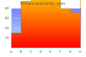
Sildigra 120 mg best
Dual-phase (early and delayed) parathyroid scan makes use of this differential washout between thyroid gland and hyperfunctioning parathyroid tissue to localize an adenoma erectile dysfunction treatment vancouver 100 mg sildigra with amex. While the majority of parathyroid adenomas are solitary and situated adjacent to the thyroid tissue purchase erectile dysfunction pump sildigra 100 mg order line, they can be multiple in quantity and/or ectopic in location (10�15%); hence, the mediastinum is included within the field of view. Frequently, parathyroid adenomas might demonstrate rapid washout of radiotracer (similar to thyroid tissue), leading to a false-negative examine since no discrete focus persists on delayed imaging. Tc99m sestamibi demonstrates nonspecific localization in tumors via passive transport throughout cell membranes and lively transport into mitochondria. False-positive research embrace thyroid adenoma (most common), thyroid carcinoma, and parathyroid carcinoma. Dual isotope imaging (Tc99m sestamibi plus both I123 or Tc99m pertechnetate) with or without subtraction can improve sensitivity and avoid a few of these pitfalls. Focal cervical activity could also be seen inside metastatic lymph nodes from any primary malignancy (including thyroid, head/neck, and breast cancers). Correlation with a scientific history and cross-sectional imaging is useful in these circumstances. Diagnosis Parathyroid adenoma P Pearls y Parathyroid scan is carried out as a localizing process previous to surgical exploration. Parathyroid imaging: technique and function within the preoperative analysis of primary hyperparathyroidism. Additionally, penile activity in a man and uterine activity in a menstruating girl can mimic rectosigmoid bleeding. A larger labeling effectivity ends in much less free or unbound technetium within the bloodstream. Imaging over the neck to detect thyroid activity can verify the presence of free technetium. Scintigraphic analysis of acute lower gastrointestinal hemorrhage: present status and future directions. Nuclear medication within the acute scientific setting: indications, imaging findings, and potential pitfalls. Presentation is usually after the age of sixty five years, and prevalence will increase with age. In general, the amount of decreased metabolism correlates with the degree of signs. In later stages, decreased exercise might be seen within the cortices and will progress along with the disease. Positron emission tomography and single-photon emission computed tomography mind imaging in the evaluation of dementia. However, extrinsic compression of the pulmonary artery is the most typical cause of a perfusion defect which is worse than ventilation. Fibrosing mediastinitis is one other entity which may lead to pulmonary artery occlusion and absence of perfusion. Cross-sectional imaging is important to decide the presence of external compression. Pulmonary artery atresia, extreme pulmonary artery stenosis, or pulmonary webs could cause unilateral absence of perfusion with perfusion defect worse than ventilation. Additionally, corrected congenital heart illness could produce comparable findings on V/Q scan. Pulmonary emboli typically current with wedge-shaped, peripheral, segmental mismatched perfusion defects. A mucous plug may hinder air flow with compensatory vasoconstriction of the pulmonary arteries. The degree of accompanying perfusion abnormality will increase with time with continued obstruction because of arteriolar constriction induced by native hypoxia. As this is primarily an airway abnormality, the air flow defect might be larger than the perfusion defect. Endobronchial lesions could cause a ventilation defect which is larger than perfusion. The relative lower in ventilation to perfusion is an indicator of the extent of luminal involvement. Diffuse air-space disease, atelectasis, or giant pneumothorax involving an entire lung will result in decreased or absent ventilation with compensatory vasoconstriction of the pulmonary arteries. The degree of lung involvement will decide the degree of perfusion and air flow defects. Unilateral hypoperfusion or absent perfusion on pulmonary scintigraphy: differential diagnosis. Unilateral absence of right-lung perfusion with regular ventilation on radionuclide lung scan as an indication of aortic dissection. The significance of unilateral absence of pulmonary artery perfusion by lung scanning. Asymmetrically increased proper renal exercise is seen on the ultimate image at 20 minutes. Renovascular hypertension accounts for about 1�4% of all hypertension instances. Narrowing of the afferent renal artery greater than 60% typically is the low-end cutoff for causing renovascular hypertension through the renin angiotensin cascade. When appropriately performed, the take a look at approaches 90% sensitivity and 95% specificity. A constructive study usually predicts improvement in hypertension upon revascularization. This impact must be bilateral and symmetric (except in circumstances of existing unilateral renal impairment or nephrectomy). In the setting of bilateral renal decline on a renogram within the presence of calcium channel blocking treatment, the research should be repeated after discontinuance of these drugs. Diagnosis Renal artery stenosis (right-sided) P Pearls y Adequate fluid hydration previous to and blood pressure monitoring during Captopril scan are necessary. Calcium channel blockers: a possible cause of false-positive Captopril renography. Cold or nonfunctioning nodules on I123 thyroid scans are nonspecific and may characterize colloid cyst (40%), nonfunctioning adenoma (40%), or thyroid carcinoma (15�20%). Hot or hyperfunctioning nodules on I123 scan, defined as uptake within the nodule suppressing the the rest of the thyroid gland, are primarily all the time benign. Treatment of localized thyroid carcinoma sometimes consists of complete thyroidectomy, adopted by I131 ablation remedy. Patients are adopted up with thyroglobulin levels, in addition to whole body I131 imaging, for at least 2 years after radioiodine ablation.
Chia Oil (Chia). Sildigra.
- Diabetes, high blood pressure, and heart disease.
- Dosing considerations for Chia.
- What is Chia?
- Are there safety concerns?
- How does Chia work?
Source: http://www.rxlist.com/script/main/art.asp?articlekey=97167
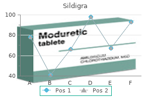
Generic sildigra 120 mg on line
Marked joint space narrowing and bony resorption are seen at the first carpometacarpal joint erectile dysfunction pump walgreens purchase sildigra 100 mg overnight delivery. Hyperparathyroidism is a systemic abnormality of calcium homeostasis that can be main (overproduction by the parathyroid glands) drugs for erectile dysfunction sildigra 25 mg cheap visa, secondary (caused by renal failure or malabsorption), or tertiary (autonomous production by the parathyroid glands due to persistent renal failure or malabsorption). Subperiosteal resorption of bone, particularly along the radial aspect of the second and third middle phalanges of the hands, is virtually diagnostic of hyperparathyroidism. Additional osseous abnormalities embrace resorption of the distal clavicles, bandlike osteosclerosis of the vertebral our bodies referred to as "rugger-jersey" backbone, brown tumors, and resorption of the terminal phalanges. Scleroderma is a systemic connective tissue disorder that ends in attribute musculoskeletal abnormalities that are most outstanding within the arms. The most typical manifestations embrace bony erosions and soft-tissue resorption alongside the distal phalanges. Severe cases end in tapering or complete destruction of the distal phalanges (acro-osteolysis). Both processes cause vascular occlusion and ischemia, resulting in soft-tissue and osseous abnormalities. Osseous manifestations include osteoporosis, periostitis, and resorption of the distal phalanges. In the setting of frostbite, the findings are sometimes bilateral with sparing of the thumbs secondary to clenched fists with the digits protecting the thumbs. Although the distribution is variable, psoriasis has a predilection for the distal interphalangeal joints of the hands. Musculoskeletal involvement of the hand consists of soft-tissue swelling, periarticular erosions, "fluffy" periostitis, and resorption of the distal phalanges. Classic radiographic findings embrace the "sausage digit" (diffuse soft-tissue swelling) and the "pencilin-cup" deformity from resorption and tapering of the distal aspect of the phalanges. Hajdu�Cheney is a uncommon syndrome that may occur sporadically or with an autosomal dominant familial inheritance. Patients have dysmorphic facies and cranial abnormalities, including an enlarged sella turcica, wormian bones, and basilar invagination. Osteolysis occurs throughout the distal phalanges of the arms and feet with traditional bandlike lucencies that isolate proximal and distal osseous fragments in the terminal tufts. Diagnosis Scleroderma P Pearls y Hyperparathyroidism ends in radial-sided subperiosteal resorption of the second and third center phalanges. Additional findings embrace scoliotic curvature of the backbone, in addition to postoperative modifications and dislocation of the proper hip. Renal osteodystrophy, primary hyperparathyroidism, hypervitaminosis D, and fluorosis are among the metabolic problems that may trigger increased bone density. Renal osteodystrophy is usually related to soft-tissue calcifications, osteomalacia, and regions of osteosclerosis preferentially affecting the ribs, pelvis and spine. Osteosclerosis can produce the basic "rugger-jersey" backbone radiographic look. Several main malignancies characteristically produce osteoblastic metastases, together with carcinomas of the prostate, breast, and pancreas; mucinous adenocarcinoma of the gastrointestinal tract; transitional cell carcinoma; carcinoid tumor; lymphoma; medulloblastoma; and neuroblastoma. Differentiation from different causes of elevated bone density can usually be made by correlation with medical historical past and the usually patchy nature of metastases. Myelofibrosis, mastocytosis, and sickle cell anemia are a few of the hematologic disorders that may show diffuse osteosclerosis. In myelofibrosis, bone marrow is replaced by fibrotic tissue, and osteosclerosis is most commonly discovered within the axial skeleton and proximal lengthy bones, particularly the humerus and femur. In sickle cell illness, abnormal red blood cells lead to bone infarcts that result in a markedly sclerotic look, especially in pelvis, backbone, and ribs. Additional findings of spinal manifestations embody "fish-mouth" vertebrae, avascular necrosis, and extramedullary hematopoiesis. The incidence of Paget disease increases with age, with the majority circumstances occurring in aged sufferers. In the blastic part of Paget illness, there are areas of sclerosis with cortical thickening, coarsened trabeculae, and bony enlargement, especially in skull, spine, and pelvis. Osteopetrosis is a rare inherited dysplasia characterized by elevated bone density, particularly in the lengthy bones, cranium, and spine. Despite the increased density, concerned bones are comparatively fragile and prone to fractures. Classic radiographic findings embrace the "sandwich" appearance of the vertebral bodies with a pointy margin between the sclerotic endplates and more lucent bone centrally, as nicely as the "bone-in-bone" appearance happens inside the pelvis and long bones. Diagnosis Osteopetrosis P Pearls y Renal osteodystrophy leads to the classic "rugger-jersey" spine, soft-tissue calcifications, and osteomalacia. Synovial (osteo)chondromatosis is a illness of the synovium resulting from synovial metaplasia, the cause for which is unknown. It is seen in men between the ages of 20 and 50 years and is mostly intra-articular but can also happen in tendon sheaths and bursae. The illness is typically monoarticular, with the knee, elbow, shoulder, and hip joints most commonly involved. Synovial metaplasia leads to the formation of synovial villonodular projections that grow to type nodules. If the nodules stay connected to the synovium, they develop a blood supply and may become ossified. Eighty-five percent have adequate calcification to be detected on radiographs, appearing as multiple, round, similar-sized calcified bodies. It can also happen extra-articularly in a bursa or tendon sheath (giant cell tumor of the tendon sheath). The intra-articular kind is characterised by villonodular proliferation of the synovium with associated hemorrhage. The hemosiderin causes susceptibility artifact and blooming on gradient-echo sequences. Treatment is synovectomy; incomplete resection is associated with high recurrence charges. Multiple small unfastened our bodies can occur inside joints affected by rheumatoid arthritis and are termed "rice bodies" because of their resemblance to polished grains of rice. The actual trigger is unknown, but one concept postulates that "rice bodies" characterize indifferent fragments of infarcted synovium. First described in affiliation with tuberculous arthritis, "rice our bodies" at the second are extra incessantly associated with rheumatoid arthritis. They can, nevertheless, also be seen in the absence of any underlying systemic disorder. Diagnosis Rice bodies P Pearls y Synovial (osteo)chondromatosis is a synovial metaplasia that leads to free our bodies; 85% are calcified. Fibrous dysplasia is a benign skeletal dysfunction characterized by replacement of medullary bone with fibrous tissue.
Syndromes
- Polymerase chain reaction (PCR) test of a sample from an ulcer
- Sarcoidosis
- History of rheumatic heart disease or previous endocarditis
- Zydone
- Congenital heart disease
- Sodium hydroxide
- A synthetic bone substitute can also be used.
Sildigra 25 mg generic without a prescription
Lacunar infarcts occur most often in aged sufferers with microvascular ischemic disease erectile dysfunction drugs natural sildigra 50 mg purchase amex. The an infection affects the meninges and spreads via the subarachnoid and perivascular areas erectile dysfunction treatment chandigarh sildigra 25 mg order without a prescription, which turn out to be distended. The commonest finding is multiple T2 hyperintense lesions within the basal ganglia with surrounding gliosis. Larger lesions are referred to as "gelatinous pseudocysts" and most frequently happen within the basal ganglia. Cryptococcomas are stable or ring-enhancing plenty which most often contain the deep gray matter. Neurocysticercosis is a parasitic an infection attributable to the pork tapeworm (Taenia solium). Lesions might involve the gray/white junction (most common), subarachnoid house, and ventricles. They most frequently occur inside the cerebral hemispheres, thalami, brainstem, and choroidal fissures. Surfactant deficiency is the most typical cause of respiratory distress in preterm infants, especially in these born earlier than 34 weeks of gestation and who weigh lower than 1,500 g. It happens because immature lungs are often unable to produce enough surfactant to hold alveoli open for effective air trade. Clinical manifestations of grunting, nasal flaring, subcostal retractions, tachypnea, and cyanosis are seen shortly after supply and nearly all the time inside the first eight hours of life. Radiographs typically show diffusely hazy and low lung volumes with air bronchograms. The granularity results from diffuse alveolar collapse, whereas the air bronchograms represent normal air-filled prealveolar airways. In extreme instances (extreme prematurity and very low start weight), assisted air flow together with surfactant utility may be wanted to achieve acceptable gasoline change. Potential issues of positive stress embody pulmonary interstitial emphysema, pneumomediastinum, and pneumothorax. Chronic intubation and oxygen administration might ultimately result in bronchopulmonary dysplasia. It is exacerbated with caesarian section due to absence of the thoracic squeeze related to vaginal deliveries, which normally helps clear fetal lung fluid. Patients show clinical enchancment within 48 hours of delivery, and radiographs normalize inside 72 hours. Radiographs typically reveal central streaky opacities and small pleural effusions (with fluid in the minor fissure). More severe circumstances show pulmonary vascular congestion and air-space opacities. Bacterial pneumonias, particularly these brought on by group B Streptococcus, predominate in the neonatal period. Radiographic findings of diffuse haziness and granularity might mimic surfactant deficiency. Pleural effusions are uncommon in surfactant deficiency however are seen in as many as two-thirds of sufferers with neonatal pneumonia. With enough antibiotic remedy, complications corresponding to empyema, pulmonary abscess, and pneumatocele are uncommon but might occur, especially with delayed therapy. Diagnosis Neonatal pneumonia P Pearls y Surfactant deficiency is the most common explanation for respiratory distress in preterm infants. Neonatal respiratory distress: a practical strategy to its analysis and administration. The lack of vaginal squeeze results in retained fetal lung fluid, which is slow to be cleared by the immature lymphatic system. Radiographic options embody regular to increased lung volumes with bilateral diffuse patchy air-space disease and normal heart measurement. Perihilar linear densities and pleural effusions (often manifested by fluid within the minor fissure) are widespread. Air-space disease, when present, is similar in look to pulmonary edema and clears inside 3 days of supply. If radiographic findings persist, various diagnoses similar to cardiac illness or neonatal pneumonia ought to be thought of. Meconium aspiration sometimes occurs in term or postterm neonates with in utero or peripartum fetal distress. The aspirated meconium causes a chemical pneumonitis and areas of airway obstruction that lead to moderate to severe respiratory distress. The diagnosis is suspected clinically when amniotic fluid is stained with meconium. Radiographic look consists of hyperinflation with patchy, asymmetric air-space illness and ropy perihilar opacities. Pleural effusions could also be current, in addition to areas of postobstructive atelectasis. Radiographs sometimes show hyperinflation, as well as patchy, perihilar, or diffuse air-space disease. Occasionally, however, chest radiographs may be regular in the setting of neonatal pneumonia. Beta-hemolytic Streptococcal pneumonia is a separate entity that usually presents with low lung volumes and is thus not included in this differential. The an infection typically resolves over a brief period of time with the correct antibiotic therapy. Congenital heart disease commonly presents with respiratory distress, parenchymal opacities, and hyperexpanded lungs. Radiographic options might include cardiomegaly and pulmonary vascular congestion, depending on the underlying etiology. Diagnostic concerns in an acyanotic infant embody shunt lesions and congestive heart failure. Considerations in a cyanotic toddler include transposition of the great arteries, truncus arteriosus, complete anomalous pulmonary venous return, tricuspid atresia, and single ventricle. The four components which outline this anomaly include right ventricular outflow tract obstruction (pulmonic stenosis), ventriculoseptal defect, overriding aorta, and proper ventricular hypertrophy. Deficiency of the principle pulmonary artery phase and elevation of the cardiac apex as the outcome of right ventricular hypertrophy lead to the traditional radiographic appearance of a "boot-shaped" coronary heart. The main anatomic defect in pulmonary atresia is underdevelopment of the best ventricular outflow tract and pulmonary valve. Although the radiographic appearance varies, severe cardiomegaly is usually seen. Associated anomalies happen in up to 30% of affected sufferers (most generally transposition of the great arteries). Double-outlet proper ventricle happens when both the aorta and pulmonary outflow tracts come up from the proper ventricle. Ebstein anomaly consists of apical displacement of the tricuspid valve, leading to atrialization of the right ventricle.
Cheap 120 mg sildigra with mastercard
Diagnosis Meningioma P Pearls y Nerve sheath tumors (specifically schwannoma) are the commonest intradural extramedullary spinal lots erectile dysfunction treatment comparison order sildigra 100 mg on-line. Patients current with acute onset of fever erectile dysfunction pump implant video purchase sildigra 50 mg otc, complications, seizures, and/or focal neurological deficits. Imaging typically reveals bilateral, uneven involvement of the cortex and subcortical white matter with sparing of the basal ganglia. There is edema with loss of gray-white differentiation and local mass impact, sometimes in a nonvascular distribution. Mild, patchy enhancement may be seen acutely, growing into gyriform enhancement often within 1 week. Patients present acutely with focal neurological deficits, altered psychological standing, and/or aphasia. Arterial etiologies embody thromboembolic disease (most common), dissection, vasculitis, and hypoperfusion. Imaging reveals cytotoxic edema with lack of gray-white matter differentiation and sulcal effacement in a vascular distribution. Both computed tomography angiography and magnetic resonance angiography could reveal the positioning of vascular occlusion. There is involvement of three or extra lobes, and there may be extension throughout white matter tracts to involve the contralateral hemisphere. Involvement of the cortex, deep gray matter, cerebellum, brainstem, and spinal twine may be seen. Limbic encephalitis is a paraneoplastic syndrome related to a major malignancy, usually lung or breast most cancers. Clinically, the onset of symptoms is usually more insidious (weeks to months) rather than acute. Treatment of the primary malignancy could end in stabilization or improvement of symptoms. Seizures result in focal elevated cerebral perfusion and disruption of the blood�brain barrier. There is related ill-defined edema involving the cortex and subcortical white matter; the temporal lobe is commonly involved. Follow-up imaging after cessation of seizures demonstrates improvement or resolution. It is necessary to do not neglect that the area of edema could also be distant from the actual seizure focus. Diagnosis Herpes encephalitis P Pearls y Herpes encephalitis is a life-threatening infection which preferentially includes the limbic system. It most often happens within the temporal lobes and is the commonest neoplastic cause of temporal lobe seizures in adolescents and young adults. Although its imaging look varies, the most common presentation is a superficial mixed cystic and stable mass; the solid elements typically present as a mural nodule and are hyperintense on T2 sequences. As the lesion is cortically based mostly, cortical growth and overlying bony transforming is often seen. Calcifications and enhancement of stable components are famous in approximately half of circumstances. It typically occurs in adolescents and younger adults; the commonest presentation is seizures. Surgical resection must embody the foci of cortical dysplasia to guarantee resolution of seizures. Calcification and enhancement are fairly unusual, occurring in roughly one-fifth to one-third of instances. As the mass is cortically primarily based, enlargement of the cortex and calvarial reworking are commonly seen. Patients commonly present with seizures, headache, and sometimes focal neurological deficits. The majority of instances contain the temporal lobe, leading to temporal lobe epilepsy. The majority of cases show overlying meningeal enhancement, which is a helpful discriminator. Taylor dysplasia is a subset of kind 2 that contains attribute balloon cells on pathologic examination. The signal abnormality is usually wedge-shaped with tapered signal pointing toward the ventricles. Regions of increased cortical signal intensity on closely T1-weighted sequences are pretty characteristic and useful in distinguishing from neoplasms. There is mass impact with sulcal effacement and medial deviation of the proper uncus. The hemorrhage is predominantly T1 hyperintense with regions of both T2 hyperintense and hypointense sign depth, in keeping with subacute hemorrhage. Hypertensive infarcts happen in adults or in younger sufferers with malignant hypertension or illicit drug use. They most frequently involve the basal ganglia/external capsule, thalamus, and posterior fossa. Hemorrhagic transformation of an arterial infarct usually occurs in the subacute section when gyral enhancement is seen or extra acutely with use of thrombolytics. Arterial infarcts are characteristically wedge-shaped and follow a vascular distribution. Hemorrhagic venous infarcts occur in sufferers with hypercoagulable states and related dural venous sinus or cortical vein (more generally associated with parenchymal hemorrhage) thrombosis. Computed tomography or magnetic resonance venography demonstrates the region of venous thrombosis. Associated aneurysms (arterial, venous, or intranidal) are a major supply of hemorrhage. Primary neoplasms prone to hemorrhage include lung, breast, renal cell carcinoma, thyroid, and melanoma. Metastases could contain the graywhite matter junction or finish arterioles throughout the deep mind parenchyma. Contusions current as patchy, superficial parenchymal hemorrhages with surrounding edema. They involve attribute places where parenchyma contacts the adjoining calvarium, including the anterior temporal, inferior frontal, and parasagittal parenchyma. Within the primary few days following trauma, contusions could broaden after which subsequently regress. Amyloid angiopathy typically presents as spontaneous, lobar parenchymal hemorrhages in aged sufferers.
Sildigra 100 mg generic without prescription
Patients typically current with fever erectile dysfunction 17 50 mg sildigra discount visa, urinary urgency and frequency erectile dysfunction oral medication purchase sildigra 25 mg amex, and flank ache. Chronic pyelonephritis occurs more frequently in diabetic patients and results in cortical scarring with underlying dilated calices. Thrombotic or traumatic infarction is normally unilateral with a segmental or subsegmental defect. In the acute setting, a wedgeshaped region of nonenhancing renal parenchyma is seen. The cortical rim sign, within the subacute part, could be seen in up to 50% of patients due to intact collateral circulation, which results from preserved capsular or subcapsular perfusion. Vasculitis leads to a quantity of bilateral wedgeshaped regions of decreased perfusion. Parenchymal scarring with capsular retraction might happen, along with microaneurysm formation. Appropriate medical history or secondary postoperative findings could be helpful to set up this prognosis. Diagnosis Chronic pyelonephritis P Pearls y Reflux nephropathy scarring favors the higher and lower renal poles. It most commonly occurs within the bladder (90%), adopted by the renal pelvis and proximal ureter. Single or a quantity of soft-tissue filling defects could be seen with stippled, serrated, or frondlike surface irregularities. Once a mass is seen within the urinary collecting system, the rest of the urothelium should be examined to evaluate for synchronous disease. The cystic mass exhibits a capsule with skinny internal septa, both of which may improve. Renal medullary carcinoma is a rare renal tumor arising in calyceal transitional epithelium. This highly aggressive tumor is seen in younger African American sufferers with sickle cell trait. Evaluation requires explicit attention for renal vein and inferior vena cava tumor extension. Transitional cell carcinoma of the urinary tract: radiologic-pathologic correlation. The opacified and medially deviated ureters are recognized with the left ureter approximating the delicate tissue density anterolaterally. Retroperitoneal fibrosis is an inflammatory course of that sometimes affects older patients (50� 60 years old) with a male predilection. It has associations with other systemic manifestations, similar to mediastinitis, sclerosing cholangitis, and thyroiditis, which has led to a proposed autoimmune foundation. Retroperitoneal fibrosis sometimes presents with an irregular, ill-defined mass centered at the aortic bifurcation. In athletic individuals, psoas muscle hypertrophy may lead to medial deviation of the ureters due to the normal shut anatomic relationship. Cross-sectional imaging can readily differentiate psoas muscle hypertrophy from different causes of medial deviation of the ureters. Pelvic lipomatosis is an idiopathic condition of increased unencapsulated fat within the pelvis. Compression of the pelvic hollow viscus, including the bladder and rectum, can occur. The distal ureters are medially deviated due to compression of the urinary bladder. Several surgical procedures might lead to postoperative medial deviation of the ureters. Common surgical procedures include retroperitoneal lymph node dissection (such as part of the remedy plan for testicular cancer), anterior abdominopelvic resections, and pelvic floor reconstructions. A retrocaval ureter occurs secondary to irregular persistence of the proper subcardinal vein. The blood clots could type round, oval, or irregular mobile filling defects, or they might kind a cast of the amassing system/ureter. Pyeloureteritis cystica is a benign condition related to continual urinary tract infection/mucosal irritation, leading to urothelial metaplasia. Multiple small, round or oval filling defects are seen projecting into the accumulating system lumen. Metastases to the higher tract mostly arise from a breast carcinoma or melanoma. Horseshoe kidney is the most typical congenital renal anomaly with an incidence of 1 in every 400 reside births. There is increased danger of a horseshoe kidney in Turner and Ellis�van Creveld syndrome. Fusion sometimes happens on the lower poles with subsequent arrest of cranial migration, due to restriction at the inferior mesenteric artery. As a outcome, the kidneys are decrease inside the stomach and the inferior renal poles appear more medial than anticipated on plain film radiography. The interconnecting isthmus may be fibrotic or composed of functioning renal parenchyma. Patients are extra vulnerable to trauma, obstruction, reflux, urinary tract infections, and stone formation. Wilms tumor is two to eight instances extra frequent in kids with a horseshoe kidney. Usually, the left kidney crosses midline and fuses with the inferior pole of the best kidney. The ureters usually insert normally on the bladder; however, vascular provide to the ectopic kidney is typically anomalous. As with horseshoe kidney, sufferers are extra susceptible to trauma, obstruction, reflux, urinary tract infections, and stone formation. Isolated renal ectopia, sometimes leading to a pelvic kidney, has a prevalence of roughly 1 in 1,000 stay births. The ectopic kidney often has an anomalous blood supply with the renal artery originating from the ipsilateral iliac artery. Although supernumerary kidneys are extraordinarily uncommon, a duplex kidney is a standard congenital anomaly and occurs alongside a spectrum from a fan of full duplication. In most instances, the affected kidney will have two ureters-one for the higher pole moiety and one for the decrease pole moiety. The decrease pole ureter inserts into the bladder usually, whereas the ectopic higher pole ureter inserts inferomedially (Weigert�Meyer rule). The upper pole moiety is vulnerable to obstruction (commonly as a outcome of an ureterocele at the ectopic insertion site) and the lower pole moiety is prone to reflux. Diagnosis Horseshoe kidney P Pearls y Horseshoe kidney and renal ectopia are susceptible to injury in blunt abdominal trauma. Infection typically happens in immunosuppressed patients (diabetes, transplant, chemotherapy, steroids, and so on.