

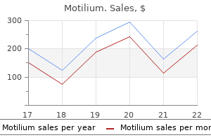
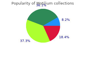
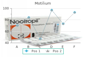
Motilium dosages: 10 mg
Motilium packs: 90 pills, 180 pills, 270 pills, 360 pills

Cheap 10 mg motilium with amex
This can happen as a outcome of gastritis symptoms livestrong buy generic motilium 10 mg an intense physiological or native inflammatory course of gastritis xarelto buy 10 mg motilium free shipping, in the absence of scarring. This could mirror native or systemic illness and in the latter could lead to temporary loss of all nails. In the long run, athletes typically develop thickened dystrophic nails matching a history of recurrent shedding. More severe trauma can lead to a degloving event removing all tissue from the tip of the phalanx. The underlying genetic abnormality of the congenital form has just lately been identified as a mutation within the Rspondin4, Frizzled6 or Wnt10a genes (see above: nail biology), which play a component in Wnt signalling within the cell [2]. There could additionally be a biological interplay with the underlying phalanx in embryogenesis (see Chapter 69) [3]. Psoriatic onycholysis can be thought of the reference point for other types of onycholysis and is typically distal, with variable lateral involvement. All the widespread causes are associated with diminished adherence of the nail to the nail mattress as a major (idiopathic) or secondary occasion: the latter embrace trauma, fungal an infection, eczema, drug reactions and photoonycholysis [3]. Reattachment is slow, and the loosened nail should be recut a quantity of instances if necessary. Where antimicrobial therapy is needed for Pseudomonas, gentamicin eye drops could be helpful. Drying beneath the onycholytic nails with a hairdryer has been advocated in order to desiccate the environment during which Pseudomonas would otherwise grow. Soaking the fingertips a quantity of nights every week in vinegar or sodium hypochlorite solution (Milton) for five min could be useful to prevent recurrence. At the upper energy it could be irritant, particularly if the world being handled is already sore. A dilution of four parts water to 1 part vinegar is more likely to avoid threat of irritancy. There may, nonetheless, be a minor traumatic element, because the situation happens quite extra typically in persons who hold their nails abnormally long. Persistent manicure is attempted to take away the particles which accumulates throughout the onycholytic house, and this can lead to a crescentic margin of onycholysis matching the onychocorneal band and showing similar in all concerned digits. If the condition persists for a quantity of months, the nail bed becomes dark and irregularly thickened. The situation is generally seen in women and heaps of cases return to normal after a quantity of months. The longer it lasts, the less likely is the nail to become reattached, because of keratinization of the uncovered nail bed. Thirty per cent of psoriatics with nail involvement will have onycholysis, with toenail involvement extra common than fingernails [9]. Onycholysis occurs in general medical circumstances, together with impaired peripheral circulation, hypothyroidism [8], hyperthyroidism [9], hyperhidrosis, yellow nail syndrome and shell nail syndrome. Minor trauma is a common trigger, and many occupational instances are as a end result of trauma [10]. Immersion of the hands in soap and water may be considered traumatic, as also might the utilization of certain nail cosmetics. It has also been described after the applying of 5% 5fluorouracil to the fingertips where it can be used therapeutically for warts [11]. There is a situation of hereditary partial onycholysis related to hard nails [12]. Changes in nail surface Longitudinal grooves Longitudinal grooves may run all or a part of the size of the nail within the longitudinal axis, and need to be distinguished from ridges which are happy with the nail floor [1]. Drugs similar to retinoids [15] and most cancers chemotherapy can be implicated, with taxanes eliciting nail adjustments in between 19 and 44% of patients, relying on the chemotherapy regimen [16]; cooling the hand with a specialised glove has been demonstrated to help diminish or delay onset of those antagonistic effects [17,18]. However, the fibrotic tissue may not at all times grossly alter the nail and can lengthen from the lateral nail fold in addition to the extra typical proximal nail fold. It most sometimes develops in trauma or lichen planus and its variants, including idiopathic atrophy of the nail [2] and graftversushost illness [3]. It can even happen in leprosy, the place it may characterize scarring secondary to neuropathic harm and secondary purulent infection [4]. Causes include trauma, systemic sclerosis [2,3], Raynaud phenomenon, lupus erythematosus, figure ninety five. The nail is break up, often in the midline, with a fir treelike look of ridges angled backwards. After a period of months or years the nails often return to normal, but relapse could occur [5] and a ridge could exchange the unique defect. Some patients give a particular historical past of trauma [1] and barely the dysfunction may be attributed to oral retinoids [6]. Although familial circumstances have been recorded, the majority of instances are sporadic and of unknown cause [7]. The distance of the groove from the nail fold is said to the time since the onset of progress disturbance. The depth and width of the groove may be related to the severity and length of disturbance, respectively. In many instances, grooves are seen on all 20 nails but are most distinguished on the thumb and great toenail, and are deeper within the midline of the nail. Fullthickness grooves may be associated with distal extension of the plane of separation of the nail plate. When numerous, they appear randomly distributed upon the nail surface or have a geometrical sample. Mild pitting may occur in affiliation with different patterns of eczema, however is usually extra subtle or localized than psoriatic pitting. Extensive pitting combined with different floor irregularities results in the looks of trachyonychia. An isolated massive pit might produce a localized fullthickness defect within the nail plate termed elkonyxis, which is present in reactive arthritis, psoriasis and following trauma. Histologically, pits represent foci of parakeratosis, reflecting isolated nail malformation [1,2] and are present in the fingernails of about half of psoriatics with nail involvement. If exogenous, similar to these as a outcome of manicure, the margin may match the proximal nail fold and the grooves may be a number of as in washboard nails associated with a behavior tic [3]. Transverse grooves could happen on isolated diseased digits (trauma, inflammation or neurological events) [4] or could additionally be generalized, reflecting an acute systemic event such as a drug response [5], myocardial infarction, measles, mumps or pneumonia. It is especially associated with alopecia areata [3], psoriasis and lichen planus, although the most common presentation is as an isolated nail abnormality. In the isolated type, histology exhibits spongiosis and a lymphocytic infiltrate [4] of the nail matrix. There is a few response to potent topical, regionally injected and systemic steroids, but this might be temporary. Nail plate pigmentation Exogenous pigment on the upper surface is simple to reveal by scraping the nail. If the proximal margin of the pigment is an arc matching the proximal nail fold, it is a additional clue confirming an exogenous supply. There is a delicate distinction between the static features, similar to forms of break up, and the subjective expertise of having brittle nails.
Motilium 10 mg cheap amex
Clinical variants Plantar and penile fibromatosis are carefully associated conditions (see later) gastritis diet ����� safe 10 mg motilium. There could additionally be a histological resemblance to fibrosarcoma nhs direct gastritis diet best motilium 10 mg, but the latter is more pleomorphic, with larger nuclei and more mitoses. Pathology Fibroblasts in affected fascia appear to be equivalent to those in regular palmar fascia however their density is increased they usually tend to be clustered around narrowed small vessels [18,19]. Later levels are characterised by the presence of myofibroblasts, which have a fibrillary cytoplasmic ultrastructure and seem to have another properties of smooth muscle. The nuclei are deeply indented, probably as a outcome of the contractile properties of the cell. The cells also have altered surface membrane properties which enable attachment to neighbouring cells and stroma. Myofibroblasts have additionally been recognized in the regular aorta and in granulation tissue, hypertrophic scars, keloids, liver fibrosis, dermatofibromata, etc. The advanced levels of palmar fascial fibromatosis are characterized by dense fibrous connective tissue with a few elongated cells. This could additionally be because of decreased degradation ensuing from elevated levels of tissue metalloproteinase inhibitors in the lesions [21]. Structural abnormalities of glycosaminoglycans, notably dermatan sulphate, may predispose to abnormal fibrillogenesis [22]. Disease course and prognosis the condition tends to progress more slowly in girls [9]. Eventually, the function of the hand is impaired due to fastened flexion of a quantity of digits. Traditionally full elimination of the palmar aponeurosis has been recommended [26], though minimally invasive subtotal fasciectomy and direct closure is more typically favoured [27,28]. Initial encouraging placebocontolled trials of collagenase injections [29] have been supported by subsequent expertise, and the method is now extra broadly practised [30]. Allopurinol may help by decreasing free radical production [31], and it has been suggested that vitamin C might stop progression of the disease by appearing as a freeradical scavenger [2]. Many other nonsurgical approaches have been tried, together with continuous gradual skeletal traction, radiotherapy, dimethyl sulfoxide, vitamin E, steroid injections and interferon, though none has been proven to be clinically useful [32]. Highdose tamoxifen following minimally invasive surgical procedure reduces the risk of recurrent fibrosis in the short term, but the effect is lost on discontinuing the drug [33]. Intriguing outcomes have been reported from the usage of relaxin gene therapy on Dupuytren myofibroblasts in vitro, with the potential to be used in vivo [34]. Genetics Palmar fascial fibromatosis is often familial, and may be inherited as an autosomal dominant trait [23], by which case the onset tends to happen at an earlier age [24]. Genomewide research show elevated expression levels of metalloproteinases 1, three and sixteen, fibroblast progress factor and several other collagen genes [25]. Environmental components Occupational publicity to handtransmitted vibration could additionally be an exacerbating issue [13]. Total excision of the lesion and the complete plantar fascia appears to give the best outcomes, with the lowest incidence of recurrence. Synonyms and inclusions � Peyronie disease � Plastic induration of the penis � Fibrous sclerosis of the penis Plantar fascial fibromatosis [1,2] Definition and nomenclature this may be a rarer condition than palmar fascial fibromatosis, though often associated; a survey from Reykjavik discovered that 15% of males with the latter had plantar fibromatosis [3]. The fibromatosis not often ends in contractures however tends to be domestically invasive and to recur. Synonyms and inclusions � Ledderhose illness pathophysiology Penile fibromatosis could happen as an isolated abnormality, or as one element of polyfibromatosis in affiliation with palmoplantar fibromatosis, keloids and knuckle pads. There may be a genetic factor, but dependable research of the mode of inheritance are lacking. The condition is rare under the age of 20 years, and the highest incidence is between forty and 60 years. Differential prognosis the differential prognosis contains keloid and fibrosarcoma. Magnetic resonance imaging might confusingly demonstrate the cerebriform pattern sometimes seen in fibromyxoid sarcoma [4]. In youthful sufferers, aggressive childish fibromatosis and aponeurotic fibroma should even be considered [5]. Complications are rare, although squamous carcinoma has been reported occurring within a lesion of plantar fibromatosis [6]. Similar nodules have been described symmetrically affecting the anteromedial elements of the heel pad in youngsters. They are asymptomatic and will resolve spontaneously [7,8]: surgery is contraindicated. Histopathology [3] the thickened plaque exhibits cellular fibroblastic proliferation surrounded by dense lots of collagen. The course of appears to begin as a vasculitis in the areolar connective tissue beneath the tunica albuginea, whence it extends to adjoining buildings. The erectile deformity could make vaginal penetration inconceivable, and pain or anxiousness about performance could trigger secondary impotence. The ache usually subsides within a number of months, but the fibrous plaque may resolve, stay unchanged or progress [5]. If needed, an erection may be induced by the intracavernosal injection of papaverine [9]. There are case reviews of success with extra aggressive remedy using pulsed dexamethasone and lowdose cyclophosphamide [10]. Clostridial collagenase injections have given promising results, as in Dupuytren contracture [11�13]. Alternatives embody plaque incision and grafting [16] and venous grafting, using the deep dorsal vein [17] A semirigid penile prosthesis may be inserted. Age Onset is usually between 15 and 30 years of age; nevertheless, lesions sometimes develop slowly and asymmetrically and may not current vital cosmetic issues for several years. Sex Knuckle pads Definition and nomenclature Knuckle pads are circumscribed thickenings overlying the finger joints. The time period is a misnomer as most lesions happen over the proximal interphalangeal somewhat than the metacarpophalangeal joints (knuckles). Synonyms and inclusions � Holoderma � Pulvinus � Subcutaneous fibroma Probably equal. Associated diseases There is a powerful affiliation with other fibromatoses similar to palmar fibromatosis [2�4]. An affiliation between Dupuytren contracture and different fibromatous lesions has been recorded in some families. In one massive family, knuckle pads were associated with sensorineural deafness and with leukonychia (Bart�Pumphrey syndrome) [5].
Syndromes
- Diziness
- Confusion
- Chills
- Allergic reaction (rare)
- Change in the sense of smell
- Hoarseness has lasted for more than 1 week in a child, or 2 weeks in an adult
- Bleeding
Discount 10 mg motilium
The use of methotrexate instead of chronic gastritis low stomach acid generic motilium 10 mg visa cyclophosphamide is associated with a better threat of relapse [39] gastritis diet ulcer motilium 10 mg discount overnight delivery. In sufferers with nonlocalized illness, cyclophosphamide types the mainstay of remedy. Cyclophosphamide can be utilized in a daily oral 2 mg/kg/day dose for 6 months or as pulsed intravenous 15 mg/kg/pulse each 2�3 weeks for six pulses. Intravenous cyclophosphamide has the advantage of a decrease cumulative dose and a lower danger of opposed events, but carries a greater threat of relapse [40,41]. Oral prednisolone in a dose of 1 mg/kg/day, to a maximum of 60 mg/day, is commonly used as an adjunct to cyclophosphamide, with the purpose of reducing the dose to 15 mg/ day at 3 months [30]. In sufferers with extreme renal disease, plasmapheresis may have a role in saving the kidney [42]. Introduction and basic description this is a uncommon systemic vasculitis characterised by bronchial asthma, peripheral blood and tissue eosinophilia (especially in the respiratory tract) and necrotizing vasculitis with extravascular granulomas. The majority of sufferers have cutaneous findings within the lively part of the disease. It was originally described by Rackemann and Greene in 1939 as an allergic illness and never classified as periarteritis nodosa; Churg and Strauss later described the syndrome and its histopathological traits in 1951 [2]. Second line Patients with contraindications to cyclophosphamide may be handled with rituximab 375 mg/m2 per week for 4 weeks as a substitute [43,44]. Third line Patients with refractory illness to cyclophosphamide and glucocorticoids can be treated with rituximab 375 mg/m2 per week for 4 weeks [45]. Remission maintenance Due to the cumulative toxicity of cyclophosphamide, azathioprine (2 mg/kg/day) is most well-liked to keep remission [48]. The switchover can occur either at the end of six pulses of Age It is most typical in these aged 15�70 years with a peak incidence around the age of 50. Associated illnesses It is associated with atopy, particularly bronchial asthma and allergic rhinitis. Allergy most likely performs a central function, but the illness is nearly certainly multifactorial. As the allergy association would possibly recommend, the inflammatory response is primarily Th2 in nature, although Th1 and Th17 responses are seen [9,10,11]. The Th2 response has been thought to be responsible for eosinophilic activation, and prolonged eosinophil survival. The merchandise of eosinophilic and neutrophilic degradation have been noticed in inflamed tissues and are probably liable for tissue damage [12,13]. The granulomas contain necrotic polymorphonuclear leukocytes, eosinophils, severe fibrinoid and fibrillar collagen degeneration, and a proliferation of granulomatous tissue. In all phases of the disease there may be cutaneous manifestations, with roughly 5% demonstrating cutaneous vasculitis [14]. Palpable purpura and infiltrated nodules (typically situated on the scalp or limbs) are the most typical pores and skin manifestations, however livedo reticularis, necrotizing livedo. The Lanham standards require asthma, peripheral eosinophilia and systemic vasculitis in two or more extrapulmonary organs [17]. Environmental elements Environmental allergens are associated with extra severe asthma. In a pooled evaluation, survival at 1 and 5 years was 94% and 60�97%, respectively [20]). By convention and worldwide consensus, sufferers are switched to azathioprine 2 mg/kg/day [23]. Current advice can be to use rituximab according to scientific want rather than each 6 months. A urine microscopy demonstrates active urinary sediment in sufferers with glomerulonephritis. Our information of its remedy comes from open labelled trials and from international consensusbased recommendations [23]. International suggestions therefore advocate that in sufferers with out organ or lifethreatening involvement the addition of methotrexate 20�25 mg/week ought to be thought-about [23]. Intravenous pulsed cyclophosphamide 15 mg/kg/pulse at 2�3weekly intervals (six pulses) with oral A condition described as periarteritis nodosa was described by Adolf Kussmaul and Rudolf Maier in 1866. In 1994, the label was uniquely applied to a condition that spared smallcalibre vessels [4]. History Although some patients with cutaneous arteritis could report constitutional symptoms, together with delicate involvement of the muscles and nerves, cutaneous manifestations are the most hanging function of the illness. Those with necrotizing lesions of livedo reticularis must be evaluated for vasculitis or vasculopathy. Other microbial associations reported have been streptococcal infections [16] and coxsackie B4 [17]. Streptococcal infections [18], erythrovirus (parvovirus B19) [19] and Mycobacterium fortuitum [20] have been reported in association with cutaneous arteritis. The concerned vessels classically show a targetlike look ensuing from an eosinophilic ring of fibrinoid necrosis. Later within the disease process, the infiltrate turns into much less neutrophilic, consisting predominantly of lymphocytes and histiocytes. Complement and IgM deposits in vessel walls of lesions of cutaneous arteritis from some sufferers may be demonstrated by direct immunofluorescence. Disease course and prognosis Gastrointestinal tract, renal, coronary heart and central nervous system involvement are related to higher mortality [26]. Similar mutations were present in a separate study of 19 patients of Georgian Jewish descent [24]. Investigations Laboratory investigations are normally nonspecific, revealing an acute part response. Skin, muscle and nerve Polyarteritis nodosa and cutaneous polyarteritis nodosa 102. Management There should be screening for infection (see the part on predisposing factors) and consideration should be given to a trial of discontinuing medicine that predates disease. If related to hepatitis B infection, antiviral therapies form the main focus of remedy together with immunosuppressive therapy. Highdose corticosteroids adopted by tapering of the dosage over 3�6 months might often be needed for some sufferers. This remedy should be supervised at a specialist centre at the aspect of a hepatologist. Associated illnesses Associated diseases embrace coronary vessel aneurysms and myocardial infarction. Pathophysiology Kawasaki illness is assumed to be as a end result of an intense inflammatory response to an unidentified infectious agent in genetically prone hosts. Pathology Third line There are case reviews for the use of rituximab in refractory disease [30,31]. Coronary arteries are sometimes involved; the aorta and enormous arteries may be concerned. Synonyms and inclusions y s � Mucocutaneous lymph node syndrome arteritis � Mucocutaneous lymph node syndrome � Kawasaki syndrome � Infantile polyarteritis t the angiitis of Kawasaki illness impacts practically all organs, with a really excessive frequency of cardiac involvement.
Motilium 10 mg purchase fast delivery
Systemic causes embrace glomerulonephritis gastritis attack 10 mg motilium order with mastercard, hypoalbuminaemia (especially nephrotic syndrome) gastritis diet ����������� motilium 10 mg discount on-line, cardiac failure, superior venocaval obstruction and thyroid disease. Some systemic infections, similar to infectious mononucleosis and scarlatina, trigger periorbital oedema. It could be the Skin diseases affecting the eyelids A large number of dermatological circumstances can affect the eyelids as part of a generalized process. Psoriasis and lichen planus can both contain the lids and cause considerable irritation. Changes in pigmentation [13�17] There are considerable racial and familial variations of the diploma of pigmentation of the eyelids. Pigmentation of the periorbital skin can be posttraumatic, postinflammatory or can accompany melanocytestimulating hormoneinduced melanosis of any trigger. Chemical pigmentation can occur from extended use of a mercurial or silver preparation, producing a slateblue or greybrown discoloration. Mauve discoloration of the eyelids and periorbital space is an early a half of chrysiasis from parenteral gold therapy. Local improve in pigmentation may also be because of cosmetics containing phototoxic brokers, usually psoralens. Increased pigmentation can even observe inflammatory dermatoses such as eczema and lichen planus. Hypopigmentation can complicate the use of topical drugs including thiotepa eye drops and mercurial ointments. Angiooedema is transient and sometimes part of a extra generalized urticarial eruption. Lymphoedema is everlasting and tends to be worse first thing in the morning and improves in the course of the day; it might be associated with underlying sinus disease or a persistent inflammatory situation such as granulomatous rosacea. Blepharochalasis is an uncommon condition which can be inherited (autosomal dominant) or sporadic; it presents in the second decade with recurrent episodes of painless lid oedema, ensuing in the growth of extra skin and thickened subcutaneous tissue which may require remedy by blepharoplasty. Anterior blepharitis describes irritation of the lid margin anterior to the grey line and concentrated round td he lashes. It may be accompanied by squamous debris or collarettes across the lashes, and inflammation could spill onto the posterior lid margin, leading to posterior blepharitis. Posterior blepharitis describes inflammation of the posterior lid margin, which can Blepharitis, meibomian gland dysfunction, rosacea, seborrhoeic dermatitis 109. It could lead to alteration of the tear movie, symptoms of eye irritation, clinically obvious irritation and ocular floor disease. Chronic blepharitis may happen in the absence of any vital dermatological association, and its classification is further difficult each by the variable affiliation of chronic blepharitis with rosacea and seborrhoeic dermatitis, and also by the time period ocular rosacea, a condition which can occur within the absence of dermatological rosacea. It is hardly shocking that this classification causes confusion amongst practitioners. This situation has arisen partly as a end result of the pathogenesis is poorly understood, with few unifying concepts. Epidemiology [6,7,8] the epidemiology of blepharitis has been hampered by difficulties of disease definition, lack of a standardized scientific evaluation and the completely different perspectives of dermatologists and ophthalmologists. Incidence and prevalence Chronic blepharitis is amongst the commonest disorders in each ophthalmic and basic medical follow. In common medical practice it makes up about 70% of ophthalmic referrals, which themselves account for between 2 and 7% of all outpatient consultations. The incidence of ocular rosacea varies amongst ophthalmic and dermatological research, ranging from 6 to 72%, being extra prevalent in ophthalmology clinics [8]. Between 3 and 58% of sufferers with rosacea have ocular involvement; this wide variation in cited frequency largely reflecting variations in disease definition. In participants with all kinds of blepharitis, the prevalence of acne rosacea ranges from 26. This classification corresponds with, and simplifies understanding of, the treatment of the different conditions, which differs between the anterior-lid and posteriorlid margin problems however not between the person circumstances within each of these two teams. In addition to these quite common types of blepharitis, there are other chronic causes in which the pathogenesis is evident. These are all unusual and are sometimes misdiagnosed as one of the types of chronic blepharitis described in Table 109. Acute blepharitis is a clearly outlined group of conditions, for which the causes are summarized in Table 109. Anterior lid margin* posterior lid margin* Anterior and/or posterior lid margin Staphylococcal blepharitis Seborrhoeic blepharitis Meibomitis/ocular rosacea other meibomian gland dysfunction Meibomian seborrhoea other blepharitis, Foamy tear film Variable according to aetiology *The anterior lid margin is the portion anterior to the meibomian gland orifices and the posterior lid margin is behind this, including the meibomian glands. Sectoral illness like this is quite widespread in staphylococcal blepharitis which can additionally be largely unilateral. It is theorized that the infestation and wastage of mites cause blockage of the follicles and glands and/or an inflammatory response. Patients with recalcitrant blepharitis have responded to therapy directed at eradicating Demodex mite, studies help its function in activation of immune mechanisms in subtypes of rosacea, The importance of cellmediated immunity within the pathogenesis of the illness was proven by experimental studies in rabbits; when these were immunized with both complete S. However, proof for a similar pathogenesis in humans is lacking; the connection between the clinical indicators of staphylococcal blepharitis and hypersensitivity to subcutaneous injections of either complete S. The pathogenesis of the, often severe, follicular and papillary conjunctivitis that accompanies this situation is assumed to be due to a mixture of transient an infection and hypersensitivity. Some researchers have hypothesized that toxins produced by the micro organism may trigger irritation, however no particular toxin has been identified [18,19]. Demodex folliculorum are small parasitic mites that reside in hair follicles, sebaceous glands and meibomian glands. They are discovered Part 10: SiteS, Sex, age Meibomian gland disease the meibomian lipids (meibum) are a fancy mixture of cholesterol esters and esterified unsaturated fatty acids. These lipids are responsible for sustaining a steady tear film, lowering tear film evaporation (and, therefore, stopping drying of the ocular surface) [23], preventing tear spill over the lid margins by lowering surface pressure and decreasing ocular surface contamination by sebum from the cutaneous floor of the lids, which otherwise forms dry spots. Bacterial lipases are produced by all the micro organism that colonize the lid margin and have the potential to break down meibum into free fatty acids, which is able to destabilize the tear movie [25]. These micro organism colonize the gland orifices and expression of lipid from deeper inside the glands can stabilize the tear film. Ocular rosacea the precise aetiology and pathophysiology of ocular rosacea stays unknown, although different theories have been proposed [27] (see also Chapter 91). Recent molecular studies propose that abnormal recognition of widespread environmental stimuli leads to activation of proinflammatory techniques as properly as innate immune responses. Factors that trigger the innate immune system lead to elevated expression of certain cytokines and antimicrobial molecules such as cathelicidin, which is vasoactive and proinflammatory [28].

10 mg motilium purchase overnight delivery
In one study of 1500 instances of thrombophlebitis gastritis diet 4 rewards 10 mg motilium sale, 31 of seventy seven occurring in affiliation with malignancy had been of a migratory type [1] gastritis treatment and diet cheap 10 mg motilium mastercard. In this study, carcinomas of the lung and pancreas have been the most typical sites for the first tumours, although carcinomas of the breast, colon and abdomen had been additionally reported [1]. Other associated illnesses are Beh�et (between 2 and 20% of all cases; see Chapter 48) and Buerger disease (one examine [2] found thrombophlebitis migrans in up to sixty five. Similar cases have been reported within the antecubital fossa, inguinal area, axilla, penis [3], abdomen and decrease limbs. Introduction and general description this is a rare, selflimiting and benign condition that was first described in 1939 [1]. Epidemiology Incidence and prevalence the incidence and prevalence of this rare situation are unknown. As with superficial venous thrombosis the mechanism is believed to be within the Virchow triad (stasis, elevated coagulability and vessel wall injury). In the chest wall type, a examine [2] of pooled instances of the disease discovered onethird of circumstances have been idiopathic, a lot of the relaxation have been associated to trauma (injury, muscular pressure, poorly becoming bras, surgical procedure, breast prosthesis, etc). Rare causes had been underlying breast most cancers, hypercoagulable states and connective tissue problems. In the penile kind, surgical trauma, extreme sexual activity, sexual vacuum practices, use of constrictive parts throughout sexual exercise, intravenous drug abuse, prolonged sexual abstinence, native or distant an infection, venous obstruction as a end result of bladder distension and pelvic tumours have all been reported [2]. A fibrous painful wire with local preputial inflammation however with out skin retraction is seen. Other attainable sites include the brachial, femoral and calf veins however, unlike chest wall Mondor illness, native irritation is current. This is a subtype described in association with axillary lymph node dissection in breast most cancers, and is characterised by retractile scarring of the fascia. They can extend into the ipsilateral arm, and even the forearm, creating linear grooves. Differential analysis the differential analysis is broad and contains cellulitis, erythema nodosum, pores and skin metastatic carcinoma, lymphangiectasia and lymphangioma. On examination, the lesions might involve any subcutaneous vein on the higher anterolateral chest wall and produce a fibrous painful wire with skin retraction. Primary varicose veins No known underlying cause Associated with valvular incompetence Secondary varicose veins Raised endoluminal venous pressure (venous hypertension) as a end result of thrombosis, pregnancy, trauma Pathogenetic features No uniform valvular abnormality discovered Intrinsic and structural abnormalities of vein walls are seen In Mondor illness on the chest wall, that is usually benign and selflimiting with spontaneous resolution normally with 2�8 weeks [2] and only 13% recurrence is reported in one sequence [4] and none in another [5]. In Mondor illness involving different venous territories, the natural historical past is much less well-known and, as with other situations with superficial venous thrombosis, could be related to deeper thrombosis. Ultrasound examination can diagnose superficial thrombus and exclude a deep thrombus. Raised venous stress is believed to cause � Stretching of the endothelium � Expression of cytokines and adhesion molecules � Activation of extracellular signalrelated kinases � Free radical production � Dysregulation of remodeling development issue � Altered fibroblast exercise Management this depends on the sort of Mondor disease. In Mondor disease on the chest wall, as this is generally benign and selflimiting, no anticoagulation is required and simple analgesia may be given if the affected person is in pain. In Mondor illness involving different venous territories, anticoagulation is beneficial and typically in penile illness the superficial dorsal vein of the penis is handled with thrombectomy or excision. If the fascial fibrous bands persist, they can be manually ruptured with immediate return of perform and reduction in pain [6]. One means valves exist in both techniques and within the perforating veins which join the 2. Incompetence in any of the valves can disrupt the conventional flow of the blood and cause venous hypertension. Valvular incompetence and venous hypertension are thought to be important and interdependent however how they occur stays to be totally elucidated. Varicose veins can be divided into primary and secondary; however, that is simplistic. The mechanical problems with valve incompetence and raised pressure can occur in each and lead to advanced molecular and histopathological alterations in the vessel wall and the extracellular matrix (Table 103. Introduction and basic description Varicose veins are quite common and their traits and administration had been described by Hippocrates and Galen [1]. Pathology the chief findings in varicose veins are intimal hypertrophy, subendothelial fibrosis, luminal dilation and wall thickening. Genetics A genetic basis has been thought to be probably relevant within the pathogenesis of varicose veins, since familial clustering of circumstances happens. The incidence of varicose veins is unknown however a quantity of studies have reported the prevalence to be excessive (varying from 10% to 50% of the adult population). Duplex scanning can be important to investigate patients with pores and skin changes attributed to venous hypertension. There is a few evidence that the incidence of recurrent varicose veins is decrease after duplex assessment has been used to plan surgical procedure. If untreated venous insufficiency in both the deep or superficial system causes the progressive syndrome of continual venous insufficiency. It arises from main valve failure or when the superficial veins become distended, causing the valves to turn out to be secondarily incompetent. Secondary perforating vein insufficiency often occurs in combination with deep vein insufficiency (postthrombotic limb). Reflux is the most typical sort of abnormality, but in about 10% of circumstances a practical obstruction Introduction and basic description Chronic venous insufficiency is widespread and can be disabling. Epidemiology Those with superficial venous insufficiency usually current with varicose veins initially. One massive study in 30 000 topics discovered a prevalence of 7% for varicose veins and 0. Serious persistent venous insufficiency resulting in venous ulcers has an estimated prevalence of roughly 0. However, it happens extra generally in Western society and this in all probability reflects life-style variations corresponding to sedentary or standing occupations, higher rates of weight problems and customarily reduced ranges of physical activity. Pathophysiology Changes occurring within the macrocirculation lead to microvascular abnormalities and persistent irritation which are thought to lead to the bodily manifestations of continual venous insufficiency. Degradation of extracellular matrix proteins leads to breakdown of extracellular matrix inflicting lowered therapeutic and promotes ulceration Pathophysiology of venous reflux and persistent venous insufficiency It is believed that there are two parts to the pathophysiology; the first is abnormal venous blood flow with reflux, and the second happens on the microvascular degree and is a persistent inflammatory process which leads to the skin adjustments seen in chronic venous insufficiency. Reflux is the presence of retrograde move in a vein in response to a stimulus corresponding to a calf squeeze. It can occur within the superficial, deep and perforating veins of the lower extremity. An elevated and sustained ambulatory venous stress (venous hypertension) is indicative of persistent venous insufficiency. This could additionally be brought on by valvular incompetence or venous outflow obstruction or poor muscle pump perform (Table 103. Such heightened strain is transmitted distally so far as the capillary system of the skin, inflicting capillary hypertension, and ultimately leading to destruction of the nutritive capillaries [7]. The present theories on the mechanisms for the pathogenesis of the persistent irritation in venous disease are outlined by Bergan et al. Environmental factors A sedentary lifestyle reduces the efficiency of the muscle pump and thus results in lowered venous return and occupations with prolonged standing act to improve the risk of higher venous pressures in the legs. Impaired calf muscle pump operate Clinical options History the medical features of persistent venous insufficiency range from delicate oedema to severe incapacitating leg ulceration (outlined in Table 103.
Purchase motilium 10 mg mastercard
Causative organisms Most episodes of cellulitis are believed to be brought on by group A streptococci gastritis symptoms from alcohol cheap motilium 10 mg. However gastritis diet ������ motilium 10 mg buy fast delivery, microbiologists think about Staphylococcus aureus to be the cause in most patients [9,10]. Clinical features History Cellulitis can range from patient to patient and episodes can range in presentation. Some episodes are accompanied by severe systemic upset, with high fever or rigors; others are milder, with minimal or no fever. Note lymphangitis crossing the watershed to the contralateral lymph node territory. Inflammatory markers are usually unfavorable and only a prompt response to a prolonged course of antibiotics confirms the diagnosis. In all circumstances of recurrent cellulitis, threat factors similar to lymphoedema, wounds, breaks in pores and skin integrity (particularly interdigital), dermatitis and fungal infections should be treated. Swollen arm Definition and nomenclature Swelling of the higher limb or extremity is invariably due to oedema however overgrowth of tissue can occur. Microbiology of any cuts or breaks within the skin or aspiration of blister fluid ought to be thought of before antibiotics are started. Lowdose prophylactic penicillin, phenoxymethylpenicillin 250 mg twice day by day, given for a period of 12 months virtually halves the danger of recurrence during the intervention period compared with placebo [1]. However, though some level of safety seems to be sustained for a number of months after the top of prophylactic therapy, this impact is lost by 36 months, a discovering that implies that longer term prophylaxis may be required. Congenital/genetic Vascular Vascular malformation Diffuse phlebectasia Klippel�Trenaunay syndrome Arteriovenous malformation Lymphatic Lymphoedema Lymphatic malformation Lymphangiomatosis other Overgrowth spectrum: Proteus syndrome Fat hypertrophy Muscle hamartoma Gigantism/ hemihypertrophy Lipoedema Dercum disease Madelung illness (benign symmetrical lipomatosis) introduction and common description Swelling of an arm may be caused by oedema, during which case pitting should be evident to a point, or it might be attributable to a rise in the volume of other tissue components, for example bone, muscle, fat or a tumour (Table a hundred and five. A swollen arm could additionally be normal but perceived to be larger if the contralateral limb has shrunk. The commonest purpose for upper limb swelling is lymphoedema following breast cancer treatment. Arm swelling can be a presentation of cancer with metastatic illness within the axilla. Upper limb swelling may due to main lymphoedema (usually related to lymphatic abnormalities elsewhere) or with a lymphatic malformation. The distribution of swelling alongside the arm varies between patients, and swelling may be confined to a selected area of the higher limb. In some patients the hand could additionally be swollen, while in others the hand may be spared despite more proximal swelling of the forearm or upper arm. By pinching up the pores and skin and subcutis of each arm between finger and thumb, the thickened ipsilateral tissues may be palpated [8]. Venous outflow obstruction could additionally be because of axillary/subclavian vein compression or stenosis (usually due to malignancy or radiation damage) or occlusion from thrombosis. Subclavian vein thrombosis is a uncommon condition that the majority usually occurs in the context of central venous catheters, pacemakers, trauma, surgical procedure immobilization, oral contraceptive tablet use, pregnancy or malignancy. It occurs notably in cancer patients receiving chemotherapy by way of central traces. Arteriovenous fistulae for haemodialysis will enhance arm dimension from an elevated blood circulate however arm oedema will solely happen with thrombosis or if lymph drainage is compromised. Differential prognosis Venous outflow obstruction due to axillary/subclavian vein compression or stenosis, or occlusion from thrombosis, will produce a discoloured (red/blue) painful swollen arm typically with parasthesia. A swollen arm as a outcome of overgrowth may be associated with lymphoedema or lymphatic malformation in which case there may be indicators of a vascular birthmark usually on the root of the limb. Rarely, systemic causes such as heart failure, superior vena caval obstruction and hypoproteinaemia can produce arm swelling. Pathology Upper extremity swelling of vascular origin will be because of oedema or increased vascular quantity. All oedema is caused by microvascular fluid filtration exceeding lymph drainage for a enough period of time. As lymph move is liable for the drainage of all tissue fluid, aside from transient durations of venous reabsorption, a chronically swollen arm due to fluid signifies lymph drainage failure. Increased filtration may be caused by high venous pressures or from enhanced vasular permeability from irritation. Causative components Recurrent infections from herpes simplex can result in upper limb lymphoedema [3]. Noninfective forms of irritation as a end result of persistent hand dermatitis [4], rheumatoid arthritis [5] or psoriatic arthropathy [6] can lead to lymphoedema. Complications the primary complication of lymphoedema anyplace is an infection and, specifically, cellulitis. Genetics Gene mutations inflicting tissue overgrowth have just lately been recognized [7]. Prognosis Cellulitis complicating upper limb lymphoedema can every so often be extreme and life threatening. In circumstances where systemic causes, for example most cancers recurrence or coronary heart failure, have lead to, or coexist with, the lymphoedema, then treatment of the medical situation have to be undertaken before embarking on particular lymphoedema therapy. The basic principle for treating a swollen limb is to restrict increased microvascular filtration and improve lymph drainage. Lymph drainage responds to train and motion done while wearing compression [9]. Where cellulitis, particularly recurrent cellulitis, happens then prophylactic antibiotics could additionally be indicated [10]. Although surgical decompression and venous angioplasty could also be thought of for thoracic outlet obstruction, the everyday therapy for major subclavian vein thrombosis is oral anticoagulation solely [11]. Synonyms and inclusions � Puffy face � Facial lymphoedema � Rosaceous lymphoedema � Morbihan illness � Solid facial oedema � Orofacial granulomatosis � Granulomatous cheilitis � Melkersson�Rosenthal syndrome Part 9: Vascular (b) figure one hundred and five. Oedema might prolong beyond the face to involve the pinnacle and neck, which occurs after surgery and/or radiotherapy for head and neck most cancers or with recurrent cancer. In instances of suspected noncancer lymphoedema, lymphoscintigraphy is the investigation of choice to confirm impaired lymph drainage. A venous duplex ultrasound examination is the primary investigation of selection in suspected venous outflow obstruction, Swollen face, head and neck desk 105. Congenital/genetic Vascular Vascular malformation Lymphatic Syndrome (neck webbing): Turner Noonan Generalized lymphatic dysplasia Mosaic with segmental lymphoedema Lymphangioma/lymphatic malformation overgrowth Macrocephaly, In one research, the most typical sites of external lymphoedema had been the neck and submental area [1]. Conditions that have to be considered include dermatomyositis, Graves illness and significantly rosacea/acne. Eyelid swelling could also be fairly simply because of acquired lax pores and skin from photoageing and other processes which have undermined tissue compliance, similar to blepharochalasis [2]. Contact allergy or angiooedema, if persistent or recurrent, could slowly compromise lymphatic function. Equally, one extreme assault of facial cellulitis may injury the lymphatics sufficiently to cause lymphoedema. Angiosarcoma or Kaposi sarcoma may infiltrate native lymph drainage, and manifest with eyelid oedema.
Dwale (Belladonna). Motilium.
- What is Belladonna?
- Are there any interactions with medications?
- Are there safety concerns?
- Dosing considerations for Belladonna.
- How does Belladonna work?
Source: http://www.rxlist.com/script/main/art.asp?articlekey=96533
Buy motilium 10 mg overnight delivery
Again gastritis diet ���������� buy motilium 10 mg mastercard, a lateral ablation may be adequate gastritis diet 1000 motilium 10 mg proven, however more advanced procedures entail altering the alignment of the matrix [2,7,8], level of the nail bed [9] and addressing any midline hypertrophy of the distal phalanx. Nail braces hardly ever produce longterm benefit, although promising outcomes have been reported [11]. This can come up via burns, surgical procedure or trauma, or be as a result of inflammatory dermatoses similar to lichen planus the place the whole nail matrix is scarred and misplaced [4]. Usually these characteristics coincide, though clinicians and patients may prefer to use one time period over the other. Variants include splitting at the lateral margins alone and a number of crenellated splits on the free edge. However, efforts at retaining hydration (gloves, emollient and base coat with nail varnish) might help reverse scientific changes. Biotin has been used as systemic therapy, however the proof for its efficacy is weak [8]. Changes in nail colour [1�7] Alteration in nail color might occur due to adjustments affecting the dorsal nail surface, the substance of the nail plate, the undersurface of the nail or the nail bed. The disruption of normal nail plate formation by illness, chemotherapy, poisons or trauma can lead to waves of parakeratotic nail cells or small splits between cells inside the nail. Both make the nail less lucent and produce the white marks of true leukonychia (see later). This disruption could occur at nail formation or subsequently within the case of fungal nail an infection, the place discoloration could begin distolaterally rather than by way of the matrix. This is a white discoloration of the nail attributable to matrix dysfunction; it happens in a variety of patterns [1,2]. In subtotal leukonychia, the proximal two thirds are white, turning into pink distally. This is attributed to a Subungual disturbances Subungual hyperkeratosis as from dermatophyte infection or psoriasis can also change the apparent colour of the nail. Subungual haemorrhage produces a variety of color adjustments ranging from shiny pink to black. Splinter haemorrhages result from leakage of blood from nail mattress capillaries and may be due to local trauma or to microemboli, classically from infective endocarditis. Nail mattress changes Vascular abnormalities can affect apparent nail colour as in blue nails from cyanosis and brilliant purple nails from carbon monoxide poisoning. In addition to such generalized vascular changes there could be localized modifications, as seen with nail mattress tumours. In white people this is abnormal and requires thorough evaluation and, in some instances, biopsy. The incorporation of heavy metals and a few drugs into the nail plate by way of the matrix can even alter nail colour, such as the grey color associated with silver or the greyblue discoloration due to antimalarials or phenothiazines. Dermoscopy can be very useful in the assessment of nail plate pigmentation and underlying nail bed modifications [7]. They are generally associated with hypoalbuminaemia, the correction of which by albumin infusion can reverse the sign. They have also recently been reported following placement of a left ventricular help system in a affected person with congestive heart failure [4]. Colour adjustments because of drugs and chemicals [1] There are a selection of colour modifications which can be caused by medication. Yellowing of the nail is a rare prevalence in prolonged tetracycline remedy, which might also produce a sample of dark distal photoonycholysis, Topical 5fluorouracil may cause yellow nails: the whole nail is affected and returns to regular when the drug is discontinued [2,3]. Other antimalarials could produce longitudinal or vertical bands of pigmentation on the nail mattress or in the nail [7]. Hyperpigmentation due to increased melanin within the nail and nail bed has been famous in youngsters after 6 weeks of remedy with doxorubicin (adriamycin) [9,10]. Other related cytotoxic medicine could trigger a selection of patterns of increased pigmentation [1]. Punctate leukonychia contains white spots of 1�3 mm diameter attributed to minor matrix trauma. Apparent leukonychia Here, adjustments in the nail bed are answerable for the white appearance [1,2]. Nail mattress pallor could additionally be a nonspecific signal of anaemia, oedema or vascular impairment. It might happen particularly patterns which have turn out to be related to sure conditions. The condition normally presents in adults, however may happen as early because the age of eight years [2]. The feature nail modifications are usually accompanied by lymphoedema [5] at a number of sites and by respiratory or nasal sinus disease. Histologically, within the nail bed and matrix, dense fibrous tissue is found changing subungual stroma, with quite a few ectatic endotheliumlined vessels [6]. It has been instructed that obstruction of lymphatics by this dense stroma leads to the irregular lymphatic function found in the affected digits in some [8] however not all [9] circumstances. In some instances, the oedema has been shown to be due to abnormalities of the lymphatics, either atresia or, in some circumstances, varicosity [10]. Other circumstances have regular lymphatics, suggesting that a functional quite than an anatomical defect could additionally be present [11], or that perhaps solely the smallest lymph vessels are faulty. The condition may be related to an increased incidence of malignant neoplasms [10,14,15]. Other associations embody dpenicillamine therapy [5] and nephrotic syndrome [16]. Attempted therapies embody oral and topical vitamin E, oral zinc, prednisolone and the therapy of continual infection at other websites [20,21,22�25]. The drug has been demonstrated to increase the rate of longitudinal development, however an open trial in eight patients demonstrated that half gained no profit with respect to nail changes [26]. It is reported that results are higher when itraconazole or fluconazole are mixed with oral vitamin E [27]. Erythema is much less intense within the distal lunula, where it could possibly merge with the nail mattress or be demarcated by a pale line, and can be obliterated by strain on the nail plate. In this setting, it typically indicates a neighborhood disturbance of vascular move, which is most probably to be a benign tumour. Glomus tumours and subungual myxoid cysts are the commonest [3] and the color could differ between blue and pink. The effect of this may be a strip the place blood within the underlying nail bed is seen extra easily not only as a end result of the nail plate is thinner but additionally as a end result of blood pools in the underlying nail mattress capillaries on account of lowered compression by the overlying nail. Such strips of thinned nail arise due to focally reduced proliferation throughout the matrix. This could be due on to matrix pathology or could also be secondary to focal strain on the matrix with secondary lack of operate. The commonest are lichen planus and Darier illness, the place thin longitudinal red streaks may terminate at the free edge with a split. Acantholytic dyskeratotic naevus and warty subungual dyskeratoma [4] could both represent localized types of Darier illness. When related to cardiac failure, it may follow the course of administration of the cardiac disease.

Order 10 mg motilium with mastercard
Some macrophages might engulf nuclear mud of neutrophils and dystrophic calcification may develop [10] gastritis zdravljenje purchase 10 mg motilium amex. Direct immunofluorescence studies have revealed deposits of complement C3 and IgM across the dermal blood vessels: these are of unsure significance [9 gastritis ginger ale generic motilium 10 mg with amex,18]. The most common disseminated fungal infections inflicting panniculitis are Candida spp. Uncommon subcutaneous fungal infections include phaeohyphomycosis, lobomycosis, rhinosporidiosis and subcutaneous zygomycosis. Presentation With the exception of the classical subcutaneous mycoses, most of those infective panniculitides occur in immunosuppressed patients and are uncommon in immunocompetent hosts. Bacterial panniculitis could seem in the setting of septicaemia, because the consequence of direct inoculation or by direct spread from an underlying an infection. In sufferers with sepsis, solitary or multiple nodules and abscesses seem as a consequence of the haematogenous dissemination of bacteria. Constitutional signs are often absent, however the basic condition of the affected person is impaired by the underlying illness. The clinical features of subcutaneous mycobacterial infections vary according to the immune state of the patient. In immunocompromised sufferers, lesions are probably to be widespread due to haematogenous dissemination. In immunocompetent sufferers, the infection is often localized and associated to trauma, Panniculitis in immunosuppressed sufferers with disseminated fungal infection presents as multiple erythematous subcutaneous nodules, pustules or fluctuant abscesses [28,30,31]. In subcutaneous mycoses, the fungus enters the skin from the soil, crops or wood through a penetrating harm and the lesions are localized largely to uncovered areas of the pores and skin, such as the face, arms, arms or feet [33]. These lesions include a solitary painless nodule that spreads slowly; with time, secondary nodules and papules could develop in adjoining skin and may be accompanied by sinuses exuding a serous or oily discharge. Apart from the neutrophilic infiltrate in the fat lobule, extra options suggestive of an infective aetiology of a lobular panniculitis are haemorrhage, proliferation of vessels, foci of basophilic necrosis and necrosis of sweat glands [1]. The histopathological findings in panniculitis attributable to mycobacterial infections differ based on the organism concerned and the immune state of the host. Ghost adipocytes harking back to pancreatic panniculitis have been documented in circumstances of mucormycosis [46] and aspergillosis [47] involving subcutaneous fats. Different histopathological patterns have been described in accordance with the inoculation route of the microorganisms into the skin. Primary cutaneous infections come up either from direct physical inoculation or on the site of an occlusive dressing over an indwelling catheter, whereas secondary cutaneous infections develop either from direct extension to the chest wall in pulmonary infections, or from haematogenous dissemination. In contrast, in secondary cutaneous infections, the epicentre of irritation is extra deeply seated and entails only the deep reticular dermis and subcutaneous fats. The blood vessels are thrombosed and dilated with plenty of organisms expanding their lumina [30]. In immunosuppressed patients microorganisms are numerous and so they may be easily recognized in tissue sections with routine H&E staining or with special stains, however in immunocompetent sufferers microorganisms are sparse they usually could additionally be troublesome to detect. Factitious panniculitis definition Factitious or artefactual panniculitides end result from exterior injury to subcutaneous fat. Aetiological components could also be mechanical trauma, chemical substances and thermal harm; the explanations for the injury could additionally be unintended, intentional or iatrogenic. In other instances, the process outcomes from iatrogenic injections of medicine or immunization agents. Biodegradable or resorbable agents could induce extreme issues however these will usually disappear spontaneously in a few months. Slowly biodegradable or non resorbable fillers might give rise to severe reactions that show little or no tendency to spontaneous improvement. Previously, factitious panniculitis incessantly resulted from subcutaneous injection of oily materials including mineral oil (paraffin) or vegetable oils (cottonseed and sesame oils) [2]. These products had been used over many years to increase the scale of breasts or genitalia however usually induced subcutaneous foreignbody reactions known as paraffinoma or sclerosing lipogranuloma. Fortunately, most such fillers have now been abandoned by medical professionals, although issues could seem a very long time after the injections, even 30 years later, and it still is feasible to see instances of paraffinoma or sclerosing granuloma [3]. In latest years, injections with Lipostabil, a phosphatidylcholinecontaining substance, have turn out to be a popular therapeutic approach for the remedy of localized fats accumulation and lipomas, inflicting factitious panniculitis of the injected fats tissue [9]. Mesotherapy injections in an try and produce discount of the thickness of hypertrophic subcutaneous fat produce a granulomatous panniculitis with some cystic fat necrosis [10]. Panniculitis has also been reported at the sites of injection of several therapeutic medication (Box 99. Cupping and acupuncture strategies for the relief of pain may induce factitious panniculitis on the limbs [26,27]. Finally, patients with psychiatric disorders could current with selfinflicted panniculitis as a result of subcutaneous injections of a variety of drugs including acids, alkalis, farming products, mustard, milk, microbiologically contaminated materials, urine and faeces [1,28]. The exact mechanisms involved in factitious panniculitis are unsure, however vasoconstriction with tissue ischaemia at injection websites, a neighborhood inflammatory response elicited by direct contact with drugs or noxious injected substances, immune mechanisms and trauma because of repeated injections may be implicated. Lesions as a outcome of blunt trauma often seem bruised and incessantly contain the arm or hand [29]. The course is chronic and recurrent, leading to progressive fibrosis of the dorsum of the hand [32]. Selfinflicted injections with contaminated materials produce an acute suppurative panniculitis, typically with systemic symptoms [33]. Early lesions manifest as inflammatory nodules and plaques secondary to fats necrosis and suppuration. A case of factitious panniculitis masquerading as florid pyoderma gangrenosum was reported in a depressed lady [34]. In most cases, early lesions show options of a predominantly neutrophilic lobular panniculitis, with severe fat necrosis and an intense inflammatory infiltrate. In some situations, eosinophils may be abundant [28], particularly in panniculitis arising on the site of cancer vaccines [20], and in sclerosing lipogranulomata of the genitalia [40]. Fully developed lesions present granulomatous infiltrates involving the fats lobule, whereas longstanding lesions are characterized by lipophagic granulomata and surrounding fibrosis. In some circumstances, polarized mild will reveal the birefringent international bodies responsible for the panniculitis. In panniculitis due to injected substances, the dermis can also be involved by the inflammatory process, which can be a clue to the correct prognosis. Sometimes, specific histopathological findings could additionally be useful in figuring out the nature of the overseas materials. Sclerosing lipogranuloma is the time period used for factitious lesions of the male genitalia secondary to injections of liquid paraffin meant to augment the dimensions of the penis [35]. Lesions much like sclerosing granuloma may appear in other locations such because the eyelids, lips or gluteal region after injection of liquid silicone [36]. Diagnosis of panniculitis secondary to injection of drugs is normally straightforward and the location of the lesions provides a clue to their cause. Pentazocine abuse has been described in patients with continual pain or dependancy: repeated injections may induce panniculitis and myositis.

Cheap motilium 10 mg without prescription
Cleft palate is extra prevalent in females gastritis diet ����� buy discount motilium 10 mg, while cleft lip is extra prevalent in males gastritis main symptoms motilium 10 mg order online. Ethnicity There are racial variations with a high incidence of cleft palate in SouthEast Asia and a low incidence in AfroCaribbean races. More than 400 syndromes could embody a facial cleft as one manifestation and cleft lip/palate may be related to many congenital syndromes. Not all cases of clefting are inherited; a number of teratogens (environmental brokers that can cause start defects) have been implicated, as nicely as defects in important vitamins. Clefts of the lip with or with out cleft palate and cleft palate alone outcome from the failure of the first branchial arches to full fusion processes and are the most common of all craniofacial anomalies. The male to feminine ratio of cleft lip/palate is 2: 1; the ratio for cleft palate alone is just the reverse, 1: 2. Failed fusion of the palatal shelves may be caused by different gene defects culminating in: � A downside within the formation of the midline epithelial seam. Predisposing factors Cleft lip/palate is more prevalent in the lower socioeconomic lessons. The teratogens incriminated include isotretinoin, which causes delivery defects such as mind malformations, learning disability, coronary heart problems, as properly as facial abnormalities. Thalidomide given to pregnant mothers was, and anticonvulsants (phenytoin, valproic acid, lamotrigine, carbamazepine) and corticosteroids may be, associated with an elevated incidence. Systemic corticosteroids have been reported to increase the danger (this is controversial) and there are additionally considerations about attainable results from topical steroids used within the first trimester. Folic acid given periconceptually might decrease the danger, but the evidence is weak [1�51]. Clinical options Presentation A person might have a cleft lip, cleft palate or both cleft lip and palate. A cleft may contain only the higher lip or could prolong to involve the nostril and the exhausting and soft palates. Lips are extra incessantly cleft bilaterally (approximately 25%) when mixed with cleft palate. Cleft lip and palate contains about 50% of the cases, with cleft lip and isolated cleft palate each comprising about 25%. About 85% of bilateral cleft lips and 70% of unilateral cleft lips Genetics Clefts could be seen in over 300 totally different syndromes (Box 110. One subgroup have cleft lip and palate with median facial dysplasia and cerebrofacial malformations; others with laryngotracheal oesophageal clefts (Opitz�Firas or G syndrome) or cranial asymmetry (Opitz or B syndrome). True median clefts have been described in affiliation with bifid nose and ocular hypertelorism. Pseudocleft of the middle of the upper lip may happen in orofaciodigital syndrome I. A somewhat related central defect, but of gentle degree, is seen in chondroectodermal dysplasia (Ellis�van Creveld syndrome). Clefts in the decrease lip are rare and usually median but could contain the mandible and generally the tongue. Cleft palate may be incomplete involving only the uvula and the muscular taste bud (velum). Clefts are often accompanied by impaired facial growth, dental anomalies, speech issues, poor listening to and psychosocial issues. Clinical variants Submucous cleft palate can be recognized by a notched posterior nasal spine, a translucent zone in the midline of the taste bud and a bifid uvula, however not all these features are necessarily current and a bifid uvula could additionally be seen in isolation. About 1/1200 births are affected and feeding difficulties, speech defects and middle ear infections might develop in 90% of affected children. Adenoidectomy is contraindicated as it could reveal latent velopharyngeal insufficiency. If the palatal defect is too extensive, it might be repaired 3 months later to allow for enough palatal progress. In any occasion, cleft palate is now usually repaired before the child speaks, between 6 and 18 months, usually at 6�12 months of age. These youngsters want a hearing evaluation, and if it is impaired, ear air flow tubes (grommets) may be indicated. Speech, if poor despite one of the best efforts by the kid and the speech pathologist, could additionally be corrected with pharyngoplasty. Palatal ulcers seen in neonates with cleft lip and palate appear to end result from trauma from the tongue and resolve if a palatal plate is fitted. Dental abnormalities embody malocclusion (almost 100%), hypodontia (50%), hypoplasia (30%) and supernumerary teeth (20%). Children could have the next prevalence of caries in both the primary and everlasting dentitions, and considerably more gingivitis, particularly within the maxillary anterior area. Adult cleft lip and palate patients might have poorer oral hygiene and more gingivitis. Prevention and continuity of care is crucial and a high rate of success may be achieved. Syndromic cleft palate Current molecular epidemiology investigations have examined both syndromic and nonsyndromic (isolated) cleft lip/palate and cleft palate. One of the common syndromic forms of cleft lip/palate, the Van der Woude syndrome, is attributable to an autosomal dominant type of inheritance at a locus on chromosome 1. Complications and comorbidities A excessive share of patients with cleft palate develop otitis media with effusion. Up to 20% have additional abnormalities that can affect management in varied ways. Investigations Health care providers that incessantly take part in a multidisciplinary cleft palate staff include: audiologists; maxillofacial, ear, nose and throat, and plastic surgeons; geneticists; neurosurgeons; nurses; dentists (paediatric dentist/orthodontist/prosthodontist); paediatricians; social workers/psychologists, and speech and language pathologists. One of the issues for the kid is feeding: a Rosti bottle with Gummi teat often helps. In common, when the lip alone is cleft, initial cosmetic restore is carried out at about 3�6 months of age, though earlier operations are becoming popular. Oral mucosal lesions could additionally be discovered in the presence or absence of cutaneous stigma [3�9]. The oral lesions are typically easy, pink or whitish benign fibromas found especially on the palatal, gingival and labial mucosae. Treatment with acitretin might result in regression of the hypertrophic lesions of the lip and mouth [14]. Down syndrome the incidence of clefts, and of angular cheilitis is elevated in people with Down syndrome, attributable to an elevated degree of Staphylococcus aureus and Candida albicans, presumably due to immune defects [1�3]. Lip fissures might seem intermittently over a interval of years or be intractable and longstanding. A long philtrum and crescent shaped mouth with downturned corners is typical [4�7].
Generic 10 mg motilium with amex
Apert syndrome is characterized by craniosynostosis and early epiphyseal closure which results in deformities of the skull gastritis upper abdominal pain motilium 10 mg buy online, hands and toes gastritis diet x program motilium 10 mg buy cheap on-line. The attribute facial abnormalities are hypertelorism, a flattened occiput, proptosis because of shallow orbits, prognathism, a parrotbeaked nose and fused shortened digits. Severely delayed tooth eruption, shovelshaped incisors and malocclusion of enamel happens. Abnormalities of the higher and decrease respiratory tracts embody cleft taste bud and bifid uvula [107]. Moderate to severe zits which usually presents early in puberty is a attribute function of Apert syndrome [108�110]. No distinction within the androgen receptor expression has been demonstrated suggesting that the underlying downside in Apert syndrome relates to abnormal sensitivity to normal circulating levels of androgens [112�114]. Apert syndrome could also be inherited as an autosomal dominant or could additionally be due to a model new mutation of paternal origin. Acne induction is partly as a outcome of androgen receptor binding leading to hypertrophy of the sebaceous glands with consequent elevated sebum output and a concomitant improve within the population density of P. Druginduced pimples embraces monomorphic inflammatory lesions with an absence of comedones, often presenting acutely on sites not commonly affected by zits. The interval between the onset of the acneform eruption and the beginning of the drug implicated depends very a lot on the agent frightening the response. Several critiques present lists of medication associated with zits or acneform rashes [120,121,122]. Steroid acne is usually more monomorphic than true zits vulgaris; nevertheless, each inflammatory and noninflammatory lesions could additionally be current on the face, back and chest [129]. Immunosuppressive medication Ciclosporin has been associated with induction of comedonal lesions 2�3 months after initiation of remedy and impartial of dose. An acnelike eruption is seen as a consequence of immunosuppressive medication utilized in transplant patients in as a lot as 25% of circumstances [134]. The reaction might lengthen beyond the face and trunk and a nodular component is common. Scalp folliculitis has been reported to occur inside a quantity of weeks of beginning sirolimus [135]. Acne on this context can impact on quality of life (QoL) and will deter sufferers from taking their medication, notably adolescent females [136]. Oral tacrolimus seems much less more likely to produce acneform reactions presumably as a outcome of the low accumulation in the skin. Localized pimples has been reported in a affected person utilizing topical tacrolimus for vitiligo after three months of use [137]. Conventional pimples therapies should be employed but if the acne is extreme and refractory modification of immunosuppressant therapy may be required. Acne presents 2�3 months after beginning remedy and severe types corresponding to pimples conglobata have been described [142]. Lithiuminduced zits is assumed to happen by way of neutrophil chemotaxis and degranulation inducing an inflammatory cascade alongside a direct effect on follicular keratinocytes resulting in follicular plugging. Comedonal lesions are probably the most regularly seen lesions and inflammatory lesions are often sparse. The mechanism has been postulated to be via selective lower in the uptake of dopamine followed by an inhibitory effect of elevated dopamine on prolactin with subsequent increase in testosterone output. The levonorgestrelreleasing intrauterine system, implants or minipills are all capable of exacerbating zits [144]. Follicular papules and sterile pustules on the face and higher trunk occur and in severe instances the limbs could additionally be affected. Histological examination of the lesions has discovered a superficial dermal inflammatory cell infiltrate surrounding a dilated follicular infundibulum. A direct correlation between efficacy to cancer remedy and severity of acneform response has been reported. Most stories recommend that topical zits therapies, oral tetracyclines and topical or oral corticosteroids are effective. Iodides could additionally be present in nonprescription preparations for asthma, expectorants, kelp and teas. Vitamins B2, B6 and B12 A monomorphic eruption consisting of small follicular inflammatory lesions on the brow and chin, higher arms and trunk has been described with B12 doses of 5�10 mg/day [148]. Women are almost exclusively affected and the onset of zits develops within the first 2 weeks postinjection. Conventional pimples therapies are often unsuccessful but withdrawal of the drug leads to resolution of the acneform rash inside 10 days. Epidermal growth factor receptor inhibitors A follicular acneform eruption often within a couple of weeks of therapy has been reported in additional than 50% of most cancers sufferers treated with Predisposing elements Genetic elements Twin research show that inherited components influence the pimples phenotype in monozygotic and to a lesser extent dizygotic twins [154]. Large cohort studies present that the risk of pimples in a firstdegree relative of someone who has had pimples is approximately 4 to five occasions larger than in relations of unaffected people [155�157]. A similar odds ratio has been found for the danger of adult (persistent or lateonset) zits in firstdegree family members of patients with pimples aged 25 years or over [158]. A variety of other research have found adolescent and adult zits to be current in first and seconddegree family members of zits sufferers at higher charges than in relatives of people without acne [21,156,159,160]. Heritability or susceptibility to adolescent pimples seems to be extra strongly linked to the maternal than the paternal line and threat will increase as extra relations are Table 90. History Secure an in depth historical past to embody: � Onset of drug therapy � Dosage regime � Duration of treatment Exclude triggers: � Hormonal therapy � Occupation � Cosmetics � Environmental components Establish the relationship between: � the beginning of the drug and the scientific signs � Improvement on withdrawal of the drug Exacerbating components Temporal relationship of the therapy acne vulgaris Table 90. Canadian Inuit solely started to develop acne and other diseases of Western civilization following the urbanization of their communities [167,168]. Leading epidemiologists have speculated that that is attributable to food regimen somewhat than race. Diet in these communities is characterized by local manufacturing, constancy, as properly as low consumption of milk, meat and processed carbohydrates. Academic interest has targeted on milk, dairy produce and high glycaemic index meals as potential triggers for acne. Ethnicity Lifestyle and environmental factors A plethora of lifestyle and environmental components which predispose to pimples or modulate its course have been reported though information are regularly contradictory (Table ninety. In distinction, zits is absent in some ethnic teams dwelling in distant communities, Studies in several settings have confirmed that zits happens earlier in patients with a constructive family historical past and should have an result on scientific presentation and therapy outcomes [159,162�164]. A recent research identified three genomewide associations in sufferers with severe acne in comparability with controls. Diet Many zits victims believe that food plan modulates their pores and skin situation [169�172] and unhealthy foods in particular are thought of to make zits worse [173�175].