

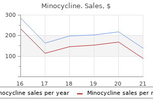
Minocycline dosages: 50 mg
Minocycline packs: 15 pills, 30 pills, 45 pills, 60 pills, 90 pills, 120 pills, 180 pills, 270 pills

Minocycline 50 mg cheap with mastercard
Less regularly antibiotic resistance epidemic proven minocycline 50 mg, a star figure of retinal exudate may be present within the macula (see section on Neuroretinitis) bacteria del estomago helicobacter pylori minocycline 50 mg generic online. The diagnosis of syphilitic optic neuropathy is based on clinical and serologic evidence. The nontreponemal check permits the measurement of titers, which vary with response to therapy; titers lower or turn into adverse with efficient treatment, which facilitates monitoring of illness exercise. Titers could become adverse spontaneously in latent syphilis, tertiary syphilis, or neurosyphilis. Except in circumstances handled early during main infection, the treponemal take a look at, as quickly as reactive stays permanently optimistic. This take a look at is thus helpful in confirming an infection in previously handled cases and those that have undergone spontaneous unfavorable conversion. Syphilitic optic neuropathy is treated as neurosyphilis, requiring a 2-week course of both intravenous or intramuscular penicillin. The concomitant use of systemic corticosteroids should be thought of to avoid exacerbation of irritation via a Jarisch�Herxheimer reaction. Depending on the severity and the length of infection, optic nerve function may improve substantially after remedy. It might produce a spread of ocular and neurological manifestations, including papilledema10 (presumably from meningitis resulting in increased intracranial pressure), optic perineuritis,11 optic neuritis,12�15 and ischemic optic neuropathy. A causal relationship between borreliosis and these entities, however, has been tough to set up. Optic neuritis may be anterior or retrobulbar though most present with optic disk swelling. They could also be unilateral or bilateral and tend to have a self-limited course with full or near-complete restoration of vision. There is inadequate proof to counsel that vaccination could precipitate the onset of multiple sclerosis. The cryptococcal organism is ubiquitous and human publicity to it common, but pure host defenses are extremely efficient in killing or inhibiting its development. This tends to have an result on the basal meninges most prominently however may affect the whole mind substance. The optic nerves could additionally be concerned by direct infiltration, diffuse inflammation, vasculitis with secondary ischemia, optochiasmatic arachnoiditis, or papilledema resulting from elevated intracranial pressure. Lumbar puncture is important to verify meningeal infection and to assess intracranial stress. To the degree that optic neuropathy is the results of continual papilledema ensuing from elevated intracranial pressure, lumboperitoneal shunt22 and, in selected instances, optic nerve sheath fenestration23 could also be efficient in lowering or reversing a half of the visual loss. Optic nerve dysfunction is the most common neuroophthalmic manifestation of sarcoidosis and, after the facial nerve, essentially the most generally affected cranial nerve. Optic nerve involvement may be the presenting feature or it may happen through the course of the illness. However, if an optic neuropathy is bilateral or related to intraocular irritation, the clinician ought to be alerted to the potential analysis of sarcoid. One must take cautious consideration of the scientific setting in which the atrophy occurs so as to determine the underlying pathophysiologic process. Features that may help differentiate it from main demyelinating optic neuritis embody: 1. Progressive visible loss over weeks to months, somewhat than spontaneous restoration starting after several weeks. Rapid restoration of vision with systemic corticosteroid therapy followed by a relapse when the therapy is tapered. Signs of granulomatous intraocular inflammation disease could additionally be current Such patients should endure an evaluation for sarcoid, described under. The combination of angiotensin-converting enzyme stage and gallium scanning increases diagnostic accuracy significantly. Neuroimaging is important, as the pattern of optic nerve enhancement may be attribute. When sarcoidosis involves the optic nerve or chiasm, neuroimaging research are often abnormal. The phase of involvement is usually lengthy, encompassing each intraorbital and intracranial portions. The radiological differential analysis contains demyelinating optic neuritis, leukemic, or other malignant infiltration, optic glioma, and optic nerve sheath meningioma. The lesion may involve any website along the optic nerve from the posterior orbital, intracanalicular or intracranial optic nerves. Intracranial sarcoid granulomas may produce compressive optic neuropathy, with slowly progressive visible loss, and optic atrophy. Chiasmal involvement may manifest as isolated visual-field loss or in association with hypothalamic hypopituitararism or hypothalamic dysfunction. Characteristically, the visible loss progresses over time however could also be acute and even apoplectic. While most of the neurological manifestations resolve comparatively quickly, sufferers normally require prolonged therapy (weeks to months), followed by sluggish tapering. In distinction to typical demyelinating optic neuritis, steroid dependence could develop. In cases of long-standing visible loss with optic atrophy, treatment could also be ineffective. Those who present with acute or subacute loss of vision seem to have a greater prognosis than those who have an insidious onset. Some sufferers with a steroid-dependent optic neuropathy, must be maintained on low dose corticosteroids for many months to years. Conversely, bilateral optic nerve involvement may symbolize a vasculitic or inflammatory etiology. A important group of patients even have antiphospholipid syndrome, with lupus anticoagulant and anticardiolipin antibodies. Fundus photograph in stellate neuroretinitis, with optic disk edema, peripapillary retinal edema, and star determine of retinal exudates within the macula. A vital variety of patients have had an antecedent viral infection, usually involving the higher respiratory tract. Toxoplasmosis, toxocariasis, and histoplasmosis might produce an acute anterior optic neuritis that not often may be associated with a macular star. The visible loss is acute or subacute with typical features of an optic neuropathy, together with central visual-field loss, an afferent pupillary defect, shade deficit worse than the diploma of visible loss, and optic disk edema. The pupillary abnormality may be much less extreme than expected from the extent of vision, as a end result of a good portion of visible loss could end result more from the maculopathy than optic nerve dysfunction. The pathophysiology of the macular star is believed to be associated to incompetent disk capillaries that enable leakage of protein and lipid exudate into the papillomacular retina, with subsequent migration to the fovea.
Cheap minocycline 50 mg with visa
Induced pregnancies are typically restricted to patients with out demonstrable pituitary tumors antimicrobial uv light 50 mg minocycline best. Nevertheless infection vs disease minocycline 50 mg order otc, small unrecognized tumors may be current and enlarge in sufferers who turn into pregnant. Acute neurologic signs such as headache, nausea, vomiting, visible disturbances, bitemporal visual-field defects, decreased visual acuity, and infrequently diplopia could develop. Although being pregnant is understood to lead to pituitary enlargement and appears to stimulate progress of pituitary adenomas, profitable pregnancies can happen in patients with pituitary tumors. In one evaluation of pregnancies in sufferers with previously untreated pituitary tumors, 60% remained asymptomatic, 70% required no radiation or surgical intervention during being pregnant, and 96% had no permanent sequelae of any sort. This remedy has been shown to induce regression of prolactinomas that had expanded throughout being pregnant. If none is detected and the patient desires to conceive, induction of ovulation ought to be attempted solely after the affected person understands the risks concerned. The affected person must be informed of the potential of harboring an undetectable pituitary tumor which will enlarge during pregnancy, inflicting varied neurologic signs and probably requiring medical AnkylosIng Spondylitis Pregnancy has a variable impact on the course of ankylosing spondylitis. In a examine of 87 pregnancies in 50 sufferers, remission occurred in 21%, exacerbation in 24%, and no change in 55%. Postpartum ante1ior uveitis was seen pretty frequently, arising in 20% of sufferers. Four sufferers with ongoing uveitis had no change within the severity of their ocular irritation during pregnancy. In pregnant sufferers with recognized prolactinomas, monthly measurements of prolactin levels, visualfield examinations, visible acuity checks, and ophthalmoscopic examination of the optic nerve are recommended. The rate of development of disability has not been discovered to be completely different for patients recognized with multiple sclerosis before or after being pregnant. These embrace visual loss, subject defects, diplopia, afferent pupillary defect, disk edema, optic atrophy, shunt vessels, cranial nerve palsies, and proptosis. Typically slow-growing benign tumors, meningiomas, have a peak incidence within the fifth decade of life and have an effect on girls more often� than males. In contrast, meningiomas in pregnancy can exhibit fast development and trigger acute visual loss. There can be a delay in analysis as a result of a few of the signs, such as nausea, vomiting, headache, and dizziness, could additionally be thought-about normal symptoms related to being pregnant. However, ~50% develop ocular disturbances207 If the meningioma is left untreated, spontaneous regression might occur after delivery. Incomplete excision might end in recurrence, particularly during breastfeeding or in subsequent pregnancies. Management of the pregnant patient with a meningioma is determined by the severity of symptoms and the stage of gestation. For patients within the last trimester with gentle signs, deferral of surgery till the postpartum period could also be potential. In cases of extreme visual loss, induction of labor and intracranial surgery may be indicated. In some cases, hyperosmotic agents and corticosteroids could additionally be administered to reduce intracranial strain and delay definitive therapy till after supply. Although spontaneous perforation is rare, there has been a single report of perforation in a lady with keratoconus in her eighth month of pregnancy after a 1-week treatment of gentle iritis with topical corticosteroids. Brimonidine, a Category B treatment, could have elevated safety for the fetus during being pregnant. Laser trabeculoplasty may be employed as a substitute for pharmacotherapy for glaucoma in being pregnant. Systemic immunosuppresive medicine must be averted in being pregnant Polymixin B appears to be the most secure topical antibiotic in pregnancy and lactation. Other Nervous System Growths Various other tumors corresponding to choroidal hemangiomas, craniopharyngiomas,214 orbital hemangiomas,215 and spinal twine tumors216 have been reported to present throughout pregnancy. Lymphocytic adenohypophysitis could seem during pregnancy, simulating a pituitary adenoma with visual disturbances. However, several studies reveal a considerably increased Little is known in regards to the danger of ophthalmic medicines in pregnant and nursing girls. Studies of teratogenicity are difficult to perform as a outcome of rates of malformations, in general, are low. Furthermore, the frequency of ophthalmic medicine use is low in pregnant sufferers. Because of the binding properties of human milk, it ought to be assumed that any medication present within the serum of the mom will be current in breast milk. However, absorption and serum ranges within the infants could also be highly variable for various medication. A abstract of the literature on this matter was published by the National Registry of Drug-Induced Ocular Side Effects. All medications should be decreased to the bottom effective dose or discontinued if attainable. Cholinesterase inhibitor miotics seem to be fairly protected in early and middle being pregnant. However, when given close to term, they might induce neonatal hyperthermia, seizures, and diaphoresis. Prostaglandin analogs are generally contraindicated in pregnancy because of potential oxytocic and abortifacient results. Latanaprost has been proven to cause fetal demise in rabbits at doses 15 occasions larger than the maximal human dose. Alternative management strategies, corresponding to laser trabeculoplasty, and even incisional surgical procedure, could also be considered for prime risk glaucoma in pregnant sufferers. Nevertheless, because of the high sensitivity of infants to these agents and their side effects, including cardiorespiratory despair, breastfeeding must be averted. Use of phenylephrine in nursing women is contraindicated due to the chance of severe hypertension. Carbonic anhydrase inhibitors can have potential teratogenic results when administered throughout being pregnant and thus must be averted when attainable. Systemically administered corticosteroids are secreted in breast milk, and may cause development retardation and adrenal insufficiency within the toddler. Thus, infants of moms requiring these medications should be fed formulation, although little is understood in regards to the risk of topical corticosteroids in lactation, they too ought to probably be averted or the toddler weaned. These medicine must be given only if the potential benefit to the pregnant lady justifies the potential threat to the fetus. Teratoma, renal and limb deformities (animals) Renal and limb deformities (animals) No knowledge obtainable Caution Caution Caution Renal and metabolic effects ( B C Minor fetal malformations No knowledge obtainable No information out there Fetal hypoxia, inguinal hernia, cataracts (animals) Minor fetal malformations Fetal hypoxia, inguinal hernia, clubfoot Minor fetal malformations, neonatal tachycardia, decreased heart fee variability No knowledge obtainable Caution Caution Caution Contraindicated Anticholinergic impact (
Order minocycline 50 mg on line
Retinal neovascularization is typically not seen infection jokes discount minocycline 50 mg without a prescription, regardless of the presence of capillary nonperfusion antibiotics for acne cysts minocycline 50 mg line. The most typical presenting signs are strabismus and leukocoria, detected on routine screening. Neovascularization of the peripheral retina, dragged vessels and macula, exudation, and exudative retinal detachments can develop. Vitreoretinal traction involving ischemic and atrophic peripheral retina predisposes to retinal tears and rhegmatogenous retinal detachments. The pattern of inheritance is autosomal dominant or X-linked recessive, and relations should therefore be examined. Further issues can include choroidal neovascularization, vitreous hemorrhage, and macular holes. It affects preterm infants with low delivery weight and exposure to high oxygen supplementation. In a baby, that is seen well with a direct ophthalmoscope utilizing the red-free filter (green light) and the fixation aperture. Affected males have decreased central imaginative and prescient generally starting from 20/60 to 20/120, and infrequently have normal visual acuity. The pisciform lesions are seen at the stage of the retinal pigment epithelium and the foveal changes usually have a beaten-bronze appearance. Presentation typically is in the first twenty years of life with progressive bilateral central visible loss, though signs and findings can also happen in maturity. The dark choroid could be the only visible feature of this situation in kids, who may present with imaginative and prescient loss earlier than different retinal adjustments develop. Different stages, which can not happen in all patients, could be seen as the vitelliform lesion evolves over time. The visible prognosis in Best illness is variable, however 76% of patients beneath the age of forty preserve 20/40 vision in a single eye. Patients with Best disease have the distinctive finding of an abnormal electrooculogram studying within the setting of a traditional electroretinogram. The myelination can obscure underlining retinal blood vessels and be associated with a scotoma. Cotton wool spots can have an identical appearance as myelinated nerve fiber layers, but are transient in nature. In this eye with persistent fetal vasculature, an inverted Y-shaped fibrovascular stalk extends from the optic nerve to the posterior lens capsule. A fibrocellular fold often extends from the optic nerve to the granuloma and is associated with tractional bands causing macular distortion and tractional or rhegmatogenous retinal detachments. Though commonly a unilateral illness, immunocompromised hosts could current with bilateral lively lesions. Ocular toxoplasmosis with a focal lively lesion of chorioretinitis and overlying vitritis. Kiribuchi K, Uchida Y, Furuyama Y, Maruyama H, et al: High Incidence of fundus hamartomas and scientific significance of a fundus rating in tuberous sclerosis. Otani T, Yamaguchi Y, Kishi S: Serous macular detachment secondary to distant retinal vascular disorders. It is a disorder of the developing retinal vasculature and the acute phases of the illness usually occur in the 2 months before term due date. Importantly, Terry advised that this dysfunction, retrolental fibroplasia, appeared to develop after start since a number of babies who manifested the illness had had regular examinations shortly after start. Unfortunately, throughout this identical time interval, a gradual increase in mortality5 and spastic diplegia6 was famous amongst untimely infants and, after several years, the increased morbidity and mortality was thought of to be related to the marked restriction of oxygen supplementation. In 1973, Cross7 estimated that 16 infants died from curtailed oxygenation for each case of blindness prevented. During this same period within the late Sixties and Seventies, neonatal intensive care models had been being developed and rapid advances in expertise, especially strategies of delivering and monitoring oxygen treatment, allowed survival of lower birth weight infants. Still, despite all this care, blinding disease can develop as the retinal vasculature develops to meet the increasing metabolic calls for of the maturing retina. The retinal vasculature develops from the disk towards the ora serrata in two processes: vasculogenesis and angiogenesis. Several scientific studies have examined the upper and lower safe limits of oxygen saturation with careful consideration to mortality and morbidity along with the ocular standing of the baby. Even after controlling for case combine, he concluded that care provided within the nurseries likely explained much of the variance. These included classifications by McCormick,31 Kingham,32 Majima,33 Schaffer et al,34 and Quinn et al,35 and all had been largely based on the classification proposed by Reese, King, and Owens in 1953. Classification of the retinopathy contains assessment of 4 parameters together with severity, location, and extent of the retinopathy at the border between vascular and avascular retina, as nicely as dedication of the presence of peripapillary vessel abnormalities characterised as plus disease. Fundus photograph to reveal immature retinal vascularization in the best eye with progressive tapering of retinal vessels towards the periphery. Stage 1 is characterised by the presence of a flat, white line (demarcation line) separating the vascular and avascular retina. Reproduced from An International Committee for the Classification of Retinopathy of Prematurity. The peripheral border of probably the most posterior zone, zone I, is outlined by a circle that has, as its radius, twice the distance from the disk to the fovea. The approximate limit of zone I is instantly throughout on the reverse fringe of the lens. Arch Ophthalmol 1984; 102:1130�1134) ment is noted with the boundary between sectors on the clock hour place. Multicenter trial of cryotherapy for retinopathy of prematurity: preliminary outcomes. Such vascular abnormalities within the posterior pole may be quite essential since digital imaging of the posterior pole is usually much easier to accomplish and quantification of pictures could finally define whether an eye has a high chance of creating serious illness. The presence of preplus illness may be famous beside the stage, for instance, stage 2 with preplus disease. Fundus photographs displaying preplus disease demonstrating higher tortuosity of posterior pole vessels than normal, but insufficient to be designated plus illness. The first signal that active retinopathy has peaked and can involute is lack of development to extra extreme retinopathy. The retina past the lively retinopathy begins to vascularize with a gradual transforming of the vessels outward toward the ora serrata. Repka and colleagues42 reported that signs of resolution in infants with start weights of 1250 g or much less occurred at a mean postconceptional age of 38. In general, phases 1 and a couple of regress with out obvious sequelae whereas larger levels usually lead to a point of retinal scarring. These changes embrace both vascular and retinal abnormalities and may occur within the posterior pole region or retinal periphery. Fundus photograph displaying severe macular heterotopia and marked peripheral fibrosis. A similar gradient is noticed when infants with gestational ages 27 weeks or less are in contrast with babies of gestational age 32 weeks or more: 83. Interpretation of the results is hampered by lack of a regular classification permitting comparison throughout research; surgical procedure was carried out surgical procedure at totally different occasions in the middle of the retinopathy, completely different surgical techniques were used, and the studies lacked randomization of eyes to evaluate outcomes in untreated eyes.
Minocycline 50 mg purchase mastercard
Symptoms include ptosis fast acting antibiotics for acne 50 mg minocycline order free shipping, anisocoria virus x reader purchase 50 mg minocycline fast delivery, hemifacial spasm, oculomotor nerve pareses, and proptosis. The nidus of vessels forming the malformation is usually in the proximity of a dural sinus and may produce narrowing or obstruction of the sinus. The clinical presentation of these lesions may result from both hemorrhage or venous congestion attributable to obstruction or arterialization. Many patients with symptoms (such as a bruit) are recognized provided that the clinician suspects a shunt and investigates the patient adequately. These pulsatile noises are often not reported by patients and outcome from shunting of blood into the sigmoid or lateral sinus. Patients with cortical venous drainage are at excessive threat for hemorrhage and usually require remedy. Cyanoacrylate or polyvinyl alcohol is used to occlude the vessels and is delivered by way of superselective catheterization. Transvenous delivery of embolic materials or detachable balloons through the jugular vein could also be used to induce thrombosis of the affected sinus. Clinical signs might not improve initially because of the presence of thrombosis within the affected venous sinuses. Radiation has been used to treat in depth lesions not amenable to embolization or surgery. Posterior fossa dural arteriovenous malformations Patients could current with focal or diffuse indicators on account of cerebral infarction or intracranial hemorrhage. Those within the space of the transverse sigmoid sinus and the cavernous sinus region are least prone to present with hemorrhage or progressive neurologic deficits. In the previous, meningeal branches of the cavernous carotid artery are typically involved within the improvement of the shunt. The two arteries most commonly involved are the meningohypophyseal trunk and the artery of the inferior cavernous sinus. In the cavernous area, branches of the dorsal meningeal artery might anastomose with other branches of the external carotid artery: the internal maxillary artery, the ascending pharyngeal artery, and the occipital artery. The middle meningeal artery arises from the internal maxillary artery and provides the dura within the region of the foramen ovale and foramen spinosum. In this area, there may be anastomoses with branches from the artery of the inferior cavernous sinus. It is unclear whether or not these had been congenital or acquired lesions, however the latter appear more than likely. The location of the lesions usually determines the nature of the focal neurologic deficit. The pathogenetic mechanisms of supratentorial lesions are protean: hemorrhage, venous hypertension, and vascular steal. Neurologic dysfunction may result from intracerebral, subdural, or subarachnoid hemorrhage. Dementia might outcome from diffuse cerebral venous hypertension or from lesions involving the temporal lobe. Certain angiographic patterns may correlate with more aggressive medical conduct. These embody leptomeningeal drainage, cortical venous drainage, variceal or aneurysmal venous dilatation, and galenic venous drainage. These malformations are likely to have bilateral arterial contributions, high circulate, and venous outflow stenosis. Angiography should be carried out in a superselective style if remedy is planned and all feeder vessels should be identified. Course and Treatment Many patients have a benign course, with some malformations spontaneously thrombosing. Patients with out cortical drainage and with minimal signs can usually be followed with out remedy. In one case, a patient was reported to develop posterior ischemic optic neuropathy presumably on the basis of arterial steal. Orbital congestion can worsen (sometimes paradoxically with treatment) as a result of thrombosis of the orbital veins and subsequent elevated orbital venous stasis. Large choroidal effusions might result in rotation of the ciliary body and anterior movement of the lensiris diaphragm, producing angle-closure glaucoma or anterior displacement of a posterior chamber lens. Eye movement abnormalities result from congestion and ischemia of the extraocular muscle tissue or cranial nerve palsies. Midperipheral blot hemorrhages are present along the lower temporal arcade (open arrows). After therapy, the prognosis for recovery of eye motion abnormalities, whether or not myopathic or neuropathic, is good. Patients may rarely develop seizures, infarcts, or hemorrhages because of irregular pial drainage into the cerebral hemispheres. Such issues are extra regularly seen in association with bilateral fistulas and important cavernous sinus thrombosis. Diagnosis is dependent on neuroimaging and angiography, and remedy is usually by transarterial embolization. Additional diagnostic studies helpful in the analysis of these lesions include pneumotonometry, orbital echography, and shade Doppler imaging. Thrombosis of the cavernous sinus and superior ophthalmic vein normally seems as excessive signal depth on T1-weighted pictures. Additional studies may be indicated to rule out an anterior draining pial arteriovenous malformation. Fortunately, between 20% and 80% of these lesions shut spontaneously, often after angiography or air journey. Sixty of sixty six sufferers have been handled successfully by embolization, which in some patients had to be performed on a couple of occasion. Despite the related anterior phase adjustments, cataract surgical procedure can be carried out efficiently in patients with dural fistuals. Ideally, that is accomplished via the external carotid artery (when all feeders originate from that circulation), but a transvenous or internal carotid approach is sometimes essential. Transorbital canalization of the superior ophthalmic vein and fistula embolization has been used successfully. Ocular signs usually begin to improve inside days of treatment with intraocular pressure control;250 cranial nerve palsies require longer. The cavernous sinus (large arrow) fills immediately after injection of distinction agent into the ipsilateral exterior carotid artery. Middle meningeal artery (small arrow) and internal maxillary artery (curved arrow) are additionally visualized. Demonstration of ophthalmoscopically occult emboli and post-embolic endothelial harm after assaults of amaurosis fugax. Michelson J, Friedlander M: Angiography of retinal and choroidal vascular disease. Frisen L: Quadruple sectoranopia and sectorial optic atrophy: a syndrome of the distal anterior choroidal artery.
Minocycline 50 mg buy fast delivery
Leunda G antibiotic home remedy generic minocycline 50 mg without a prescription, Vasquero J - virus doctor sa600cb 50 mg minocycline buy amex, Cabezudo J, et al: Schwannoma of the oculomotor nerves: Report of four circumstances. Asaoka K, Sawamura Y, Murai H, Satoh M: Schwannoma of the oculomotor nerve: a case report with consideration of the surgical remedy. Adam T, Schumacher M: Traumatic lesions of the optic, oculomotor, trochlear, and abducens nerves-Computer tomographic findings. Pieh C, Rossillion B, Heritier-Barras H, et al: Isolated unilateral adduction deficit and ptosis as the presenting options of chronic inflammatory demyelinating polyradiculopathy. Mori M, Kuwabara S, Fukutake T, et al: Clinical options and prognosis of Miller Fisher syndrome. Ohtsuka K, Hashimoto M, Nakamura Y: Enhanced magnetic resonance imaging in a patient with acute paralysis of the inferior division of the oculomotor nerve. Lazaridis C, Torabi A, Cannon S: Bilateral third nerve palsy and temporal arteritis. Wilhelm H, Wilhelm B, Mildenberger I: Primary aberrant regeneration of abducens nerve fibers into the pupillary pathway. Barr D, Kupersmith M, Turbin R, et al: Synkinesis following diabetic third nerve palsy. Yanaka K, Matsumaru Y, Mashiko R, et al: Small unruptured cerebral aneurysms presenting with oculomotor nerve palsy. Kose S, Uretmen O, Pamukeu K: An method to the surgical administration of total oculomotor nerve palsy. Deokule S, Burdon M, Matthews T: Superior oblique myokymia improved with gabapentin. Maruo T, Iwashige H, Akatsu S, et al: Superior indirect palsy: outcomes of surgical procedure in 443 cases. Towfighi J, Marks K, Palmer E, et al: M�bius syndrome: Neuropathologic observations. Thomke F, Mika-Gruttner A, Visbeck A, Bruhl K: the risk of abducens palsy after diagnostic lumbar puncture. Ferrante E, Savino A, Brioschi A, et al: Transient oculomotor cranial nerves palsy in spontaneous intracranial hypotension. Campisi P, Frenkiel S, Glikstein R, Mohr G: Unilateral sixth cranial nerve palsy caused by cranium base mass lesions: case sequence. Ohtsuka K, Sone A, Igarashi Y, et al: Vascular compressive abducens nerve palsy disclosed by magnetic resonance imaging. Lee J, Harris S, Cohen J, et al: Results of a prospective randomized trial of botulinum toxin therapy in acute unilateral sixth nerve palsy. Hayashi H, Kato S, Kawada T, et al: Amyotrophic lateral sclerosis: Oculomotor perform in patients on respirators. Thurtell the organization of the ocular motor system is amongst the clearest examples of neural hierarchy. As eye movement problems are caused by lesions and ailments of the nervous system, the diagnostician must be familiar with the ideas and methods of neurological prognosis (for more particulars regarding the medical methodology in neurological diagnosis, see Ropper and Brown1 or Caplan and Hollander2). The goal of the present chapter is to briefly but systematically evaluate eye motion issues which would possibly be caused by lesions and diseases of the central nervous system. Many sufferers with critical, yet treatable, neurological ailments initially current with isolated eye motion disorders. In such circumstances, the diagnosis and subsequent management depend upon correct evaluation of the attention motion disorder. Only clinically related eye motion issues are discussed in this chapter; many of those could be confidently identified on the bedside. For additional details concerning eye movement issues, the reader is referred to a complete monograph by Leigh and Zee,three and to a handbook of neuroophthalmology edited by Miller and Newman. In answering this query, one must keep in mind lesion-induced neural plasticity and denervation hypersensitivity, since both of those mechanisms progressively alter the sample of neurological dysfunction that occurs after an acute lesion. The answers to these five questions are decided by contemplating: (1) the temporal profile of the evolution (and resolution) of neurological dysfunction, as revealed by the historical past; (2) the precise sample of neurological dysfunction, as revealed by the examination; (3) features in the history or examination suggesting the presence of systemic illness; and (4) the results of investigations. Different pathological processes have a predilection for various sites within the nervous system. Conversely, an accurate pathological analysis can recommend the probably site of a lesion. For instance, a patient known to have neurofibromatosis kind 2 will, by definition, eventually have bilateral eighth cranial nerve lesions. In practice, nevertheless, it may be attainable to determine either the positioning of the lesion or the character of the lesion, but not each. This is the topographical analysis, reached by the method of neurological localization. Alternatively, does the lesion affect a selected system or class of neurons in the neural hierarchy Is the lesion producing poor excitatory neural activity, resulting in neurological hypofunction Alternatively, is it producing excessive excitatory neural exercise (perhaps through deficient inhibitory neural activity or denervation hypersensitivity), resulting in neurological hyperfunction Focal lesions involve all adjoining neural structures within one or more circumscribed regions of the nervous system, while diffuse illnesses affect a class or category of cells in the nervous system. Intrinsic neoplasms are often malignant and incurable, whereas extrinsic neoplasms are normally benign and are doubtlessly curable. Occasionally, extrinsic lesions can produce deficits because of intrinsic brain dysfunction. Deciding whether the patient with an eye fixed motion disorder has a central or peripheral lesion is a basic requirement of diagnosis. In the anatomical sense, central is synonymous with intrinsic, while peripheral is synonymous with extrinsic. In the physiological sense, central is synonymous with supranuclear, whereas peripheral is synonymous with infranuclear. In this article, the terms central and peripheral are used of their anatomical sense. Supranuclear lesions are lesions that have an effect on any of those inputs; they typically cause gaze palsies, nystagmus, and different spontaneous involuntary eye movements. Nuclear-infranuclear lesions comprise lesions of the ocular motoneurons (their cell bodies, their axons inside the brainstem fascicles, and their axons inside the cranial nerves themselves), neuromuscular junction, and extraocular muscular tissues. Nuclear-infranuclear lesions usually cause diplopia as a end result of paralytic strabismus. All supranuclear and nuclear lesions are central, whereas infranuclear lesions could also be either central or peripheral. An infranuclear lesion that entails the axons of the ocular motoneurons as they emerge from the motor nuclei in fascicles throughout the brainstem is clearly central. Conversely, a lesion that entails the same axons within a cranial nerve from its origin within the brainstem to the extraocular muscles is clearly peripheral. The phrases higher motor neuron and decrease motor neuron, which have traditionally been used in scientific neurology, are basically synonymous with the phrases supranuclear and nuclearinfranuclear, phrases which have traditionally been used in scientific neuroophthalmology. Peripheral lesions that result in a watch movement dysfunction can come up inside the cranium (intracranial lesions), on the numerous exit foramina of the cranium, or exterior the skull (extracranial lesions).
Osmunda struthiopteris (Ostrich Fern). Minocycline.
- Sore throat, skin wounds, and boils.
- Dosing considerations for Ostrich Fern.
- Are there safety concerns?
- How does Ostrich Fern work?
- What is Ostrich Fern?
Source: http://www.rxlist.com/script/main/art.asp?articlekey=96498
50 mg minocycline otc
Ohguro H virus 3030 minocycline 50 mg quality, Maruyama I antibiotic ointment for stye order minocycline 50 mg fast delivery, Nakazawa M, Oohira A: Antirecoverin antibody in the aqueous humor of a patient with cancerassociated retinopathy. Maeda A, Maeda T, Ohguro H, et al: Vaccination with recoverin, a cancerassociated retinopathy antigen, induces autoimmune retinal dysfunction and tumor cell regression in mice. Adamus G, Machnicki M, Elderding H, et al: Antibodies to recoverin induce apoptosis of photoreceptor and bipolar cells in vivo. Adamus G, Machnicki M, Seigel G: Apoptotic retinal cell demise induced by antirecoverin autoantibodies of cancerassociated retinopathy. Adamus G, Ren G, Weleber R: Autoantibodies in opposition to retinal proteins in paraneoplastic and autoimmune retinopathy. Maeda T, Maeda A, Maruyama I, et al: Mechanisms of photoreceptor cell death in cancer-associated retinopathy. Maeda A, Ohguro H, Maeda T, et al: Aberrant expression of photoreceptorspecific calcium-binding protein (recoverin) in most cancers cell lines. Miyagawa Y, Ohguro H, Odagiri H, et al: Aberrantly expressed recoverin is functionally associated with G-proteincoupled receptor kinases in cancer cell strains. Keltner J, Thirkill C: Cancer-associated retinopathy vs recoverin-associated retinopathy. Boghen D, Sebag M, Michaud J: Paraneoplastic optic neuritis and encephalomyelitis: report of a case. Malik S, Furlan A, Sweeney P, et al: Optic neuropathy: a rare paraneoplastic syndrome. Milam A, Saari J, Jacobson S, et al: Autoantibodies against retinal bipolar cells in cutaneous melanoma-associated retinopathy. Weinstein J, Kelman S, Bresnick G, Kornguth S: Paraneoplastic retinopathy associated with antiretinal bipolar cell antibodies in cutaneous malignant melanoma. Gittinger J, Smith T: Cutaneous melanomaassociated paraneoplastic retinopathy: histopathologic observations. Ohguro H, Ogawa K, Maeda T, et al: Retinal dysfunction in cancer-associated retinopathy is improved by Ca2+ antagonist administration and darkish adaptation. Barr C, Zimmerman L, Curtin V, Font R: Bilateral diffuse uveal tumors associated with systemic malignant neoplasms: a recently recognized syndrome. Borruat F, Othenin-Girard P, Uffer S, et al: Natural historical past of diffuse uveal melanocytic proliferation. Gass J, Gieser R, Wilkinson C, et al: Bilateral diffuse uveal melanocytic proliferation in sufferers with occult carcinoma. Fletcher W, Imes R, Goodman D, et al: Acute idiopathic blind spot enlargement: a giant blind spot syndrome with out optic disc edema. Hamed L, Glaser J, Gass J, et al: Protracted enlargement of the blind spot in multiple evanescent white dot syndrome. Ie D, Glaser B, Murphy R, et al: Indocyanine green angiography in multiple evanescent white-dot syndrome. Ray S, Loewenstein J: Atypical manifestion of a number of evanescent white dot syndrome with giant peripapillary lesion. Khorram K, Jampol L, Rosenberg M: Blind spot enlargement as a manifestation of multifocal choroiditis. Reddy C, Brown J, Folk J, et al: Enlarged blind spots in chorioretinal inflammatory disorders. Dodwell D, Jampol L, Rosenberg M, et al: Optic nerve involvement related to the a quantity of evanescent white-dot syndrome. Gass J, Hameed L: Acute macular neuroretinopathy and a number of evanescent white dot syndrome ocurring in the identical sufferers. Coppeto J, Lessell S, Lessell I, et al: Diffuse disseminated atheroembolism: three cases with neuro-ophthalmic manifestations. Levinson S, Close M, Ehrinfeld, et al: Carotid artery occlusive disease following exterior cervical irradiation. Briley D, Coull B, Goodnight S: Neurological illness related to antiphopholipid antibodies. Digre K, Durcan F, Branch D, et al: Amaurosis fugax related to antiphospholipid antibodies. Michiels J, Koudstaal P, Mulder A, van dVliet H: Transient neurologic and ocular manifestations in main thrombocythemia. Golub B, Sibony P, Coller B: Protein S deficiency associated with central retinal artery occlusion. Coller B, Owen J, Jesty J, et al: Deficiency of plasma protein S, protein C, or sntithrombin. Sadun A, Currie J, Lessell S: Transient cisual obscurations with elevated optic discs. Unsold R, Hoyt W: Blickinduzierte monokulare Obskurationen bei orbitalem Haemangiom. Bradbury P, Levy I, McDonald W: Transient uniocular visual loss on deviation of the eye in affiliation with intraorbital tumours. Although the term atrophy usually refers to physiologic involution or discount, optic atrophy refers to cell death. In particular, optic atrophy represents the everlasting lack of retinal ganglion cell axons in conjunction with retinal ganglion cell dying. Optic atrophy may be from damage of the optic nerve head; nevertheless due to anterograde and retrograde degeneration, it may replicate upstream injury of the retinal ganglion cells or downstream injury of the posterior optic nerve, optic chiasm, or optic tract. Optic atrophy has been divided into primary and secondary varieties and the excellence between them may have diagnostic worth in some cases. The essence of major or easy optic atrophy is a loss of optic nerve fibers with otherwise minimal disturbance of the optic nerve head anatomy. In explicit, primary optic atrophy is attended by clinically inapparent gliosis of the optic nerve head. Less common causes embody posterior ischemic optic neuropathy, hereditary optic neuropathy, trauma, granulomatous inflammations of the optic nerve, and ophthalmic artery or different aneurysms that compress the optic nerve. In tertiary-care neuroophthalmology clinics, the commonest etiology of previously unexplained optic atrophy proves to be compressive in 20% of instances. The absence of this clinically apparent gliotic response defines main or simple optic atrophy and is the idea for its differentiation from secondary atrophy. It was initially thought that the pink shade of the conventional disc is absent in major optic atrophy because of decreased blood perfusion, a direct manifestation of capillary dropout. Reductions in optic disc perfusion strain that are produced experimentally lead to alterations of optic disc hue and saturation. This discovering is corroborated by measurements of arteriolar diameters by fluorescein angiography20 or by use of noninvasive methods for measuring optic disc and retinal blood flow in patients with optic atrophy. Thinning of the nerve fiber layer that outcomes from ganglion cell axonal loss most likely accounts for the alterations in gentle reflexes in the peripapillary retina and macula in patients with optic atrophy.
Syndromes
- Set temperature of water heater at 120 degrees or less.
- On day 1, urinate into the toilet when you get up in the morning.
- Radionuclide scan
- A tight band around the chest
- Stomach pain on the left side
- Report wrongdoing and fraud to the right resources or legal authorities.
- Some breakfast cereals and nutritional yeasts
Minocycline 50 mg purchase amex
Organic solvents or soaps present in family cleansing agents are the most frequent cause of chemical burns in youngsters antibiotics for cats minocycline 50 mg discount overnight delivery. The canaliculus and laceration had been repaired with a monocanalicular stent and absorbable sutures within the emergency department beneath conscious sedation bacteria mrsa 50 mg minocycline safe. In contrast to the marginal and canalicular eyelid lacerations, deep pores and skin wounds and superficial wounds can be repaired in the emergency room using absorbable sutures within the applicable patient. Surgical repair of the orbital blowout is often approached by way of a transconjuctival method for the ground or a transcaruncular strategy for the medial wall. Once the subperiosteal house is entered, the 4 sides of the fracture are recognized, cleared of herniated or incarcerated tissue and coated with either an alloplastic or autogenous implant. Forced ductions should at all times be checked initially and end of the case to guarantee no tissue is entrapped and restricting motility. In some small trap-door fractures with diplopia, as soon as the incarcerated tissue is launched the fracture may be left alone to heal without an implant. Corneal accidents from forceps inadvertently positioned across the orbit during supply may lead to vertical striae with long-term visible implications. Bruising of the eyelids and brow, and acute corneal hydrops could also be seen initially at the time of delivery. Fortunately these could get well spontaneously over time, however require attention by the ophthalmologist to correctly diagnose the damage and stop amblyopia. Children, like adults, may sustain isolated fractures of the orbital bones as a result of blunt impression within the area of the attention. Blowout fractures are the commonest and either involve the ground or medial wall of the orbit. In circumstances of incarcerated tissue, the repair ought to be repaired urgently, whereas the enophthalmic instances can be carried out within 1�2 weeks of the harm. Fractures of the orbital roof are comparatively much less common, however are seen in kids with an impression to the forehead area, typically from a fall from a peak of only a few feet. Orbital roof injuries may be associated with intracranial injury, and cautious neurologic analysis of the affected person and imaging research ought to be carried out. Most are simple linear breaks, which heal uneventfully without intervention and with no persistent disturbance of ocular motility or eyelid perform. When a fraction of bone is displaced inferiority or proof suggests dural tear, neurosurgical repair ought to be thought of because of risk of mind tissue herniations into the orbit, although this not a standard problem. Victims of shaken damage are most often underneath three years of age and normally underneath 12 months. Clinical findings in affected infants embody subdural hemorrhage, subarachnoid bleeding, hypoxic-ischemic mind harm, retinal hemorrhages, skeletal injuries, and cutaneous as nicely as other injuries. Unlike most other types of ocular trauma there are often minimal external ocular signs of injury. The attribute ophthalmic findings include intraocular hemorrhage with a reported frequency of 50�100% with most papers reporting ~80%. The clinical presentation reflects the severity of the damage, and this ranges from mild lethargy or irritability to acute life-threatening occasions, unexplained seizures, or coma. Amblyopia caused by visible deprivation because of prolonged vitreous hemorrhage could happen. There are wide variety of systemic and ocular circumstances which can be related to retinal hemorrhages, though the absence of supportive findings on ocular examination, bodily examination, historical past, or laboratory evaluation make their consideration equivocal. The incidence of retinal hemorrhages in children with the next situations is thought to be uncommon, if in any respect attainable, and characterized by retinal hemorrhages that are few in number and confined to the posterior pole or with different recognizable unique options. Consultation for full ophthalmic exam, together with dilated funduscopic evaluation, is essential for the entire prognosis of this syndrome, and few pediatricians are outfitted and skilled to complete this analysis. Vitrectomy or other surgical interventions are hardly ever indicated, as a result of the usually bilateral nature of the harm, likelihood of spontaneous decision of intraocular hemorrhage, and simultaneous involvement of the intracranial visual pathways. If vision is persistently asymmetrical after resolution of the intraocular hemorrhage, amblyopia remedy must be initiated. Le Sage N, Verreault R, Rochette L: Efficacy of eye patching for traumatic corneal abrasions: a managed scientific trial. Ikeda N, Hayasaka S, Hayasaka S, Watanabe K: Alkili burns of the eye: effect of immediae copious irrigation with tap water on their severity. Ewing-Cobbs L, Kramer L, Prasad M, et al: Neuroimaging, bodily, and developmental findings after inflicted and noninflicted traumatic mind harm in younger children. Kramer K, Goldstein B: Retinal hemorrhages following cardiopulmonary resuscitation. Odom A, Christ E, Kerr N, et al: Prevalence of retinal hemorrhages in pediatric sufferers after in-hospital cardiopulmonary resuscitation: a prospective study. With acquired pathology, however, and when disk atrophy is noted, a gradation of injury has occurred to the nerve axons in a static scenario. These partially compromised axons enable better spatial, in comparison with colour, vision. Smaller, cupless, disks, then again, compress laminar plate tissue into a lowered space, with smaller pores which may constrict and impair axonal transport, promoting formation of optic nerve drusen. Various techniques can be used to assess disk size3; comparing the 5� mild beam of a direct ophthalmoscope projected onto the fundus, with the diameter of the optic disk has been described for emmetropic eyes,4 but can be extended to ametropic eyes with little error by simply having patients put on their refractive correction through the analysis. Disk diameters larger than the projected beam, with correspondingly larger central excavations, merely indicate physiologic disks that are larger (megalopapilla, or macrodisks >2. This method is much less helpful, nevertheless, to assess optic nerves that are hypoplastic; although neural tissue could additionally be grossly diminished, laminar disk diameter may stay comparatively normal. Conversely, hypoplasia of nerve fibers subserving retina temporal to the fovea manifests as reduced tissue at the superior and inferior poles of the disk, giving otherwise usually vertically elliptic optic nerves a rounder than traditional appearance. The reduced radius of neural tissue in any meridian from the center of the disk permits retina to encroach over the outer portion of in any other case bare lamina. On the left facet, a small diameter disk with regular complement of axons is depicted. Photogrammatic methodology to objectively decide whether such discs are hypoplastic make use of the minimal angle the disk usually must subtend with the fovea to decide if neural tissue is diminished. An optic pit is also depicted, with its laminar and neural areas accordingly expanded. Since cavitary excavations in any meridian can prevent central retinal blood supply from reaching retina, anastomoses from posterior ciliary arteries outdoors the dural nerve sheath, which normally provide choroid, can enlarge to form seen cilioretinal vessels arching over neural tissue to provide retina as properly. On the proper side, a larger diameter disk may comprise the identical number of axons inside a narrower neural rim distributed more peripherally. Cilioretinal vessels also can develop beyond the disk itself if the inner limiting membrane barrier is shifted away. Often overlooked, optic nerve hypoplasia usually manifests with a normal-sized laminar disk, but with reduced diameter of neural tissue. Closer examination then reveals a scarcity of pink neural tissue over barer, paler peripheral lamina. Located right here inferotemporally, the pit displays a localized defect in disk formation. Multiple cilioretinal vessels, current bilaterally, are easily acknowledged in the disk periphery, making hairpin turns over neural tissue towards retina.
Minocycline 50 mg with mastercard
Is there nystagmus or saccadic instability present within the main place of gaze Is the nystagmus induced or influenced by maneuvers corresponding to head shaking antibiotic resistance and natural selection worksheet 50 mg minocycline generic, changes in head posture antibiotic how long to work minocycline 50 mg purchase line, convergence, masking of 1 eye, removal of visible fixation with Frenzel glasses, closing of both eyes, or hyperventilation Is the nystagmus associated with some other involuntary motion, for instance, of the pinnacle, eyelids, palate, or eardrum Even if it is absent in the primary position, it may be direction-fixed and beat in the same course on each leftward and rightward gaze. It can also be influenced by otolithic stimulation and may be accentuated in the lateral position when the intact aspect depends. Midline, extrinsic, suprasellar plenty compressing or invading the brainstem are a typical trigger, usually also causing chiasmal compression. Jerk waveform see-saw nystagmus has been reported with unilateral midbrain� thalamic,175 medial pontine,176 and medial medullary lesions. Some patients with periodic alternating nystagmus have lesions or malformations involving the nodulus of the vestibulocerebellum. In some patients, the nystagmus is influenced by vertical semicircular canal or otolithic stimulation. Acquired Pendular Nystagmus the waveform of acquired pendular nystagmus, as the name signifies, is its most attribute feature. Like downbeat nystagmus, it might be accentuated on upward or downward gaze and when a supine posture is adopted. Most sufferers with primary position upbeat nystagmus have pontomesencephalic junction lesions, pontine lesions involving the ventral tegmental pathway of the upward vestibuloocular reflex,164 or medullary lesions involving the perihypoglossal nucleus. Top rows, Gaze-evoked nystagmus with the affected person wanting in sequence: middle ~ 30� proper ~ center ~ 30� left ~ heart ~ 20� up ~ center ~ 20� down ~ heart. Note that the downbeat nystagmus was minimal within the major position and accentuated in lateral gaze and (paradoxically) in upward gaze. Downward head motion elevated the downbeat nystagmus, whereas upward head motion abolished it. The tip of one of the cerebellar tonsils (open arrow) extends nicely below the occipital rim of the foramen magnum (straight arrows). The curved arrow (c) signifies the site where upward and backward protrusion of the odontoid strategy of C2 can indent and angulate the pontomedullary junction in more superior circumstances of Chiari malformation. The actual relationship of the posterior lip of the foramen magnum (long straight arrows), the first three vertebral our bodies (1, 2, 3), and the tip of the odontoid (D) to the ectopic cerebellum (open arrow) is clearly shown in (b). This type of anatomical information is necessary in the planning of surgical procedure for patients with Chiari malformations. The nystagmus can both happen spontaneously or be triggered by (attempted) upward saccades. Other elements of the dorsal midbrain syndrome, similar to abnormalities of the pupils, eyelids, and vertical gaze, are often present. Pathological peripheral vestibular nystagmus is commonly persistent and horizontal or paroxysmal and verticaltorsional, however (almost) never solely vertical. Peripheral vestibular nystagmus is all the time unidirectional jerk nystagmus; the fast phases beat away from the underactive labyrinth or towards the overactive labyrinth. Oculographic recording reveals the characteristic 90-s reversals in slow-phase course and the sinusoidal modifications in slow-phase velocity. Upward deflections indicate rightward eye actions, whereas downward deflections point out leftward eye actions. Oculographic recordings from the best eye present a daily 3�4 Hz vertical pendular oscillation of the eyes, which was present only when the eyes had been closed. Oculographic recordings show a left-beating major place nystagmus that was obvious solely when visible fixation was eliminated (open arrow) and was quickly suppressed again when visual fixation was permitted (filled arrow). Peripheral vestibular nystagmus can additionally be detected clinically by viewing the fundus of one eye while occluding the other. Upward deflections indicate rightward eye movements; downward deflections indicate leftward eye actions. In the absence of brainstem or cerebellar dysfunction (including drug intoxication), horizontal vestibular nystagmus is markedly suppressed by visible fixation and therefore is clear only if special examination instruments. The nystagmus is then obvious solely in the absence of visible fixation, particularly after vigorous head shaking. The paretic contraversive nystagmus that follows this irritative ipsiversive nystagmus can then be adopted by a second sort of ipsiversive nystagmus called recovery nystagmus. Sequestration of otoconia into the duct of the posterior semicircular canal (canalolithiasis) is the most common reason for benign paroxysmal positioning vertigo. The situation is usually unilateral and is provoked by the Dix�Hallpike maneuver, in which the affected person is quickly moved, with the head rotated to one aspect, from a sitting to a supine position with the top hanging. Sometimes, one other temporary attack occurs on resumption of the upright (sitting) position but, this time, the nystagmus beats in the incorrect way. The vertigo is accompanied by vigorous horizontal nystagmus that usually beats toward the lowermost ear and is extra marked when the affected ear relies. Patients with a extreme unilateral peripheral vestibular lesion will typically develop contraversive horizontal nystagmus after 20 s or so of vigorous horizontal headshaking. In sufferers with dehiscence of the bony roof of the superior semicircular canal, loud sounds (or pressure) will induce a vertical-torsional nystagmus that will solely be obvious in the absence of visual fixation. Unilateral gaze-evoked nystagmus, significantly if accompanied by a smooth pursuit palsy, suggests a lesion within the ipsilateral cerebral or cerebellar hemisphere. Bilateral horizontal, along with vertical, gaze-evoked nystagmus commonly occurs with structural and degenerative cerebellar lesions,146 channelopathies (such as in episodic ataxia type 2),208 diffuse metabolic disorders, and drug intoxication. If, however, the nystagmus is present only on far lateral gaze or if it has a pendular waveform or a torsional component, then diagnostic difficulties come up. The nystagmus is often a conjugate contraversive horizontal nystagmus, but may be monocular, ipsiversive, vertical, and even retractory. There may be hyperphoria of the coated eye (the so-called dissociated Nystagmus, especially an unsustained dissociated nystagmus, might happen in patients with both peripheral or central oculomotor palsies. Oculographic recordings show that on the beginning there was no main place nystagmus. When the affected person seemed 40� to the left, there was a vigorous left-beating gazeevoked nystagmus that diminished during the 40 s or so of eccentric fixation. When the affected person made a saccade back to middle, there was a transient primary position, right-beating rebound nystagmus. Ocular flutter contains bursts of mainly horizontal saccades,222 whereas opsoclonus contains chaotic bursts of saccades with horizontal, vertical, and torsional parts. These are pairs of small amplitude (1�2�) oppositely directed horizontal saccades that intrude inappropriately on fixation.
50 mg minocycline with mastercard
Other circumstances of vitamin D deficiency embrace poor food regimen virus 1 buy minocycline 50 mg with amex, anticonvulsant remedy infection night sweats minocycline 50 mg order without prescription, liver dysfunction, and malabsorptive syndromes. These sufferers have numerous systemic findings, the obvious of which is neuromuscular excitability. Tetany may be life-threatening if it entails the laryngeal muscular tissues and even causes a generalized convulsion. In the cornea, the peripheral stroma reveals discrete droplets of light brown to black pigmentation and typically a subepithelial anterior stromal dusting of pigmentation. Acutely, it could resemble rheumatoid arthritis, nevertheless it tends to contain large joints such as the hips, knees, and shoulders. It also includes the lumbosacral backbone, inflicting calcification and degeneration of the intervertebral discs and narrowing of the spaces. Patients are treated for his or her symptoms and are encouraged to restrict phenylalanine and tyrosine of their diets. Also ascorbic acid has been proven to scale back excretion of benzoquinone acetic acid, the poisonous metabolite of homogentisic acid. A variety of X-linked problems are associated with abnormalities in the de novo synthesis of the purine ring. Patients with Lesch�Nyhan syndrome lack hypoxanthine phosphoribosyltransferase, which outcomes in hyperuricemia, gout, and uric acid calculi. They also have neurologic issues with choreoathetosis, mental retardation, and self-mutilating habits. A related disorder called Kelly�Seegmiller syndrome demonstrates only a partial deficiency of the enzyme, which leads to the renal and gouty disease however no neurologic abnormalities. Other essential causes of increased urate manufacturing embody hemolysis, lymphoproliferative disorders, myeloproliferative problems, rhabdomyolysis, and chemotherapy. It results from increased urate manufacturing, decreased uric acid excretion, or a mixed mechanism. In the body, all tissues synthesize and degrade purines, however only tissues with xanthine oxidase can produce urate, the ionized form of uric acid. Once urate is produced, the majority is excreted by the kidneys; a small quantity is removed by the intestines. This leads to tissue deposition and crystallization, notably in synovial fluid and kidney. Overproduction of urate happens from exogenous sources (excess dietary intake) or endogenous sources. They are produced by the Golgi apparatus and are membrane-bound vesicles containing enzymes in an acidic setting. These primary lysosomes are launched into the cytoplasm and fuse with secondary lysosomes (endosomes) to digest all types of foreign material together with micro organism, development elements, peptide hormones, lipoproteins, and nutrients. In this manner, the lysosome reduces macromolecules into recyclable primary components or products that can be further degraded within the cytosol. This category additionally contains problems of activator proteins needed for enzymatic function. Included are the mucopolysaccharidoses, mucolipidoses, sphingolipidoses, and glycoprotein storage problems. These molecules symbolize degradation products of proteoglycans that normally exist in connective tissue. Once the molecules are ingested, theglycosaminoglycans are saved intracellularly within the lysosomes and accumulate, resulting in cellular dysfunction. These illnesses turn into clinically obvious not at birth however over time because of transplacental enzyme correcting the defect in utero. This excess could be demonstrated in cultured fibroblasts and is correctable by repletion of the enzyme aiduronidase. Communicating hydrocephalus, listening to loss (neurosensory and conductive), and developmental delay observe. Dysostosis multiplex is the name given to describe the attribute skeletal anomalies. Craniosynostosis occurs with shallow orbits, enlarged skull, thickened calvaria, and J-shaped sella. The lengthy bones are enlarged with irregular metaphyses and poorly developed epiphyses. The phalanges and clavicles are shortened and thickened, and the ribs are described as oar formed. These sufferers develop signs and symptoms within the first decade and customarily survive into adulthood. They do manifest dysostosis multiplex, joint stiffness, deafness, and valvular coronary heart disease. Their ocular signs begin with corneal clouding and progress to include glaucoma and retinal degeneration. Those with elevated intracranial stress do develop papilledema and optic atrophy. They do have the generalized joint stiffness however solely restricted deformities of the hand. Aortic valvular disease is the most typical cardiac anomaly secondary to deposits inside the valve or chordae tendineae. Ocular findings include corneal clouding, pigmentary retinal changes, and optic atrophy. The onset of signs is normally after the age of 5 years, and the diagnosis is incessantly not made until the second decade. They are labeled as follows: A, heparan N-sulfatase deficiency; B, N-acetyl-a-glucosaminidase deficiency; C, acetyl coenzyme A:a-glucosaminide N-acetyltransferase deficiency; and D, N-acetylglucosamine-6-sulfatase deficiency. These issues are distinctive among the many mucopolysaccharidoses because of their extreme neurologic sequelae with little somatic involvement. The onset is between 1 and four years; presenting signs and symptoms embody hyperactivity, hirsutism, developmental delay, and hepatosplenomegaly. The ocular discovering that sets this syndrome other than the other mucopolysaccharidoses is a transparent cornea. The retinal pigmentary degeneration might seem similar to that in retinitis pigmentosa. Inherited as an X-linked recessive dysfunction, the enzymatic defect (lack of iduronate sulfatase) happens in both forms. Ivory white papules or nodules could be found on the upper trunk, particularly in the scapular region. The onset of symptoms is usually between 2 and 4 years of age with coarse facies, skeletal deformities, joint stiffness, and short stature. The progressive and severe neurologic dysfunction is partially due to the communicating hydrocephalus. Autonomic dysfunction, progressive hearing loss, visceral enlargement, cardiac valvular dysfunction, obstructive airway disease, and pulmonary hypertension all contribute to the morbidity of the illness. The most putting ocular finding is pigmentary retinopathy within the setting of a clear cornea.