Diclofenac Gel
Diclofenac Gel dosages: 20 gm
Diclofenac Gel packs: 4 1% gels, 6 1% gels, 8 1% gels, 10 1% gels, 12 1% gels, 14 1% gels, 16 1% gels
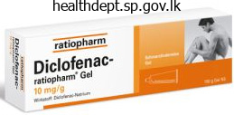
Diclofenac gel 20 gm cheap with amex
Some experts support this counter argument (Box 4) particularly in groups exhibiting resilience and positive coping mechanisms arthritis in knee fish oil 20 gm diclofenac gel cheap mastercard. Another problem associated to the well being of street children arthritis pain associates pg county discount diclofenac gel 20 gm mastercard, is their health-seeking behavior, or somewhat, the lack of it (Box 5). On one hand, multiple reasons predispose street children to be hesitant in seeking out well being care companies, whereas on the other, the conventional system is tentative in dealing with this unconventional group of patients. The major components of this program, as outlined by the Ministry, are summarized in Box 7. This program, however, concentrates more on the runaway or deserted avenue child and never poor, working kids as a complete. Thus, these initiatives have fallen prey to the exclusive definitions of street youngsters, taking cognizance of the problems of just one a part of the whole group of youngsters dwelling a lifetime of excessive vulnerability. Street youngsters view typical health-care system as unhelpful, inaccessible, unaffordable, unfriendly, and regard health-care professionals with suspicion-may be a part of the pure coping mechanism. Lack of empathy, and an impersonal service provider could further alienate these youngsters, who view everyone outside their safety circle with suspicion. Health-care suppliers may worry about problems with consent and compliance whereas treating unaccompanied minors, especially in crucial cases with unpredictable or unfavorable outcomes. Drug-seeking behavior of drug-using children could modulate treatment or prescribing decisions. Early intervention for repatriation of runaway or abandoned youngsters with their families. E · nrolment of shelter/drop-in-center-visiting youngsters in vocational E training applications. Objectives · Early identification of road children at railway stations, bus terminals, and market locations. Working with Street Children: Selected Case Studies from Africa, Asia and Latin America. Children in particularly tough circumstances: Supporting annex, exploitation of working and avenue children. This should be bolstered with abolition of child labor with stringent implementation of legal provisions. Traditionally, higher focus has been on the achievement of kid wants as determined by the policy-making adults; the main focus needs to shift from paternalistic fulfilling perceived baby needs to assured provision of inalienable rights (see remark in Box 8). Establish the requisite variety of drop-in-centers and shelter houses with adequate skilled workers to ensure easy functioning. There should be periodic assessment to guarantee high quality efficiency by the functionaries. Expansion of childline and youngster protecting providers on a national level, and then increase it in phases, primarily based on pilot project experiences. Develop a cadre of counselors and social workers to ensure psychosocial rehabilitation of abused kids. Fulfill the provisions of the Juvenile Justice Act of 2000 and create a Juvenile Justice Board and Child Welfare Committee in each district of the nation. The most accepted version classifies them as children on the street (those who work on the streets however return to their families at night) and youngsters of the road (those for whom the streets have turn into the houses and sources of livelihood). The estimated global tally of street youngsters is between 10 million and a hundred million, with 40 million of them being within the Latin American countries, and 25 million in Asia. Most road youngsters are boys, aged 612 years, and initially from matrifocal families. Four major themes underlie the etiologic issues for children being pushed out of their houses: urbanization, city poverty, aberrant households, and medical causes. The course of by which a poor, working-family youngster is pushed out into the streets is a gradual process, comprising of four sequential stages. Street youngsters usually have a tendency to undergo from a quantity of medical circumstances, usually resulting from poverty or neglect. Poor nutrition, trauma and injuries, infectious illnesses, especially sexually transmitted ailments, are the commonest afflictions. Street kids adopt a quantity of coping methods to take care of the cruel street life. Some maladaptive coping methods embody alcohol, tobacco and drug use; and group, business, and survival intercourse. Some research have advised that kids living and working on the streets might enjoy a greater health profile than their housed, poor, working friends. Street kids avoid contact with the traditional health care system, and regard it with appreciable suspicion and scorn. An Integrated Program for Street Children has been envisioned in India, the primary focus of which is early identification, intervention, and repatriation of avenue kids with their estranged households. As citizens, children have rights that entitle them to the sources required to protect and promote their development. India has the largest number of baby labor on the earth and constituted around three. But the alarming characteristic of the problem in recent years was the enormous improve of kid staff in urban settings, especially in various kinds of work conditions in unorganized urban sector. As a end result these children are disadvantaged of their right to childhood, and are relegated to a life of drudgery. Child in the Social Milieu not matured and their physiological processes and reflexes are nonetheless undergoing growth. This refers to financial exploitation by means of low wages and bodily and mental exploitation in phrases of lengthy hours of work, hazardous working situation, and lack of health-care, schooling and leisure amenities which finally threaten the well being and total improvement of children. From the traditional instances, youngsters have been working in agriculture and as apprentices to artisans. Child labor underwent major enlargement and restructuring in the course of the 1700s as a consequence of the need created by the industrial revolution for giant variety of staff. In that era, mill homeowners most well-liked to have children as workforce rather than adults. Children, as younger as eleven years, particularly ladies, were sent to work in the mills by their households as a result of the wages they could earn far exceeded the earnings of their mother and father working of their rural farms. Thus, in the preindustrial revolution period, the phenomenon of child labor was prevalent all over the world although having a special nature and magnitude altogether. During the postindustrial period, the kid labor turned a growing phenomenon which is constant to grow in growing international locations. In India, baby labor was recognized as a major downside as early as 19th century and children have been employed in cotton or jute mills and coal mines. As early as 1881, during the British period, legislative measures for the safety of kid staff employed in hazardous jobs had been adopted. Inspite of protective laws, the social evils of child labor persisted in India from the early days of commercial interval.
Generic diclofenac gel 20 gm with amex
Chapter thirteen Nontraumatic Abdominal Emergencies 411 Endovascular Repair of Abdominal Aortic Aneurysm Endovascular stent grafting of the aorta has turn out to be a great possibility for poor surgical candidates definition of arthritis pain diclofenac gel 20 gm order otc, changing open restore arthritis in dogs hips diclofenac gel 20 gm purchase otc. Ultrasonography typically requires using contrast air bubbles to diagnose an endoleak. Plain films are helpful within the evaluation of stent-graft migration, part separation, and structural abnormalities. Magnetic resonance angiography time-resolved techniques can be utilized to characterize endoleaks. Digital subtraction angiography is usually reserved for therapeutic interventions. Wound problems such as hematoma, an infection, necrosis, and lymph fistulas occur generally. Imaging of an contaminated graft will reveal periaortic fluid collections, perigraft gentle tissue thickening, and surrounding or inner fuel. Infection might spread to contain the adjoining backbone, paraspinal musculature, adjoining bowel, and even lead to pseudoaneurysm formation. Most happen in the third portion of the duodenum just proximal to the graft typically 3 to 5 years after surgical procedure. These require immediate restore to forestall irreversible hyperdynamic (high output) heart failure. Patients present with tachycardia, heart failure, decrease extremity edema, belly thrill, renal failure, pelvic venous hypertension, and peripheral ischemia. Seventy percent occur in the widespread segment, 20% are internal, and 10% are seen in the exterior artery. In addition, once a bleeding lesion has been recognized, therapeutic interventions 412 Section iV AbdominAl emergencieS mL of distinction media (350 mg of iodine per millimeter or higher) at four mL/sec, adopted instantly by a 40 mL zero. Hyperattenuating materials within the noncontrast scan usually indicates latest, nonactive bleeding. However, care must be taken to differentiate these hyperattenuating areas from potential pitfalls, including suture material, clips, overseas our bodies, orally administered pharmaceutical medication, cone beam artifacts, and fecaliths or retained contrast materials inside a diverticulum. Despite the relatively low yield, colonoscopies are routinely performed in patients with melena and a normal upper endoscopy findings as a result of many right-sided colonic lesions may present with melena. Computed tomography angiography can demonstrate the presence and site of lively bleeding or current hemorrhage, in addition to the potential trigger, within the majority of sufferers. The anatomic coverage includes from the diaphragm to the inferior pubic ramus to encompass the complete rectum. A preliminary unenhanced examination is carried out to depict any preexisting intraluminal hyperattenuating material. The initial scan is followed by a biphasic acquisition in the arterial and portal phases. Sulfur colloid imaging may be very sensitive and might detect bleeding charges as low as zero. However, the imaging time is markedly restricted at no more than 10 minutes because of fast clearance from the intravascular space by the reticuloendothelial system. Unless the affected person is actively bleeding on the time of the examine, the location of hemorrhage will doubtless not be detected. Liver and spleen activity is visualized but to a lesser diploma than with sulfur colloid imaging and infrequently obscures a bleeding focus. B, Subsequently confirmed with angiography (arrow) and handled with embolization (not shown). Small bowel bleeds incessantly show solely temporary accumulation with fast dissipation due to marked small bowel motility. Glucagon may generally be helpful in aiding detection of small bowel bleeding by producing easy muscle leisure. Such sufferers may require angiographic intervention to locate and treat the supply of bleeding. Contrast extravasation into the bowel lumen is considered definitive proof of a bleeding web site. Vasopressin causes generalized vasoconstriction and produces a speedy discount in native blood circulate that progressively returns to normal a quantity of hours after the infusion has been terminated. The objective is to decrease the perfusion strain and allow secure clot formation on the bleeding site. However, rebleeding as quickly as vasopressin has been stopped is widespread, and its use could be related to vital ischemic events. Embolization works by mechanically occluding the arterial blood provide to the bleeding web site. Materials used for embolization are both short-term (Gelfoam) or permanent (microcoils). Acute renal vein thrombosis is discussed in Chapter 13, section 5, "Renal and adrenal emergencies"; acute hepatic vein thrombosis and portal/mesenteric vein thrombosis are discussed in Chapter 13, section eight, "hepatic inflammation and infections". In instances of suspected acute primary adrenal insufficiency, diagnostic imaging can be performed to consider sufferers for adrenal hemorrhage and an infection, the 2 most typical causes. Mazza the adrenal glands and kidneys are paired retroperitoneal organs that serve critical physiologic capabilities. When the physiologic functions of both the adrenal glands or kidneys are disrupted, the results may be catastrophic and life threatening. Adrenal Hemorrhage Bilateral adrenal hemorrhage is essentially the most frequent explanation for acute adrenal insufficiency. This is most commonly due to Waterhouse-Friderichsen syndrome, a syndrome related to overwhelming sepsis, classically from meningococcal an infection though other pathogens have been implicated (including Staphylococcus species), and anticoagulation therapy (heparin, warfarin). Thrombocytopenia is a complication of heparin remedy that can trigger adrenal vein thrombosis, leading to the leakage or rupture of quite a few small venous channels in the adrenal glands with resultant adrenal hemorrhage and subsequent hemodynamic collapse. Antiphospholipid antibody syndrome is caused by circulating antibodies to a plasma glycoprotein and is characterized by recurrent arterial and venous clots that can result in adrenal venous thrombosis and resultant adrenal hemorrhage. Patients with antiphospholipid antibody syndrome can also hardly ever develop adrenal infarctions that can result in acute primary adrenal insufficiency, within the absence of adrenal hemorrhage. Although adrenal tumors can hemorrhage, these are unlikely to end in acute adrenal insufficiency. Most (80%) adrenal tumors are unilateral, and higher than 90% of adrenal tissue needs to be destroyed to ensure that the signs of adrenal insufficiency to turn out to be manifest. The physique of the adrenal gland is made up of principally cortical tissue that produces glucocorticoids and mineralocorticoids, whereas the limbs are mostly medullary tissue and produce norepinephrine and epinephrine. The adrenal glands are important endocrine organs that produce hormones that assist the physique deal with infections, hypotension, and stresses corresponding to surgery. The commonest nontraumatic emergencies that affect the adrenal glands are hemorrhage and infection, and both can lead to acute major adrenal insufficiency. Adrenal Insufficiency Primary adrenal insufficiency is a life-threatening condition which will lead to severe hypotensive crisis and decreased mentation and represents essentially the most catastrophic of all attainable nontraumatic adrenal emergencies. Timely prognosis is critical so that substitute glucocorticoids and mineralocorticoids can be instantly began to avert hemodynamic collapse. If time permits, a corticotropin simulation take a look at could additionally be Adrenal Infection Adrenal infections are the second most common explanation for primary adrenal insufficiency in the United States.
Syndromes
- Low back pain
- Have you had an injury or accident involving the knee?
- Sensitivity to light
- Large amounts of protein in the urine
- Hypothyroidism
- How severe is it?
- Blood tests can measure the levels of allergy-related substances, especially one called immunoglobulin E (IgE).
- Cushing syndrome
- Bleeding?
Diclofenac gel 20 gm order free shipping
The parascapular flap may be raised as a fasciocutaneous flap with out disrupting any of the shoulder musculature arthritis in fingers nz cheap diclofenac gel 20 gm without a prescription. Donor site defects as broad as 7 cm may be closed primarily after harvesting of a musculocutaneous latissimus dorsi flap arthritis hands medication cheap 20 gm diclofenac gel with amex. The ascending department of the deep circumflex iliac artery supplies the interior oblique muscle. When an iliac crest flap is harvested, the exterior oblique muscle is harvested with the bone. The pores and skin paddle for the iliac crest may be extended from the anterosuperior iliac spine to roughly 9 cm posteriorly. The primary blood supply to the gracilis is from the adductor artery, a branch of the profunda femoris. The nerve provide to the gracilis is from the anterior branch of the obturator nerve, which enters the muscle distal to the entry point of the vascular pedicle. Patient 2 is present process temporal bone resection, parotidectomy, and complete neck dissection. Anterior neck pores and skin defects can be reconstructed with pectoralis major musculocutaneous pedicle flaps. Both of these defects can be reconstructed with parascapular fasciocutaneous free flaps. Providing adequate bulk and offering epithelial protection are equally essential goals in reconstruction of the tongue. The radial forearm free flap is the popular free flap for oral tongue reconstruction in most situations. Defects bigger than half of the taste bud are higher served by secondary intention therapeutic than reconstruction. Most oropharyngeal defects after transoral resection of the tonsil and tumors of the bottom of the tongue may be left to granulate without the need for reconstruction. Lower lip defects as much as half the size of the lip can be repaired with main closure. The dimension of the Abbe flap ought to be designed as half the width of the defect for the lower lip and the identical size of the defect for the higher lip reconstruction. The Gillis fan flap will end result in superior functional results compared with the Karapandzic flap. Very giant defects of buccal mucosa are better left to granulate to avoid trismus. The resultant defect can be reconstructed with primary closure with equally good practical outcomes. The radial forearm osteocutaneous free flap is the most suitable choice for bony reconstruction. For left-sided defects, the left fibula is the higher possibility by means of the position of the skin paddle and the pedicle. A fibula osteocutaneous free flap and an iliac crest free flap will give the best probability for placement of dental implants and dental rehabilitation. It is finest to do nerve grafting from the stump of the facial nerve to all the branches. If a couple of wall of the orbit is missing, reconstruction is required to protect good operate within the eye. The V-Y development flap is an efficient choice for cephalad nasal dorsum defects bigger than 2 cm. Large defects of the nasal tip are finest reconstructed with a paramedian brow flap. A jejunal free flap will be the best option for voice rehabilitation through tracheoesophageal puncture. The radial forearm free flap can be utilized as a patch for restore of enormous pharyngeal defects. Can be higher protected with harvesting of a cuff of soleus or flexor hallucis longus muscle. Larger palatal defects are better fit for a palatal obturator than are smaller defects. In V wedge resection, an angle wider than 45 levels will give better aesthetic outcomes. Limiting the wedge resection to the boundaries of lip tissue will obtain higher aesthetic results. Both latissimus dorsi and serratus anterior muscle flaps may be raised based on the thoracodorsal artery. The distal portion of the latissimus dorsi muscle has the most effective blood provide for designing the pores and skin paddle. To cut back shoulder morbidity, the surgeon ought to protect the lower portion of the serratus anterior muscle when harvesting the serratus flap. The size of a rotation flap should be approximately 4 instances the width of the defect in face reconstruction. The loss of function of the muscle will trigger a noticeable practical deficit in adduction of the thigh. Peripheral vascular illness within the superficial femoral artery is a contraindication to use of this flap. The blood supply to the gracilis enters the muscle on the junction of the upper two thirds and lower one third. Have the greatest rigidity perpendicular to the proximal border of the flap (the base). The danger of a pathological fracture can be considerably decreased with prophylactic plating. The harvested bone is normally thick sufficient to accept dental implants in oromandibular reconstruction. The defect after maxillectomy with orbital preservation without an inferior orbital wall defect C. The defect after maxillectomy with orbital preservation and lack of the inferior orbital wall D. Synkinesis can happen if all the branches of the facial nerve are reanastomosed to the primary trunk. If a zonal approach to facial reconstruction is used, the upper branch is rehabilitated with nerve grafts. In salvage laryngectomy after definitive chemoradiation therapy failure, major closure is probably the most profitable method in limiting incidence of fistulas. If the skin of the neck has to be excised with the tumor, the pectoralis major myocutaneous flap with split-thickness skin grafts is a viable choice.
20 gm diclofenac gel quality
An intraperitoneal bladder rupture will require surgical repair to keep away from urinary peritonitis arthritis in knee dla diclofenac gel 20 gm generic amex. High-attenuation contrast material from the ruptured bladder inside the peritoneum can mimic bowel injury with extravasation of oral or rectal contrast arthritis in feet and running diclofenac gel 20 gm without prescription. Cystography can be falsely unfavorable for bladder rupture when contrast is blocked from leaking by detrusor contraction, a small tear, a blood clot, or a balloon of the Foley catheter. Extraperitoneal bladder rupture is triggered both by bone spicules from a pelvic bone fracture perforating the bladder wall or by pulling of fascial connections between the bladder and pelvis throughout pelvic trauma. The contrast appears to be coming from the bladder dome and likewise is seen outlining bowel loops (arrows). Clots arising in the area of the ureteral orifices may be due to renal or ureteral trauma somewhat than bladder harm. Complications of a missed bladder damage include urinary tract infection, pelvic abscess, bladder fistula formation, and incontinence. Most circumstances of extraperitoneal bladder rupture could be handled successfully with transurethral or suprapubic bladder catheterization. Combined intraperitoneal and extraperitoneal ruptures are seen in approximately 5% to 10% of all ruptures, and cystography will show options typical of both accidents. An intravesicular filling defect (small arrow), likely representing blood clot, lies adjacent to the location of bladder injury and may be partially blocking distinction egress. Streaky, flame-shaped contrast extravasation is seen extending from the lower abdomen to the scrotum. Urethral accidents are unusual in girls as a end result of the protective configuration of the female pelvic ground and shorter urethral length. Approximately 10% of patients with main pelvic fractures will maintain a urethral damage, sometimes involving the posterior (proximal) portion. In males, blood on the urethral meatus, incapability to void, elevation of the prostate gland on rectal examination, or perineal swelling or hematoma ought to elevate the suspicion for this damage. In women with urethral harm, pelvic fractures, the presence of vaginal bleeding, labial edema, dysuria, blood on the urethral orifice, hematuria, or urine leak through the rectum have all been described. Anteroposterior pelvic radiography will virtually always reveal pubic symphysis diastasis in a affected person with a urethral harm. Imaging assessment of the urethra ought to precede cystography but ought to be delayed till after pelvic arteriography. A retrograde urethrogram is carried out using 30 mL of 60% contrast medium by way of a Foley balloon catheter positioned in the distal urethra and inflated with 2 mL of saline. Urethrography results can be utilized to classify the injury utilizing the Goldman system Table 11-13), which helps in remedy planning. Anterior (distal) urethral injury is extra generally attributable to iatrogenic or penetrating quite than blunt trauma. The harm may be limited to the corporal bodies if the Buck fascia stays intact or, if disrupted, could unfold all through the scrotum, perineum, and anterior belly wall. Differentiating testicular hematoma, rupture, or torsion is exceeding difficult by medical examination. Rapid and correct assessment is vital as a result of a ruptured testis can be salvaged in 90% of sufferers if repaired within seventy two hours, however salvage drops to 55% with increasing time since damage. Ultrasound examination has 100% sensitivity and 65% specificity in detecting testicular rupture. Testicular rupture indicates tearing of the tunica albuginea with extrusion of the testis into the scrotal sac. Acute hematomas are sometimes isoechoic to normal testicular parenchyma and could be troublesome to establish. Although a hematoma will often be managed conservatively, follow-up to resolution is really helpful because of the chance for infection and necrosis, which can require orchiectomy. Dislocation is most commonly seen in a patient involved in a motorcycle collision with impaction of the scrotum against the fuel tank. Prompt analysis is essential as a result of a missed dislocation puts the affected person at risk for growth of intratesticular cellular modifications that will predispose to malignant degeneration. The "fracture" is precise rupture of the tunica albuginea, often accompanied by a cracking sound from the erect penis, with pain and detumescence. Corporal laceration may be seen with direct trauma corresponding to a kick to the flaccid penis. Color Doppler might present blood flush through the tunica defect upon squeezing of the penile shaft. Ovarian and adnexal torsion: spectrum of sonographic findings with pathologic correlation. Potential errors in the analysis of pericardial effusion on trauma ultrasound for penetrating injuries. Sonography in a scientific algorithm for early analysis of 1671 sufferers with blunt stomach trauma. Diagnosis and preliminary management of blunt pancreatic trauma: tips from a multiinstitutional review. Importance of evaluating organ parenchyma during screening belly ultrasonography after blunt trauma. Revision of current American Association for the Surgery of Trauma renal damage grading system. Chapter eleven Blunt Abdominal and Retroperitoneal Trauma Catalano O, Aiani L, Barozzi L, et al. Sexually transmitted illnesses remedy guidelines, 2010: pelvic inflammatory disease. What are the specific computed tomography scan criteria that can predict or exclude the necessity for renal angioembolization after high-grade renal trauma in a conservative management strategy? Computed tomography grading methods poorly predict the necessity for intervention after spleen and liver accidents. Classification and treatment of pooling of distinction materials on computed tomographic scan of blunt hepatic trauma. The status of ultrasonography training and use generally surgical procedure residency applications. Abdominal ultrasound is an unreliable modality for the detection of hemoperitoneum in patients with pelvic fracture. Blunt trauma of the pancreas and biliary tract: a multimodality imaging approach to prognosis. Diagnosis and classification of pancreatic and duodenal accidents in emergency radiology. Institutional and particular person studying curves for targeted stomach ultrasound for trauma: cumulative sum evaluation. Appearance of stable organ injury with contrast-enhanced sonography in blunt abdominal trauma: preliminary expertise. Radiographic predictors of need for angiographic embolization after traumatic renal injury.
20 gm diclofenac gel discount visa
Neonatal mortality contributes to nearly 2/3rd of infant deaths and most of these deaths occur through the first week of life rheumatoid arthritis headache diclofenac gel 20 gm buy cheap on-line. Specifically targeting these frequent issues and assessing a toddler for other associated issues supplies a holistic approach to handle common childhood sicknesses arthritis knee support diclofenac gel 20 gm discount without prescription. Integrated strategy is another effective step as a result of typically a young toddler or baby suffers from multiple illness and customary childhood sicknesses on this age group have similar presenting symptoms. Moreover, poor access to health-care and delay in referral additional compound the problem. Assessment of Sick Young Infant or Child Sick young infants or sick kids are assessed utilizing selected reliable scientific indicators and signs (Annexure 1). All sick younger infants are checked for very extreme illness and jaundice, feeding problems and malnutrition, immunization standing and if they produce other problems. All sick youngsters are checked for malnutrition, anemia, immunization status, prophylactic vitamin A and iron folic acid supplementation standing, and different issues. The technique contains three main parts: Improvements within the case-management skills of well being employees; Improvements in the total well being system; and Improvements in household and community health-care practices. Classification tables are used beginning with the pink rows, adopted by yellow and green rows. If the sick younger toddler or sick child has no pink classifications, have a glance at the yellow rows. A sick younger infant or sick child is classified in a single shade solely beneath each group of symptoms. If the young toddler has signs from more than one row, the most extreme (pink) classification is selected. Assess the young infant/child Classify the illness Identify therapy Treat the young infant/child Counsel the mother Follow-up care mom or caretaker must be taught how to give an oral antibiotic at residence. To return instantly if any of the hazard indicators (breastfeeding/drinking poorly, becomes sicker, develops a fever or feels cold to contact, fast/difficult breathing, yellow palms and soles if younger infant has jaundice, presence of seen blood in stools in young infants with diarrhea) For local bacterial an infection, jaundice, diarrhea, any feeding drawback or oral thrush follow-up after 2 days, and for low weight for age, advise follow-up after 14 days. Pink classification requires hospital referral or admission within the in-patient part of the hospital, yellow for therapy under supervision within the health facility, and green implies that the child can be despatched house with careful recommendation on when to return. Community Pediatrics Identify Treatment After classifying a sick younger infant or sick youngster the next step is to determine remedy. All the treatments required are listed in the identify therapy column of the assess and classify the sick young toddler or youngster chart (Annexure 1). If a sick younger toddler or sick baby has more than one classification, treatment required for all of the classifications should be identified. Referral can also mean admission to the inpatient department from the outpatient or casualty of the same facility. Treatment of Sick Children Referral All sick youngsters with a severe classification (pink) are referred to a hospital as soon as evaluation is accomplished and needed pre-referral treatment is run. Treatment of Sick Young Infants Referral All sick younger toddler with a extreme classification (pink) are referred to a hospital as quickly as evaluation is completed and needed prereferral treatment is administered. Treatment in Outpatient Clinic Give the first dose of the antibiotics Amoxicillin for 5 days for pneumonia and acute ear infection, ciprofloxacin for 3 days for dysentery, and a single dose of doxycycline for cholera. For malaria, give choloroquin or artesunate depending upon threat space for malaria and fast blood check or blood smear report. Instructions may be given about the way to deal with eye infection with tetracycline eye ointment; dry the ear by wicking to deal with ear an infection; and deal with mouth ulcers with gentian violet. Feeding assessment consists of questioning the mom or caretaker about: (1) breastfeeding frequency and night time feeds; (2) kinds of complementary meals or fluids, frequency of feeding and whether feeding is lively; and (3) feeding patterns during the current illness. The mother or caretaker must be given acceptable recommendation to assist overcome any feeding issues discovered. One should take heed to the mom and praise her for correct feeding practices; and train and encourage her to change these practices which have to be changed. If a child with no pneumonia: cough or cold has fast/ troublesome breathing and a child with diarrhea has blood in stools or drinks poorly. Counseling the Mother or Caretaker Counseling a mother/caretaker is critical not only for successful referral of a severely ill younger infant but additionally for therapy in the outpatients clinic in addition to for residence care. Good communication skills have to be used from the start of the go to significantly whereas counseling the mother/caretaker for therapy. The success of house therapy is decided by how nicely the mom or caretaker knows how to give treatment, understands its significance and is aware of when to return to a health-care supplier. Some advice is easy; other recommendation requires instructing the mother or caretaker tips on how to do a task. When a mother is taught tips on how to treat her youngster at home, three fundamental teaching steps must be used: give info; present an instance; let her practice. Moreover, for the administration of very sick younger infants or children the guidelines stop short at referral of pink classifications to a hospital. These pointers are directed at small hospitals where fundamental laboratory services and cheap essential drugs are available. These evidence based mostly guidelines additionally enhances standard complete pediatric textbooks. Integrated administration of childhood illness by outpatient well being employees: technical basis and overview. Integrated Management of Neonatal and Childhood Illness: Training Modules for Physicians. World Health Organization Management of the Child with a Serious Infection or Severe Malnutrition: Guidelines for care on the first-referral degree in developing countries. Normal values refer to those measurements which are often central and around which most of the values lie in healthy individuals. Almost all normal laboratory values are represented and expressed in a reference range and infrequently in phrases of a single measurement. For example, a large group of healthy children in the age group of 612 years might have a median hemoglobin level of 14 g/dL with the person values ranging from 11. In this case, the conventional hemoglobin values in 612 years old are mentioned to vary between eleven. The normal or reference range is an integral part of all laboratory values due to appreciable magnitude of inter-individual variability within the normal population. Variations in regular topics occur as a outcome of a number of causes, other than age and intercourse, which may include biological, genetic, environmental and chance elements other than the interobserver and laboratory variability. In addition, the analytic method used within the laboratory is a serious factor affecting the value obtained. All these sources are instrumental in making a somewhat wide selection of values that could be referred to for classifying a person as having a traditional or irregular laboratory check. Fortunately, these three measures usually coincide in a lot of the laboratory measurements in wholesome people. In such scenario, the conventional values are expressed by way of a spread masking 95% of values from a set of values present in a wholesome population, excluding 2.
Diclofenac gel 20 gm buy lowest price
In the ascending aorta the true lumen is often contiguous with the aortic valve orifice and is the smaller of the 2 lumens rheumatoid arthritis in feet and knees 20 gm diclofenac gel buy with amex. The true lumen tends to be on the left of the false lumen (closer to the pulmonary artery) arthritis in feet images 20 gm diclofenac gel cheap otc. When a fenestration is present in the ascending aorta, the flaps on each side of the defect point away from the true lumen, a finding generally referred to because the intimomedial rupture sign. In the arch and descending aorta the most dependable approach to distinguish the true from the false aortic lumen is the flexibility to instantly trace the suspected true lumen to the intact "normal" lumen of the proximal aorta. Other findings embody the scale, as a outcome of the true lumen is normally smaller than the false lumen, and the presence of calcifications, which are usually in the intima surrounding the true lumen. Care should be utilized in continual dissections as a end result of the false lumen can often calcify. The arrow denotes a area the place the false lumen creates an acute angle with the flap, known as the beak sign. Although variable, classically the celiac artery, superior mesenteric artery, and inferior mesenteric artery come up from the true lumen, and the left renal artery tends to arise from the false lumen. As a outcome, left renal ischemia with resultant left flank ache may be the presenting symptom of an acute aortic dissection. Another useful finding within the false lumen is the presence of skinny strands, representing fibers of the media that remain as blood has dissected on this false area. These are sometimes referred to as aortic cobwebs and are indicative of the false lumen. The angle created by the flap and the outer aortic wall can be used for separating the true from the false lumen. A mainstay of aortic dissection remedy is blood strain management, but clearly some patients require surgical intervention. At our institution, we use the Stanford classification, during which involvement of the ascending aorta is taken into account a Stanford kind A dissection, requiring surgical procedure. Arch and descending aortic involvement alone are considered Stanford kind B dissections and could also be amenable to medical therapy alone. Of note, although some authors have beforehand described the left subclavian artery because the landmark for delineation between Stanford A and Stanford B, increasingly ascending aortic involvement is used. In different words, only when the ascending aorta is concerned is the dissection thought of type A. Ascending aortic involvement prompts surgical intervention due to the chance for acute aortic valve regurgitation, coronary artery involvement, or tamponade from hemopericardium. The risk for dying from the dissection exceeds the potential threat from surgical procedure (including paralysis). Although most isolated descending aortic dissections are considered medical lesions, often these dissections can turn into surgical. In this case the intima and internal media have completely separated from the rest of the aorta. The report must also include details about end-organ ischemia and the nature of the ischemia. If the ischemia is from propagation of the flap into the vessel for that organ, the patient may be stented. If the flap occludes the orifice of the feeding artery with out instantly involving it, the patient might undergo fenestration at that stage. Most aortic dissections will current with a linear filling defect within the aortic lumen that corresponds to the intimomedial flap. Rarely the dissection will involve 360 levels of the aorta so that the false lumen completely surrounds the true lumen. In this occasion the intimomedial flap could turn out to be freed from the rest of the aorta and type an irregular filling defect. Occasionally this circumferential flap will start to infold or intussuscept much like bowel intussusception. If the stress in the media exceeds luminal stress, the media might create a fenestration with the true lumen. This course of has been sometimes referred to as a reverse dissection, though the imaging will mimic a true aortic dissection that begins on the luminal aspect. This occasion can lead to a very uncommon appearance of the pulmonary arteries and may be fairly confusing to the unaware reader. These sufferers might do as properly (if not better) than patients present process median sternotomy. This might arise from the lumen and a tiny ulcerlike projection or from retrograde move from an intercostal artery. These blushes can result in confusion in the categorization of which entity of acute aortic syndrome is present. Some of the literature has shown that a blush from an ulcerlike projection is at increased risk for progressing to a real aortic dissection. In another variation a dissection involving the ascending aorta may rupture simply above the valve plane right into a shared sheath with the pulmonary artery. The blood in the wall of the pulmonary artery will compress the pulmonary artery and may occlude it. Sometimes the blood will dissect around the pulmonary artery to the segmental arterial stage. A third uncommon manifestation of aortic dissection is a focal dissection on the degree of the aortic root near the sinotubular junction. This localized process has been referred to as an incomplete aortic dissection and is type of at all times related to cystic medial necrosis and aortic root enlargement. As a common guideline, one ought to have a low threshold for reimaging the aortic root when a linear defect is encountered within the setting of annuloaortic ectasia or in a affected person with known Marfan syndrome or bicuspid valve. Focal dissections of the belly aorta are rare in the absence of prior trauma or aortic catheterization. However, imaging of an aortic dissection ought to embrace the belly aorta and the iliac vessels to the level of the frequent femoral arteries. Inclusion of the stomach and pelvis is carried out to look for any end-organ ischemia and to delineate the extension of the dissection. Increasingly surgeons are counting on peripheral cannulation for initial bypass in aortic restore. The function of imaging is to assist distinguish true from false lumen in order that any peripheral cannulation for bypass avoids cannulating the false lumen, which might preclude sufficient bypass and will probably be deadly. The perception is that the macrophages within the plaque weaken the intima and allow for this focal ulceration. These sufferers shall be carefully adopted and if possible, might bear endoluminal remedy. Vascular Chest Emergencies 237 promote an space of relative stasis along the lesser curve of the aorta on the stage of the isthmus. These filling defects are normally encountered throughout the aorta at the degree of the isthmus and within the descending thoracic aorta. Radiologists should be aware of the acute aortic thrombus to keep away from confusion with an aortic tumor. Treatment for an acute aortic thrombus begins with anticoagulation but may include a thrombectomy and rarely endoluminal stent placement to entice the thrombus.
Insam (Ginseng, Panax). Diclofenac Gel.
- Depression, anemia, fluid retention, stomach inflammation and other digestive problems, chronic fatigue syndrome (CFS), fibromyalgia, breast cancer, ovarian cancer, lung cancer, liver cancer, skin cancer, fever, bronchitis, cancer, common cold, influenza, and other conditions.
- Thinking and memory.
- Dosing considerations for Ginseng, Panax.
- Improving mood and sense of well-being.
- What is Ginseng, Panax?
- Male impotence (erectile dysfunction).
Source: http://www.rxlist.com/script/main/art.asp?articlekey=96961
20 gm diclofenac gel buy otc
Ultrasonography of the child exhibits a big complex mass of the neck with secondary tracheal compression footwear for arthritic feet cheap diclofenac gel 20 gm without a prescription. The report states that a homogenously enhancing tumor with intermediate sign depth was identified arthritis and diet uk diclofenac gel 20 gm buy generic on line. A 44-year-old woman presents with a tumor histologically much like adrenal, sympathetic ganglionic neuroblastomas and retinoblastomas. On immunohistochemical analysis, optimistic staining of neoplastic cells with antibodies against desmin was famous. The many medicine that have been used embody vincristine, dactinomycin, doxorubicin, cisplatin, ifosfamide, and 5-fluorouracil. The operative approach should include all of the lymphatic malformation in a monobloc style. Approximately 75% of all lymphatic malformations occur within the head and neck area. Stage V (a) Bilateral infrahyoid lymphatic malformation (b) Unilateral suprahyoid (c) Bilateral infrahyoid and suprahyoid (d) Unilateral infrahyoid (e) Unilateral infrahyoid and suprahyoid 60. Conventional osteosarcoma can be categorized into osteoblastic, chondroblastic, and fibroblastic subtypes. Head and neck osteosarcoma has a lowered likelihood of distant metastases in contrast with extremity osteosarcoma. A threefold improvement within the diseasefree survival fee was realized within the Eighties with the introduction of neoadjuvant chemotherapy in the remedy of extremity osteosarcoma in the pediatric population. The anatomical presentation of lipomas of the neck is between the superficial and middle layers of the deep cervical fascia. The anatomical presentation of lipomas of the neck is between the middle and deep layers of the deep cervical fascia. In common, high-grade sarcomas and other sarcomas exceeding 4 to 5 cm require adjunctive postoperative radiation therapy. All high-grade sarcomas require postoperative radiation therapy to enhance the overall survival fee. All high-grade sarcomas require postoperative radiation therapy to cut back distant metastases. Which surgical strategy for a plunging ranula is associated with the bottom recurrence rates? The most common site of presentation of osteosarcomas of the head and neck is the A. Still to be decided; no formal consensus exists on what constitutes "greatest" therapy for grownup osteosarcoma. Patients older than 60 years Which of the next statements about angiosarcomas of the top and neck is correct? D Core Knowledge · Sarcomas are categorised according to their tissue of origin, which could be bone or delicate tissue, whether or not the tumor is high-grade or low-grade, and the anatomical subsites of presentation within the head and neck. In the United States, more than 10,000 new cases are actually reported yearly, and the mortality rate yearly consists of 4000 sufferers. This may be difficult in an anatomically confined region corresponding to the pinnacle and neck. When these pathological varieties are identified, this data must be thought of within the operative technique or adjunctive radiation fields. Chondrosarcomas, fibrosarcomas, liposarcomas, leiomyosarcomas, neurogenic sarcomas, and hemangiopericytomas require individualized grade characterization. The medical group is predicated on the extent of residual tumor after surgery with consideration of regional lymph node status. Subtypes include osteoblastic, chondroblastic and fibroblastic, multifocal, telangiectatic, small cell, intraosseous well-differentiated, intracortical, periosteal, parosteal, high-grade floor, and extraosseous. Predisposition to this tumor is expounded not solely to deletion of chromosomal region 13q14, which inactivates the retinoblastoma gene, but additionally to Paget illness, fibrous dysplasia, enchondromatosis, Li-Fraumeni syndrome, and Rothmund-Thomson syndrome. The Codman triangle and a sunburst appearance are the classic radiological features. The alveolar ridge, sinus flooring, and palate are traditional maxillary tumor locations. Neoadjuvant chemotherapy within the pediatric population has resulted in a threefold improved survival price. Complete surgical excision to achieve adverse surgical margins is the main aim of remedy. A constructive margin carries with it a significant lower in survival price from 75% to 35%. Memorial Sloan Kettering Cancer Center stories 3-year general, disease-specific, and recurrence-free survival charges of approximately 81%, 81%, and 73%, respectively. Combined modality therapies are utilized, including surgical extirpation with a large 2-cm margin and adjunctive radiation remedy of 60 to 66 Gy to extensive treatment fields. Neoadjuvant or adjuvant taxane chemotherapy can be thought of in choose instances. Well-differentiated and myxoid types are considered low-grade, whereas pleomorphic and round cell sorts are high-grade tumors. Survival probability is set by the subtype of tumor, grade, measurement, and anatomical web site of presentation. Radiation remedy has been proven to improve local recurrence rates; nevertheless, it could not affect overall survival charges. Patients with low-grade lesions and adequate surgical margins receive surgery alone. Patients with highgrade lesions or optimistic surgical margins ought to receive adjuvant radiation therapy. Intensity-modulated radiation therapy permits for the delivery of high-dose conformal photons with the use of both commonplace fractionation, as with protons, or hypofractionated regimens. Outcomes after definitive remedy for cutaneous angiosarcoma of the face and scalp. Head and neck rhabdomyosarcoma: a important evaluation of population-based incidence and survival knowledge. Prognostic components for sufferers with localized soft-tissue sarcoma handled with conservation surgical procedure and radiation remedy: an analysis of 1225 patients. Dentigerous cysts frequently contain the maxillary canine and mandibular third molar. Dentigerous cysts develop when fluid accumulates between the lowered enamel epithelium and tooth crown. Basal cell nevus syndrome usually presents with odontogenic keratocysts because the preliminary presenting element of the syndrome. Basal cell nevus syndrome is associated with multiple basal cell carcinomas, odontogenic keratocysts, and skeletal abnormalities. Malignant ameloblastomas are defined by metastatic spreading to lymph nodes, distant sites, or both. Ameloblastic carcinomas have cytopathological options much like these of benign ameloblastomas.
Diclofenac gel 20 gm generic mastercard
Treatment of atlantoaxial rotatory displacement is predicated on the underlying etiology and length of signs arthritis diet recipes discount 20 gm diclofenac gel overnight delivery. Patients seen inside a couple of days of the onset of signs may be managed with analgesics and a delicate collar arthritis use heat or cold 20 gm diclofenac gel generic fast delivery. For sufferers with signs lasting higher than 1 week however less than 1 month, head halter cervical traction is utilized to right the deformity and the neck immobilized in a cervical collar for 46 weeks. Patients with a fixed deformity or with recurrent displacement may require an atlantoaxial fusion. Many of these anomalies are asymptomatic and therefore undiagnosed and infrequently picked up as incidental findings or as a outcome of trauma. There seems to be no distinction in scientific options or management between syndromic and nonsyndromic anomalies. Cervical instability could occur in the presence of a congenital anomaly, extreme ligament laxity (connective tissue disorders), metabolic diseases, infections and inflammatory situations. Basilar Impression In this condition, the floor of the cranium is indented by the higher cervical backbone. The tip of the dens is extra cephalad and sometimes protrudes into the opening of the foramen magnum. This might encroach on the brain stem, risking neurologic injury from direct damage, vascular compromise, or alterations in cerebrospinal fluid move. Basilar impression can be major or secondary and the common causes are listed in Table 1. Asymmetry of the skull and face (68%), painful cervical motion (53%), and torticollis (15%) can even occur. Symptoms are of raised intracranial pressure (because the aqueduct of Sylvius turns into blocked) and or myelopathy (because of direct compression or ischemia of the cord). Secondary basilar impression is a developmental condition attributed to softening of the osseous constructions on the base of the cranium. Treatment Patients with basilar impression and no neurological signs or with minimal signs and no signal of progressive neurological injury ought to be noticed and examined periodically. Treatment of sufferers with neurological deficits involves surgical decompression and stabilization. Odontoid anomalies could be identified on routine lateral view and open-mouth odontoid view radiographs. This raises the controversy of whether or not prophylactic surgical remedy of asymptomatic sufferers ought to be routinely performed or not. Patients with pain or torticollis usually enhance with cervical traction or immobilization. Odontoid Anomalies Congenital anomalies of the odontoid though rare are of significance as they can lead to atlantoaxial instability. Partial improvement of the odontoid as hypoplasia; the bone size varies from a small, peg-like projection to nearly regular measurement. In os odontoideum, the odontoid is a separate ossicle with a clean, sclerotic border, which is separated from the axis by a transverse gap, leaving the apical segment without its basilar help. Disorders of Bones and Joints Diagnosis these anomalies often are detected as incidental findings in a routine cervical backbone radiograph following trauma or when signs happen because of atlanto-occipital instability. Patients could present with ache or torticollis, or neurological compressive signs such as transient paralysis, myelopathy or sphincter disturbances. Atlantoaxial instability might compress the vertebral artery resulting in cervical and brain stem ischemia, presenting as vertigo, visible disturbances, syncope or seizures. Absence of involvement of cranial nerves is a vital clue to differentiate os odontoideum from different occipitovertebral anomalies as a result of the spinal cord impingement occurs below the foramen magnum. This is especially important in patients present process surgical procedure because the atlantoaxial joint could subluxate underneath anesthesia. Vertebral fusion outcomes from a failure of segmentation of the cervical somites through the third to eighth weeks of life. The most constant function is limitation of neck movement; the extent of limitation depends on the extent of the anomaly. Symptoms are inclined to come up in the second or third decades, not from the fused segments however from the hypermobile adjoining segments. There may be ache as a end result of segmental hypermobility or neurological signs from instability. Management and Prognosis the pure history of these kids primarily depends on the occurrence of extreme renal or cardiac issues, which may limit life expectancy. Instability of the cervical spine can develop with neurologic involvement, especially within the upper segments or in these with iniencephaly. Later in life, degenerative joint and disc illness might develop in these with decrease segment instabilities. Because youngsters with massive fusion areas are at excessive risk for creating instabilities, contact sports could also be avoided. Mechanical signs brought on by the hypermobile segment usually respond to traction, a cervical collar, and analgesics. Neurological signs usually require surgical stabilization with or without decompression relying on the compressive etiology. The real dilemma is whether or not or not prophylactic stabilization should be undertaken for asymptomatic hypermobile segments. Surgery solely for cosmesis is unwarranted and positively fraught with dangers and problems. The different anomalies related to the syndrome are scoliosis (60%, either congenital or idiopathic), cervical ribs (1215%), congenital limb deficiency, hand deformities (syndactyly, thumb hypoplasia and additional digits), genitourinary anomalies, deafness (30%), synkinesis (mirror actions in 20%), pulmonary dysfunction, and congenital heart disease. Goldenhar syndrome, Mohr syndrome, and fetal alcohol syndrome are also associated with congenital fusion or anomalies of the cervical backbone. The organism, usually Staphylococcus spreads hematogenously and most often localizes within the adjacent vertebral end plates of a disc space. Bony destruction first occurs on the finish plates and then spreads to the rest of the vertebral body. End plate involvement leads to loss of diet to the intervertebral disc with resulting necrosis. Abscess formation happens and pus might spread into the spinal canal or alongside the soft tissue planes of the neck. A widespread sample is fusion of C12 and C34, leading to a excessive danger of instability at the unfused C23 level. Flexion and extension lateral radiographs are helpful to assess for cervical instability. Flexion-extension lateral radiographs should also be obtained earlier than any general anesthetic to rule out any occult instability of the cervical backbone.
Buy diclofenac gel 20 gm with mastercard
Blunt thoracic accidents are the third commonest accidents in polytrauma patients arthritis in dogs meloxicam 20 gm diclofenac gel overnight delivery, following these of the top and extremity arthritis young dog cheap 20 gm diclofenac gel mastercard. Although 50% of blunt chest injuries are minor, 33% would require hospital admission. A chest radiograph is generally the primary modality of radiologic evaluation of the chest trauma affected person. It is essentially a screening examination and is used to identify life-threatening circumstances such as a big hemothorax, rigidity pneumothorax, dangerously malpositioned lines and tubes, and mediastinal hematoma. A pulmonary contusion results from injury to the alveolar wall and pulmonary vessels, permitting blood to leak into the alveolar and interstitial areas of the lung. The causes are thought to embrace compression of the lung towards the chest wall, shearing forces, rib fracture, or beforehand formed pleural adhesions tearing peripheral tissue because the lung separates from the chest wall at impact. The appearance of pulmonary contusion on the chest radiograph is determined by its severity. Air bronchograms may be seen in contused lung if there has not been filling of the airways with blood. Pulmonary contusion is often nonsegmental and geographic, and it readily crosses pleural fissures, in distinction to pneumonia or atelectasis. Contusions are a danger factor for improvement of pneumonia or acute respiratory misery syndrome. Pulmonary Laceration the mechanism of damage causing a pulmonary laceration is thought to be similar to that of pulmonary contusion: tearing of lung parenchyma as a result of compression, shearing forces, direct damage from rib fracture(s), or at the web site of previously shaped pleural adhesions. Computed tomography is far more delicate in detection of pulmonary lacerations compared with radiography. There is patchy, ill-defined opacity within the peripheral aspect of the best midlung (arrow). There is diffuse air-space opacification all through the whole right lung, indicating extreme, widespread pulmonary contusion. Note the 1 to 2 mm of subpleural sparing, a standard discovering in pulmonary contusion however not in other air-space processes such as pneumonia. An uncommon complication of a pulmonary laceration is formation of a bronchopleural fistula. This condition is extra prone to happen if the laceration is within the periphery of the lung, with only the skinny pseudomembrane between it and the pleural area. Air may enter the pleural house during trauma because of penetrating harm from a knife or gunshot, puncture from a fractured rib, speedy deceleration forces causing pulmonary laceration, or alveolar rupture from all of a sudden elevated intrathoracic strain at the time of influence. The appearance of a pneumothorax on the chest radiograph will depend tremendously on the patient place. The preliminary chest radiograph of the trauma affected person is normally a supine movie obtained in the emergency division or trauma bay. There is intensive pulmonary contusion all through the whole visualized proper lung. A, Minimal patchy opacity and a large oval lucency (arrows) in the midportion of the left lung. Chapter eight Blunt Chest Trauma air will acquire in the anterior and medial elements of the pleural area. The "double diaphragm" sign, seen when air in the pleural space outlines each the dome and the anterior insertion of the diaphragm, may also be recognized. Note that pulmonary lacerations could additionally be filled with blood (short arrow), air (arrowhead), or both (long arrow), in which case a meniscus sign is seen. A, Extensive pulmonary contusion all through the best lung and upper medial side of the left lung. B, There are a number of small foci of high density alongside the proper lateral margin of the laceration (arrow) that enhance in measurement and stay dense on the portal venous images. A, Multiple pulmonary lacerations in left decrease lobe (arrows) with surrounding pulmonary contusion. A, There is a skinny rim of lucency above the best hemidiaphragm (arrow), suspicious for inferior pneumothorax. Air in the gentle tissues of the proper lateral chest wall is one other clue that pneumothorax may be current (arrowheads). There is elevated lucency of the left higher quadrant (long arrows), indicating presence of a left pneumothorax. Also observe a small residual right inferior pneumothorax after placement of right-sided chest tube (short arrow). Generally a chest tube is placed for all pneumothoraces seen on the supine chest radiograph or in any unstable affected person with a potential pneumothorax. It was previously thought that an occult pneumothorax required a prophylactic chest tube due to threat for enlargement or improvement of pressure pneumothorax, particularly in patients on mechanical air flow. There is rising evidence that a small occult pneumothorax can be safely managed in the majority of sufferers with shut remark, with out thoracostomy tube placement. However, there are lung markings distal to this line and two further linear densities medial to the first (arrowheads). A, Only a small quantity of subcutaneous air in the delicate tissues of the left chest wall (arrows). This could be considered an occult pneumothorax as a end result of there were no definite indicators of pneumothorax on the supine radiograph. Note that pressure pneumothorax is a clinical analysis but could be instructed by radiologic signs of elevated lucency of the affected hemithorax, contralateral shift of the mediastinum, widened ipsilateral rib interspaces, despair of the ipsilateral hemidiaphragm, and lung collapse towards the hilum. Rapid reexpansion of the lung after therapy of rigidity pneumothorax can lead to pulmonary edema. This occurs immediately after remedy of the pneumothorax and may be unilateral or bilateral. Sources of bleeding may be the intercostal arteries, thoracic spine, lung, great vessels, or heart. On chest radiography a minimal of 150 to 200 mL of blood are needed to detect pleural fluid on an upright chest radiograph. A meniscus signal is usually seen with pleural fluid of any sort and appears as a concave sloping of fluid in the costophrenic angle toward the lateral chest 178 Section iii thoracic emergency radiology wall. Because most trauma chest radiographs are carried out with the patient in the supine position, blood will acquire in the dependent portion of the thorax. A hemothorax will be seen on the supine film as a diffuse increase in density over the complete hemithorax. Slightly greater density in the dependent portion of the hemothorax could additionally be seen because of settling of blood products. The actual web site of bleeding must be decided whether or not surgical procedure or catheter embolization is the chosen therapy. Simple or serous effusion, bile, chyle, and urine can also accumulate within the pleural space.
Diclofenac gel 20 gm cheap overnight delivery
Serial axial photographs (E to G) present an asymmetric injury with right "dissolving pedicle" and left "bare side" signs arthritis pain relief 20 gm diclofenac gel generic with amex. Certain situations arthritis health associates patient portal diclofenac gel 20 gm buy with mastercard, together with ankylosing spondylitis and diffuse idiopathic skeletal hyperostosis, lead to unusually brittle spines. In these sufferers, even minor trauma, corresponding to a fall from standing, can end result in devastating hyperextension damage. Osteoporosis and poorly outlined disk spaces within the ankylosed spine contribute to diagnostic difficulty. Lateral radiographs can reveal anterior disk house widening or distraction of anterior vertebral physique fracture fragments. The three column spine and its significance in the classification of acute thoracolumbar spinal accidents. Validity of a set of medical standards to rule out damage to the cervical backbone in patients with blunt trauma. Imaging Severe comminution, translation, and/or rotation are hallmarks of those grossly unstable accidents, and multiplanar reconstructions are essential in their analysis. Hyperextension Injuries An essential variant of spinal fracture dislocation, the hyperextension fracture constitutes less than 3% of all thoracolumbar backbone fractures. Severe hyperextension forces trigger distraction of the backbone with subluxation and canal compromise. In the conventional nonfused backbone, chapter 7 Nontraumatic Spine Emergencies Thomas Ptak Infection of the vertebral column represents up to 20% of all osteomyelitis and is the commonest an infection of the axial skeleton. If ignored or misdiagnosed, an infection of the backbone can have a chronic, harmful, and significantly debilitating course. Infection is usually unfold hematogenously, though direct extension from adjoining an infection similar to pneumonia or pyelonephritis is also attainable. In 60% to 70% of patients the most typical infectious brokers encountered are gram-positive cocci, particularly pores and skin flora corresponding to Staphylococcus (in about 60% of positive cultures), and less commonly Streptococcus with Peptostreptococcus, Escherichia coli, and Proteus. Although typically implied by examination findings, a definitive infectious source is identified in lower than 50% of patients with pyogenic spinal osteomyelitis. In uncommon circumstances, such as immunodeficiency or patients from underdeveloped international locations, one should consider atypical brokers such as fungi or Mycobacteria. Patients at risk embrace older adults, intravenous drug abusers, those with persistent illness, and people with a latest history of surgery, especially of the back or genitourinary system. Insidious onset of diffuse again ache (in 90%) and fever (in 50% to 70%) ought to recommend spinal an infection. A historical past of latest back or genitourinary surgery, intravenous drug use, or infection of the gentle tissue or respiratory or genitourinary system ought to improve the likelihood of spinal an infection. Infection entails the vertebral body in 95% of instances, with the posterior components being involved in lower than 5%. Weight loss can also be present, but this constitutional symptom is less helpful in that it may even be associated with each inflammatory and neoplastic illness. The nucleus pulposus of the disk receives nutrients usually by diffusion from the subchondral vascular bed of the vertebral end plate. In addition, the outer annulus fibrosis has a direct capillary supply by way of spinal canal arteries, additionally contributing vitamins across the concentric fibrous layers of the annulus. The bloodborne organism is able to set up itself at the extra vascular outer free margin of the disk and on the finish plate within the capillaries of the subchondral vascular mattress. From right here the organism can spread contiguously into the more avascular central disk and into the adjoining bony vertebrae. Advanced infection is characterised by an irregular intervertebral disk and irregular adjacent vertebral finish plates. Both are directed at figuring out bony adjustments which will indicate an infection of the vertebral our bodies. Plain film has limited sensitivity because of many overlying confluent shadows and different technical difficulties in acquiring an image. Gross findings such as vertebral sclerosis, vertebral collapse with acute angulation of the backbone. Contrast administration might produce enhancement of the actively inflamed tissue with nonenhancing areas of central liquifactive debris in early abscess formation, if present. Attention must also be directed to adjacent organ techniques such as the kidneys or lungs to provide clues to the origin of an infection. Typical findings suspicious for acute or energetic infectious involvement of the bone embrace focal or regional one hundred sixty five 166 Section ii Spine emergencieS Table 7-1 Common Locations and Characteristics of Spinal Infections Infection Vertebral osteomyelitis Spinal epidural abscess Diskitis Characteristics Elderly and debilitated Male predominance Hematogenous gram-positive cocci Adult postdiscectomy commonest Rare in pediatrics; often accompanies chronic infections. Also note involvement of the superior end plate of the T6 vertebra, not appreciated on the plain movie. It is crucial to embrace fat suppression within the analysis as a outcome of irregular signal indicating infection on T2-weighted and postcontrast T1-weighted sequences may be confused with hyperintense signal produced in the normal fats planes of the epidural area, marrow compartment, and in the interstitial spaces of spinal ligaments. Precontrast T1-weighted pictures are typically carried out with out fat suppression in order that anatomic detail is preserved (and to save time), with T2-weighted sagittal and postcontrast T1-weighted images being fat suppressed in all planes. Chemical fats suppression is often employed however is vulnerable to inhomogeneity as a end result of off-resonance excitation ensuing from local susceptibility effects and static subject inhomogeneity. More homogeneous suppression is attained, and the signal-to-noise ratio of water and edema with respect to the encompassing tissue is optimized. Starting with a T2-weighted, fat-suppressed, large field-of-view picture could also be helpful and efficient for figuring out the extent and involvement of irritation. Once inflammation is identified, additional analysis may be carried out with extra centered T1-weighted precontrast and postcontrast imaging in further planes optimized to greatest present the lesion(s) identified. Inflammation and/or enhancement identified adjoining to or within potential spaces should embody additional orthogonal views (typically axial if sagittal was already used to survey the region) alongside the full size of the gentle tissue house concerned to guarantee identification of the full extent of involvement. Enhancement adjoining to the central spinal canal should result in a careful analysis of the extradural house to determine any infection or abscess formation. This evaluation could be carried out utilizing two orthogonal planes via the region of interest. Evaluation of the bony buildings is especially necessary, particularly in areas of adjoining delicate tissue swelling or enhancement. Again, fat-suppressed T1-weighted imaging in the optimum airplane for visualization is crucial. Precontrast T1 weighting with fat-suppressed imaging ought to show the normally hypointense densely calcified bony cortex as very hypointense, however infected edematous cortical mantle could appear relatively hyperintense or nearly isointense to adjacent marrow sign, making it indistinguishable from adjoining marrow. On postcontrast T1-weighted imaging, areas of infectious involvement of the bony cortex will enhance, producing a hyperintense disruption within the usually sharply outlined, skinny hypointense sign of the normal bony cortical stripe. As discussed earlier, one might expect diskitis to begin in the periphery at the outer annulus or alongside the diskend plate margin; these are the areas of vascular provide and are the most probably factors of origin for a blood-borne infection. Infection can then take maintain within the avascular portion of the disk, the place the immune response could be minimal. Fatsuppressed T2-weighted imaging might show hyperintensity in the thickened outer annular fibers, doubtlessly extending to the disk-vertebrae margin. Do not be concerned if the central liquifactive debris seems barely roughly intense Chapter 7 Nontraumatic Spine Emergencies than expected. The central debris is often highly concentrated with cellular parts and other lipoproteinaceous milieu, resulting in shortened T1 rest and inflicting the central debris to seem slightly more hyperintense than expected on nonenhanced T1-weighted photographs. Similarly, part dispersion caused by the relatively proteinaceous materials could trigger elevated T2 leisure.

