

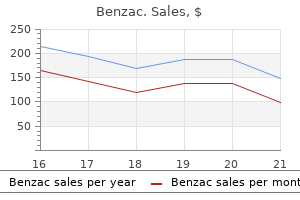
Benzac dosages: 20 gr
Benzac packs: 3 tubes, 6 tubes, 9 tubes, 12 tubes, 15 tubes, 18 tubes, 21 tubes, 24 tubes

Benzac 20 gr order otc
Superficial veins: the cephalic vein programs alongside the cranial (cephalic) margin of the limb acne 30s 20 gr benzac for sale, whereas the basilic vein programs alongside the caudal (basic) margin of the limb skin care anti aging 20 gr benzac generic with mastercard. Deep veins: Deep veins in the limbs usually take the form of paired accompanying veins, bearing the identical name as the artery they accompany. Lymphatic vessels: the superficial lymphatic vessels generally converge on and comply with the superficial veins, and the deep lymphatics follow the deep veins. Dermatomes: As a consequence of plexus formation, two patterns of cutaneous innervation happen within the upper limb: (1) segmental innervation (dermatomes) by spinal nerves and (2) innervation by multisegmental peripheral (named) nerves. The former sample is easiest to visualize if the limb is positioned in its initial embryonic position (abducted with the thumb directed superiorly). Cutaneous innervation: Like the brachial plexus, which forms posterior, lateral, and medial (but no anterior) cords, the arm and forearm have posterior, lateral, and medial (but no anterior) cutaneous nerves. Myotomes: Most upper limb muscular tissues embody components of multiple myotome and thus receive motor fibers from several spinal cord segments or spinal nerves. Anterior Axio-Appendicular Muscles 448 a the spinal twine segmental innervation is indicated. The pectoralis major is a big, fan-shaped muscle that covers the superior a half of the thorax. The sternocostal head is much bigger, and its lateral border types the muscular mass that makes up a lot of the anterior wall of the axilla. The pectoralis major and adjoining deltoid muscular tissues type the slim deltopectoral groove, by which the cephalic vein runs. Producing highly effective adduction and medial rotation of the arm when acting collectively, the two components of the pectoralis major also can act independently: the clavicular head flexing the humerus, and the sternocostal head extending it again from the flexed place. To check the clavicular head of pectoralis main, the arm is kidnapped 90�; the individual then moves the arm anteriorly against resistance. To take a look at the sternocostal head of pectoralis main, the arm is kidnapped 60� after which adducted towards resistance. Its base (proximal attachment) is fashioned by fleshy slips connected to the anterior ends of the 3rd�5th ribs close to their 449 costal cartilages. Of the anterior axio-appendicular muscle tissue forming the anterior wall, solely parts of the pectoralis main (attaching ends, a central part overlying the pectoralis minor, and a dice of muscle mirrored superior to the clavicle), the pectoralis minor, and the subclavius remain. All the clavipectoral fascia and axillary fats have been eliminated, as has the axillary sheath surrounding the neurovascular bundle. This enables remark of the medial wall of the axilla, shaped by the serratus anterior overlying the lateral thoracic wall, and of the latissimus dorsi contributing to the posterior wall. It also assists in 450 elevating the ribs for deep inspiration when the pectoral girdle is fastened or elevated. The pectoralis minor is a helpful anatomical and surgical landmark for structures within the axilla. With the coracoid course of, the pectoralis minor types a "bridge" underneath which vessels and nerves must move to the arm. The subclavius lies virtually horizontally when the arm is in the anatomical place. This small, spherical muscle is situated inferior to the clavicle and affords some safety to the subclavian vessels and the superior trunk of the brachial plexus if the clavicle fractures. The subclavius anchors and depresses the clavicle, stabilizing it throughout movements of the upper limb. The serratus anterior overlies the lateral part of the thorax and forms the medial wall of the axilla. This broad sheet of thick muscle was named due to the sawtoothed look of its fleshy slips or digitations (L. The muscular slips cross posteriorly after which medially to connect to the whole length of the anterior floor of the medial border of the scapula, including its inferior angle. The robust inferior part of the serratus anterior rotates the scapula, elevating its glenoid cavity so the arm can be raised above the shoulder. It additionally anchors the scapula, keeping it closely applied to the thoracic wall, enabling different muscles to use it as a exhausting and fast bone for actions of the humerus. To test the serratus anterior (or the function of the lengthy thoracic nerve that provides it), the hand of the outstretched limb is pushed against a wall. If the muscle is performing usually, several digitations of the muscle could be seen and palpated. Posterior Axio-Appendicular and Scapulohumeral Muscles Posterior axio-appendicular muscular tissues (superficial and intermediate groups of 451 extrinsic back muscles) connect the superior appendicular skeleton to the axial skeleton (in the trunk). Posterior Axio-Appendicular Muscles aThe spinal twine segmental innervation is indicated. Superficial posterior axio-appendicular (extrinsic shoulder) muscles: trapezius and latissimus dorsi. Scapulohumeral (intrinsic shoulder) muscle tissue: deltoid, teres main, and the four rotator cuff muscular tissues (supraspinatus, infraspinatus, teres minor, and subscapularis). Trapezius the trapezius supplies a direct attachment of the pectoral girdle to the trunk. This giant, triangular muscle covers the posterior side of the neck and the superior half of the trunk. The trapezius attaches the pectoral girdle to the skull and vertebral column and assists in suspending the upper limb. The fibers of the trapezius are divided into three elements, which have totally different actions at the physiological scapulothoracic joint between the scapula and thoracic wall. Arrows indicate the path of pull; the muscular tissues (and gravity) producing every motion are recognized by numbers, that are listed in Table three. Descending and ascending trapezius fibers act together in rotating the scapula on the thoracic wall in numerous directions, twisting it. The trapezius additionally braces the shoulders by pulling the scapulae posteriorly and superiorly, fixing them in place on the thoracic wall with tonic contraction; consequently, weak spot of the trapezius causes drooping of the shoulders. If the muscle is appearing normally, the superior border of the muscle can be simply seen and palpated. This giant fan-shaped muscle passes from the trunk to the humerus and acts instantly on the glenohumeral joint and not directly on the pectoral girdle (scapulothoracic joint). In combination with the pectoralis major, the latissimus dorsi is a robust adductor of the humerus and performs a serious function in downward rotation of the scapula in association with this motion. It can additionally be useful in restoring the higher limb from abduction superior to the shoulder; therefore, the latissimus dorsi is essential in climbing. In conjunction with the pectoralis main, the latissimus dorsi raises the trunk to the arm, which happens when performing chin-ups (hoisting oneself so the chin touches an overhead bar) or climbing a tree, for instance. These actions are also used when chopping 457 wooden, paddling a canoe, and swimming (particularly during the crawl stroke). To check the latissimus dorsi (or the operate of the thoracodorsal nerve that supplies it), the arm is abducted 90� after which adducted in opposition to resistance supplied by the examiner. If the muscle is regular, the anterior border of the muscle may be seen and simply palpated within the posterior axillary fold (see "Axilla"). These muscle tissue provide direct attachment of the appendicular skeleton to the axial skeleton. The superior third of the strap-like levator scapulae lies deep to the sternocleidomastoid; the inferior third is deep to the trapezius.
Valeriana rhizome (Valerian). Benzac.
- Are there any interactions with medications?
- How does Valerian work?
- What other names is Valerian known by?
- Depression, anxiety, restlessness, convulsions, mild tremors, epilepsy, attention-deficit hyperactivity disorder (ADHD), chronic fatigue syndrome (CFS), muscle and joint pain, headache, stomach upset, menstrual pains, menopausal symptoms including hot flashes and anxiety, and other conditions.
- Insomnia.
- Are there safety concerns?
- What is Valerian?
- Is Valerian effective?
- Dosing considerations for Valerian.
Source: http://www.rxlist.com/script/main/art.asp?articlekey=96840
Benzac 20 gr buy generic on line
Thus skin care test order benzac 20 gr on line, tapering lateral extensions of the spinal dura surround every pair of posterior and anterior nerve roots as dural root sheaths acne 50s benzac 20 gr low price, or sleeves. Nerve fibers are distributed to the spinal dura by the (recurrent) meningeal nerves. In a lumbar spinal puncture, the needle traverses the spinal dura and arachnoid concurrently. Bleeding into this layer creates a pathological space on the dura�arachnoid junction in which a subdural hematoma is fashioned. Delicate strands of connective tissue, the arachnoid trabeculae, span the subarachnoid space connecting the spinal arachnoid and pia. The spinal pia additionally immediately covers the roots of the spinal nerves and the spinal blood vessels. Inferior to the conus medullaris, the spinal pia continues because the filum terminale. The spinal cord is suspended within the dural sac by the filum terminale and the right and left denticulate ligaments (L. The denticulate ligaments include a fibrous sheet of pia extending halfway between the posterior and anterior nerve roots from the lateral surfaces of the spinal wire. The 20�22 sawtooth-like processes attach to the inner surface of the arachnoidlined dural sac. The most superior process of the best and left denticulate ligaments attaches to the cranial dura immediately superior to the foramen magnum, and the inferior course of extends from the conus medullaris, passing between the T12 and the L1 nerve roots. The lateral projections point out extensions of the subarachnoid space into the dural root sheaths around the spinal nerve roots. The spinal dura and arachnoid mater have been split and pinned flat to expose the spinal cord and denticulate ligaments between posterior and anterior spinal nerve roots. Dural root sheaths, enclosing spinal nerve roots in extensions of the subarachnoid space, protrude from the perimeters of the lumbar cistern. These arteries run longitudinally from the medulla of the brainstem to the conus medullaris of the spinal cord. Three longitudinal arteries provide the spinal cord: an anterior spinal artery and two posterior spinal arteries. Radicular arteries are shown at only the cervical and thoracic ranges, however in addition they happen at the lumbar and sacral levels. The pattern of the arterial provide of the spinal twine is from three longitudinal arteries: one anterior mendacity in the anteromedian place and the other two lying posterolaterally. These vessels are strengthened by medullary branches derived from the segmental arteries. The sulcal arteries are small branches of the anterior spinal artery coursing within the anterior median fissure. Sulcal arteries arise from the anterior spinal artery and enter the spinal twine through this fissure. The sulcal arteries provide approximately two thirds of the cross-sectional space of the spinal wire (Standring, 2016). Each posterior spinal artery is a branch of both the vertebral artery or the posteroinferior cerebellar artery. The posterior spinal arteries generally kind anastomosing channels within the pia mater. By themselves, the anterior and posterior spinal arteries can supply solely the short superior part of the spinal cord. The circulation to much of the spinal wire depends on segmental medullary and radicular arteries working along the spinal nerve roots. The anterior and posterior segmental medullary arteries are derived from spinal branches of the ascending cervical, deep cervical, vertebral, posterior intercostal, and lumbar arteries. The segmental medullary arteries occur primarily in association with the cervical and lumbosacral enlargements, areas where the necessity for an excellent blood supply is greatest. The nice anterior segmental medullary artery (of Adamkiewicz), which is on the left facet in about 65% of people, reinforces the circulation to two thirds of the spinal twine, including the lumbosacral enlargement. The posterior and anterior roots of the spinal nerves and their coverings are provided by posterior and anterior radicular arteries (L. Most radicular arteries are small and supply solely the nerve roots; however, some of them could assist with the availability of superficial components of the grey matter within the posterior and anterior horns of the spinal wire. The spinal veins are arranged longitudinally, talk freely with one another, and are drained by up to 12 anterior and posterior medullary and radicular veins. The veins of the spinal cord be part of the interior vertebral (epidural) venous plexuses within the epidural area. The internal vertebral venous plexuses cross superiorly through the foramen magnum to talk with dural sinuses and vertebral veins in the skull. The inner vertebral plexuses also communicate with the external vertebral venous plexuses on the exterior floor of the vertebrae. Consequently, the L5 spinal nerve roots are the thickest and their foramina, the narrowest. This increases the possibility that these nerve roots shall be compressed if osteophytes (bony spurs) develop. Myelography Myelography is a radiopaque distinction process that allows visualization of the spinal cord and spinal nerve roots. This method exhibits the extent of the subarachnoid space and its extensions across the spinal nerve roots within the dural root sheaths. They develop as a single layer from the mesenchyme surrounding the embryonic spinal twine. Fluid-filled spaces kind inside this layer and coalesce to produce the subarachnoid area (Moore et al. The origin of each pia and arachnoid from a single membrane is mirrored by the numerous arachnoid trabeculae passing between them. The delicate pia mater provides a shiny appearance to the floor of the spinal twine but is barely seen to the unaided eye as a distinct layer. Flexion of the vertebral column facilitates insertion of the needle by spreading apart the vertebral laminae and spinous processes, stretching the ligamenta flava. The pores and skin covering the decrease lumbar vertebrae is anesthetized, and a lumbar puncture needle, fitted with a stylet, is inserted within the midline between the spinous processes of the L3 and L4 (or L4 and L5) vertebrae. Recall that a airplane transecting the very best points of the iliac crests-the supracristal plane- often passes via the L4 spinous process. After passing 4�6 cm in adults (more in overweight persons), the needle "pops" through the ligamentum flavum, then punctures the dura and arachnoid, and enters the lumbar cistern. Epidural Anesthesia (Blocks) An anesthetic agent is injected into the epidural house utilizing the position described for lumbar spinal puncture, or through the sacral hiatus (caudal epidural anesthesia/block) (see medical box "Anesthesia for Childbirth" in Chapter 6, Pelvis and Perineum). Fractures, dislocations, and fracture�dislocations could intervene with the blood supply to the spinal wire from the spinal and medullary arteries. Deficient blood supply (ischemia) of the spinal twine impacts its perform and can result in muscle weakness and paralysis. The spinal wire may also undergo circulatory impairment if the segmental medullary arteries, notably the good anterior segmental medullary artery (of Adamkiewicz), are narrowed by obstructive arterial disease.
20 gr benzac generic fast delivery
Consequently skin care summer purchase benzac 20 gr free shipping, in the proximal forearm skin care associates buy benzac 20 gr with visa, the "anterior" flexor�pronator compartment truly lies anteromedially, and the "posterior" extensor�supinator compartment lies posterolaterally. The radial artery (laterally) and the sharp, subcutaneous posterior border of the ulna (medially) are palpable options separating the anterior and posterior compartments. No motor nerves cross either demarcation, making them helpful for surgical approaches. At the extent of the wrist, 9 tendons from three muscular tissues (and one nerve) of the anterior compartment of the forearm traverse the carpal tunnel; eight of the tendons share a standard synovial flexor sheath. The medial epicondyle and supra-epicondylar ridge present attachment for the forearm flexors, and the lateral formations present attachment for the forearm extensors. Thus, rather than lying strictly anteriorly and posteriorly, the proximal parts of the "anterior" (flexor�pronator) compartment of the forearm lie anteromedially, and the "posterior" (extensor�supinator) compartment lies posterolaterally. Spiraling gradually over the length of the forearm, the compartments turn into truly anterior and posterior in position within the distal forearm and wrist. These fascial compartments, containing the muscle tissue in practical groups, are demarcated by the subcutaneous border of the ulna posteriorly (in the proximal forearm) after which medially (distal forearm) and by the radial artery anteriorly 548 and then laterally. These buildings are palpable (the artery by its pulsations) throughout the forearm. Because neither boundary is crossed by motor nerves, additionally they provide websites for surgical incision. The flexors and pronators of the forearm are in the anterior compartment and are served mainly by the median nerve; the one and a half exceptions are innervated by the ulnar nerve. The extensors and supinators of the forearm are within the posterior compartment and are all served by the radial nerve (directly or by its deep branch). The anterior compartment is phenomenal in this regard as a result of it communicates with the central compartment of the palm by way of the carpal tunnel. Muscles of Forearm There are 17 muscle tissue crossing the elbow joint, some of which act on the elbow joint solely, whereas others act at the wrist and fingers. In the proximal a half of the forearm, the muscles type fleshy lots extending inferiorly from the medial and lateral epicondyles of the humerus. The tendons of those muscle tissue pass by way of the distal part of the forearm and proceed into the wrist, hand, and fingers. The flexor muscular tissues of the anterior compartment have approximately twice the majority and power of the extensor muscle tissue of the posterior compartment. The tendons of most flexor muscular tissues are situated on the anterior floor of the wrist and are held in place by the palmar carpal ligament and the flexor retinaculum (transverse carpal ligament), thickenings of the antebrachial fascia. Muscles of Anterior Compartment of Forearm 551 a the spinal twine segmental innervation is indicated. A superficial layer or group of 4 muscular tissues (pronator teres, flexor carpi radialis, palmaris longus, and flexor carpi ulnaris). These muscular tissues are all connected proximally by a common flexor tendon to the medial epicondyle of the humerus, the common flexor attachment. A deep layer or group of three muscles (flexor digitorum profundus, flexor pollicis longus, and pronator quadratus). All muscles within the anterior (flexor�pronator) compartment of the forearm are provided by the median and/or ulnar nerves (most by the median; just one and a half exceptions are provided by the ulnar). Therefore, the brachioradialis is a serious exception to the rule that (1) the radial nerve provides only extensor muscular tissues and (2) that all flexors lie within the anterior (flexor) compartment. The lengthy flexors of the digits (flexor digitorum superficialis and flexor digitorum profundus) also flex the metacarpophalangeal and wrist joints. This action is bolstered by the flexor digitorum superficialis when pace and flexion towards resistance are required. When the wrist is flexed at the identical time that the metacarpophalangeal and interphalangeal joints are flexed, the long flexor muscular tissues of the fingers are working over a shortened distance between attachments, and the action ensuing from their contraction is consequently weaker. Extending the wrist will increase their working distance, and thus, their contraction is extra efficient in producing a strong grip. Tendons of the lengthy flexors of the digits cross through the distal part of the forearm, wrist, and palm and continue to the medial 4 fingers. The flexor digitorum superficialis flexes the middle phalanges, and the flexor digitorum profundus flexes the center and distal phalanges. The following dialogue supplies additional details, beginning with the muscle tissue of the superficial and intermediate layers. The pronator teres, a fusiform muscle, is probably the most lateral of the superficial forearm flexors. If acting usually, the muscle is distinguished and can be palpated at the medial margin of the cubital fossa. In the center of the forearm, its fleshy belly is replaced by a long, flattened tendon that turns into cord-like because it approaches the wrist. To check the flexor carpi radialis, the particular person is requested to flex the wrist in opposition to resistance. It has a short belly and a long, cord-like tendon that passes superficial to the flexor retinaculum and attaches to it and the apex of the palmar aponeurosis. The tendon lies deep and slightly medial to this nerve before it passes deep to the flexor retinaculum. To test the palmaris longus, the wrist is flexed and the pads of the little finger and thumb are tightly pinched collectively. The ulnar nerve enters the forearm by passing between the humeral and ulnar heads of its proximal attachment. This muscle is outstanding among muscle tissue of the anterior compartment, being totally innervated by the ulnar nerve. To test the flexor carpi ulnaris, the individual places the posterior facet of the forearm and hand on a flat desk and is then asked to flex the wrist in opposition to resistance while the examiner palpates the muscle and its tendon. The median nerve and ulnar artery enter the forearm by passing between its humero-ulnar and radial heads. The four tendons are enclosed (along with the 4 tendons of the flexor digitorum profundus) in a synovial frequent flexor sheath. To take a look at the flexor digitorum superficialis, one finger is flexed on the proximal interphalangeal joint in opposition to resistance and the other three fingers are held in an extended place to inactivate the flexor digitorum profundus. The fascial plane between the intermediate and deep layers of muscles makes up the first neurovascular plane of the anterior (flexor�pronator) compartment; the main neurovascular bundles exclusive to this compartment course inside it. Each tendon is capable of flexing two interphalangeal joints, the metacarpophalangeal joint and the wrist joint. The part of the muscle going to the index finger normally separates from the remainder of the muscle relatively early in the distal part of the forearm and is able to independent contraction. To check the flexor digitorum profundus, the proximal interphalangeal joint is held in the prolonged place whereas the individual makes an attempt to flex the distal interphalangeal joint.
Buy benzac 20 gr with amex
Discussion Building partially on the Human Genome Project skin care for swimmers generic 20 gr benzac fast delivery, scientific advances in understanding the molecular basis of oncology have spurred substantial trade activity in developing new oncology therapies since 1995 acne diagram benzac 20 gr cheap overnight delivery. As quantified here by the number of new molecular entities in development and the variety of molecular targets being explored, exercise has more than doubled over the interval with almost three quarters (71 percent) of late stage growth candidates now attacking specific targets, typically with related segmented affected person sub-populations. To date, the smallest incidence most cancers indications have usually obtained as little as one quarter as many distinct therapeutic investigations as the biggest incidence cancers (5 versus 23 to forty six for prostate, lung and breast cancer) and even decrease percentages of unique, single cancer therapeutics. While the volume of candidates in late stage development continues to rise, since 2009 the early stage growth pipeline has skilled its first declines, approaching ten percent, each in the number of lively unique candidates and distinct molecular targets being explored. Compared to the millions of patients with heart problems or diabetes, even the most important oncology indications, such as breast cancer with 209,000 new U. Our trend evaluation of the oncology pipeline from 1995 to the present, emphasizing oncology segmentation by organ of origin and therapeutic molecular target in addition to web product approvals, supplies an objective quantification of oncology drug therapy evolution. Future work may try to correlate elements starting from unmet medical need, market dimension and scientific understanding to funding availability, development processes, regulatory requirements and economic incentives which, whereas not proof of causation, may nonetheless recommend the relative importance of those numerous components. The segmentation of oncology therapies creates each alternatives and challenges. Additional benefits of such improved efficacy may embrace faster, smaller clinical trials and better success rates of candidate therapies in those trials, leading to more fast broad patient entry and more effective care as fewer patients receive therapies that present limited benefits. To the extent that the segmentation ends in medication having elevated performance improvements over other therapies, the segmentation into extra stratified medicines might facilitate premium pricing, reflecting in part the higher fixed prices of growth of small-population medication and their superior performance in treating life-threatening illnesses, thereby incentivizing their growth, though putting larger pressures on payers. Without changes within the procedures by which we develop, evaluate, disseminate and finance new medicines, even when science generates promising intervention hypotheses, if the affected person subpopulation falls beneath a certain threshold�whether a few thousand or a number of hundred�it could prove clinically impractical and economically infeasible for developers to create the efficacy, security and clinical profit evidentiary data bundle required at drug prices payers can afford. The opinions expressed herein are these of the authors, and not essentially these of the research sponsor or the Massachusetts Institute of Technology. Acemoglu D, Linn J [2004], "Market dimension in innovation: principle and proof from the pharmaceutical industry". American Cancer Society [2012, "Learn About Cancer: the History of Cancer: Nineteenth Century. The effect of castration, of estrogen and of androgen injection on serum phosphatases in metastatic carcinoma of the prostate". Mukherjee, Siddhartha [2010], the Emperor of all Maladies: A Biography of Cancer, New York: Scribner, A Division of Simon & Schuster, Inc. Cancer Statistics Working Group [2010], "United States Cancer Statistics: 1999�2006 Incidence and Mortality Web-based Report", Table A. Vanchieri C [2007], "National Cancer Act: a look back and forward", J Natl Cancer Inst 99(5):342-345. English Translation [1860], "Cellular pathology as based mostly upon physiological and pathological histology twenty lectures delivered within the Pathological Institute of Berlin during the months of February, March and April, 1858 archive. Yin, W [2008], "Market Incentives and pharmaceutical innovatoin", Jour Health Econ 27(4):1066-77. Annals of Agricultural and Environmental Medicine 2017, Vol 24, No 2, 254�260 Among free-living amoebae that are broadly distributed in nature solely 4 genera/species are known as agents of human infections: Acanthamoeba spp. Infections due to these amoebae, despite low morbidity, are characterised by comparatively high mortality price and pose critical scientific issues. This evaluate research presents and summarizes present knowledge about infections as a result of pathogenic and opportunistic free-living amoebae targeted on epidemiology, scientific manifestations, analysis and therapy primarily based on international literature. All 4 genera have been recognized as etiologic factors of fatal central nervous system infections and other serious ailments in humans. Amoebic ailments are difficult to diagnose which outcomes in delayed therapy, and end in a excessive mortality rate. These type of infections, regardless of low morbidity, are characterised by comparatively excessive mortality price, which is a huge challenge for environment friendly diagnosis and remedy [1, 2]. From many genera of free-living amoebae that exist in nature, solely 4 are involved in human and animal infections. Among them there are several of Acanthamoeba species; however, only one species of each genera: Balamuthia (B. These amoebae are ubiquitous, and have been found in water, soil and air, but additionally in sewage, swimming swimming pools, flowerpots, water tubs, humidifiers, aquaria, eye wash solutions and hospital environment. These protozoa are referred to as amphizoic due to their capability to complete their life cycle inside host organism as nicely as in the surroundings. Human infections with these amoebae are rare however have been reported everywhere in the world [3]. Of greatest concern is the comparatively excessive Address for correspondence: Katarzyna Kr�l-Turmiska, Chair and Department of Medical Microbiology Medical University of Lublin E-mail: katarzyna. Early analysis of these infections is very important though it may be difficult because of non-specific signs. Balamuthia mandrillaris (the solely recognized species of Balamuthia), Naegleriafowleri, Sapiniapedata and several species of Acanthamoeba. Naegleriafowleri is an thermophilic, free-living amoeboflagellate spread worldwide in most types of heat water reservoirs. Since then, new cases have been reported also from Germany, France, Belgium, Hungary and Iran [3, 8]. Balamuthia mandrillaris Sappiniapedata Insufficient data Immunocompetent and immunocompromised Immunocompetent and immunocompromised Immunocompetent and immunocompromised Immunocompromised Acanthamoeba spp. Increased contact of the nasal mucosa with infested water predispose humans to an infection. Most casualties are younger adults and kids with a history of contacts with water containing the amoebae, about a week prior to the onset of neurological symptoms. For that cause, any information about previous contact with warm water is essential [1, 9]. The amoebic trophozoites penetrate the olfactory mucosa and migrate alongside the olfactory nerve, crossing the cribriform plate and reach the olfactory bulbs. Trophozoites induce an inflammatory response associated with lytic necrotic haemorrhage. Lesions are noticed mainly across the base of the orbitofrontal and temporal lobe, base of the mind, hypothalamus, midbrain, pons, medulla oblongata, and the higher a half of the spinal twine [3, 11]. The earliest symptoms are bifrontal or bitemporal headaches, high temperature (38. Subsequently, photophobia and neurological abnormalities develop, including lethargy, seizures, confusion, coma, diplopia or bizarre behaviour may occur. Cranial nerve palsies (third, fourth and six cranial nerves) might indicate cerebral oedema and herniation. Increased intracranial strain and herniation normally lead to dying [1, 9, 10, 11]. In addition, protein focus might range from 100mg/100mL to 1,000mg/100mL, and glucose may be 10 mg/100mL, or lower. Prognoses are weak, demise happens typically inside 7�10 days following infection [9, 11, 12, 13]. The sera collected from several individuals with a historical past of swimming in freshwater revealed not only IgM but in addition IgG antibodies. A part distinction microscope is beneficial to optimize visualization of the amoebae [4, 5, 9].
Benzac 20 gr generic
To test the triceps (or to determine the level of a radial nerve lesion) acne 26 year old female benzac 20 gr purchase visa, the arm is kidnapped 90� after which the flexed forearm is extended towards resistance supplied by the examiner acne zeno 20 gr benzac purchase with mastercard. Its strength should be comparable with the contralateral muscle, given consideration for lateral dominance (right or left handedness). The anconeus assists the triceps in extending the forearm and tenses the capsule of the elbow joint, stopping its being pinched during extension. It can be said to exert an abducting drive on the ulna during pronation of the forearm. Brachial Artery the brachial artery provides the main arterial provide to the arm and is the continuation of the axillary artery. Functionally and clinically necessary peri-articular arterial anastomoses encompass the elbow. The resulting collateral circulation allows blood to reach the forearm when flexion of 533 the elbow compromises move via the terminal a part of the brachial artery. In this deep dissection, part of the biceps is excised and the cubital fossa is opened widely by retracting the forearm extensor muscles laterally and the flexor muscles medially. The radial nerve, which has simply left the posterior compartment of the arm by piercing the lateral intermuscular septum, emerges between the brachialis and brachioradialis and divides right into a superficial (sensory) and a deep (motor) department (details are shown in. The brachial artery, relatively superficial and palpable throughout its course, lies anterior to the triceps and brachialis. At first, it lies medial to the humerus the place its pulsations are palpable within the medial bicipital groove. It then passes anterior to the medial supra-epicondylar ridge and trochlea of the humerus. The radial nerve and accompanying profunda brachii artery wind posteriorly round, and immediately on the surface of, the humerus in the radial groove. The radial nerve and radial collateral artery then pierce the 535 lateral intermuscular septum to enter the anterior compartment. The ulnar nerve pierces the medial intermuscular septum to enter the posterior compartment after which lies in the groove for the ulnar nerve on the posterior side of the medial epicondyle of the humerus. The main named branches of the brachial artery arising from its medial side are the profunda brachii artery and the superior and inferior ulnar collateral arteries. The collateral arteries assist kind the peri-articular arterial anastomoses of the elbow area. Other arteries involved are recurrent branches, sometimes double, from the radial, ulnar, and interosseous arteries, which run superiorly anterior and posterior to the elbow joint. These arteries anastomose with descending articular branches of the deep artery of the arm and the ulnar collateral arteries. The profunda brachii accompanies the radial nerve alongside the radial groove because it passes posteriorly across the shaft of the humerus. The profunda brachii terminates by dividing into middle and radial collateral arteries, which take part within the peri-articular arterial anastomoses across the elbow. Here, it anastomoses with the posterior ulnar recurrent and inferior ulnar collateral arteries, taking part in the peri-articular arterial anastomoses of the elbow. It then passes inferomedially anterior to the medial epicondyle of the humerus and joins the peri-articular arterial anastomoses of the elbow area by anastomosing with the anterior ulnar recurrent artery. Veins of Arm Two units of veins of the arm, superficial and deep, anastomose freely with one another. The superficial veins are in the subcutaneous tissue, and the deep veins accompany the arteries. Their frequent connections embody the artery, forming an anastomotic network inside a common vascular sheath. The 537 pulsations of the brachial artery assist move the blood through this venous network. The brachial vein begins at the elbow by union of the accompanying veins of the ulnar and radial arteries and ends by merging with the basilic vein to kind the axillary vein. Not uncommonly, the deep veins be a part of to type one brachial vein during part of their course. Nerves of Arm Four main nerves move by way of the arm: median, ulnar, musculocutaneous, and radial. Their origins from the brachial plexus, courses within the upper limb, and the constructions innervated by them are summarized in Table three. After supplying all three muscles of the anterior compartment of the arm, the musculocutaneous nerve emerges lateral to the biceps because the lateral cutaneous nerve of the forearm. It turns into actually subcutaneous when it pierces the deep fascia proximal to the cubital fossa to course initially with the cephalic vein within the subcutaneous tissue. After crossing the anterior side of the elbow, it continues to provide the pores and skin of the lateral facet of the forearm. The radial nerve enters the arm posterior to the brachial artery, medial to the humerus, and anterior to the long head of the triceps, where it gives branches to the lengthy and medial heads of the triceps. The radial nerve then descends inferolaterally with the profunda brachii artery and passes around the humeral shaft in the radial groove. The department of the radial nerve to the lateral head of the triceps arises within the radial groove. When it reaches the lateral border of the humerus, the radial nerve pierces the lateral intermuscular septum and continues inferiorly in the anterior compartment of the arm between the brachialis and the 538 brachioradialis to the level of the lateral epicondyle of the humerus. Anterior to the lateral epicondyle, the radial nerve divides into deep and superficial branches. The deep department of the radial nerve is totally muscular and articular in its distribution. The superficial branch of the radial nerve is totally cutaneous in its distribution, supplying sensation to the dorsum of the hand and fingers. The median nerve then descends into the cubital fossa, where it lies deep to the bicipital aponeurosis and median cubital vein. Around the center of the arm, it pierces the medial intermuscular septum with the superior ulnar collateral artery and descends between the septum and the medial head of the triceps. The ulnar nerve passes posterior to the medial epicondyle and medial to the olecranon to enter the forearm. Posterior to the medial epicondyle, the place the ulnar nerve is referred to in lay phrases as the "funny bone. Cubital Fossa the cubital fossa is apparent superficially as a despair on the anterior facet of the elbow area. Medially, the mass of flexor muscles of the forearm arising from the common flexor attachment on the medial epicondyle; most particularly, the pronator teres. Laterally, the mass of extensor muscles of the forearm arising from the lateral epicondyle and supra-epicondylar ridge; most particularly, the brachioradialis. The floor of the cubital fossa is fashioned by the brachialis and supinator muscles of the arm and forearm, respectively.

Benzac 20 gr buy generic on-line
Identify individuals who should be targeted for turberculin skin testing to diagnose latent tuberculosis infection skin care vitamins and minerals 20 gr benzac buy fast delivery. Identify the optimal pharmacologic regimen for remedy of latent tuberculous an infection skin care 2013 purchase benzac 20 gr overnight delivery. Discuss the public well being implications of figuring out and treating latent tuberculous an infection. Cardiovascular, Pulmonary, Renal Rev 7/22/2019 Page 33 of 35 Tobacco Prevention & Intervention 1. Describe the primary courses of immunosuppression utilized in medical kidney transplantation. State the magnitude and regulated vary of NaCl and water handling by the kidneys. Describe the main epithlelial transport mechanisms for NaCl and water reabsorp on in each major tubular section. Describe the general position of each major tubular phase within the regula on of NaCl and water reabsorp on. Identify the main hormones that regulate tubular reabsorption of NaCl and water and their tubular and cellular web site of ac on. State the Starling equation for the flow of resolution from the renal interstitium to the peritubular capillaries. Give values for each of the Starling forces and the web strain driving the circulate in (7). Describe qualitatively the results of increasing and decreasing tubular move on water and sodium excretion. Define "glomerulotubular balance" and "tubuloglomerular feedback" and describe the roles these processes play in the regulation of NaCl and water reabsorption. List the incidence, gross and microscopic options of the next benign renal tumors: Renal Papillary Adenoma, Angiomyolipoma, and Oncocytoma. For renal cell carcinoma variants, including clear cell carcinoma, papillary carcinoma and chromophobe carcinoma, list their: a. Describe the fundamental genetic differences between spontaneous and familial renal tumors. Cardiovascular, Pulmonary, Renal Rev 7/22/2019 Page 34 of 35 Upper Airways & Larynx 1. Define the major symptom complexes that indicate issues of the higher airway and larynx. Define how the advanced of symptoms referred to as hoarseness is characterised and evaluated. Describe the different types of urinalysis and understand their corresponding clinicalpathological correlation, together with macroscopic examination and chemical evaluation (use and interpretation of dipstick for: glucose; bilirubin; particular gravity; blood; pH; protein; urobilinogen; nitrite; leukocyte esterase; microscopic examination; and cytology). Interpret a few of the most common chemical and cytological adjustments in urine samples in the commonest inflammatory and neoplastic illnesses of the kidney and the urinary tract. Describe the pathogenesis of urinary tract an infection in phrases of routes of an infection, organism virulence elements, host protection mechanisms, predisposing elements, scientific manifestations, and problems. Compare and contrast the features and pathogenesis of the two major causes of persistent pyelonephritis (urinary tract obstruc on and vesicoureteral reflux). Describe the mechanisms of air flow (neural, chest wall, diaphragm and different muscular tissues, airways). Identify differences in air flow all through the lung associated to anatomic location/gravity. Identify fundamental lung pressurevolume relationships and perceive lung compliance. Describe the mechanisms of dynamic airflow resistance (and its inverse, conductance), and the way resistance (conductance) and compliance determine ventilation. Classification methods have been developed for numerous manifestations of neuropathy. Among kind 1 diabetes patients, one potential research found a 29% cumulative incidence after approximately 5 years of follow-up, while another found a cumulative incidence of 35% over a follow-up of 13�14 years. However, associations have been discovered with different traits, together with peak, blood stress, and lipid levels. Although glycemia is a risk issue among people with kind 1 diabetes, it has not clearly been recognized as such for individuals with sort 2 diabetes. In an evaluation performed for Diabetes in America, 3rd version, heart price (beats/minute) was considerably greater in adults with identified diabetes (mean 75. Heart price was additionally greater in those who had been recognized at the study go to with diabetes (mean seventy three. Of those with diabetes, the heart price was considerably larger amongst diabetic individuals with glycosylated hemoglobin (A1c) eleven. The basis for the higher coronary heart rate among diabetic sufferers and the relation of coronary heart rate to A1c are unknown. A number of questions must be answered with regard to diabetic neuropathy, such as whether glucose variability influences its improvement past the results of the diploma and duration of hyperglycemia. Such information ought to finally result in a better understanding of the method to treat and stop the dysfunction. Neuropathies have been described in sufferers with diabetes of differing causes, suggesting a common etiologic mechanism based mostly on continual hyperglycemia. The hallmark of the diabetic neuropathies is a progressive lack of all populations of nerve fibers, which can be assessed in a selection of methods. The late sequelae of neuropathy are well recognized, with foot issues, including ulceration (7), gangrene, and Charcot neuroarthropathy (8), representing the most typical reason for hospitalization among diabetic patients in most Western countries. Not surprisingly, diabetic neuropathy often has an antagonistic effect on quality of life (9,10,11). The exclusion of nondiabetic causes has additionally been emphasised, as as much as 10% of peripheral neuropathy in diabetic patients could also be of nondiabetic etiology (13,15,16). It is incessantly related to microvessel retinal and kidney disease, however different causes of neuropathy have to be excluded (18). Therefore, findings offered in this chapter are mainly pertinent to these two types of neuropathy. It is often of gradual onset and may be present at the diagnosis of kind 2 diabetes in >10% of subjects (15,24). Mononeuropathy (mononeuritis multiplex) (atypical forms) Isolated cranial or peripheral nerve. The signs normally progressively improve with establishment of stable blood glucose levels (26). However, its relationship to diabetes is controversial, since it might be an opportunity incidence of two common problems. This would possibly end in an array of signs and indicators, similar to postural hypotension, decreased bowel motility, decreased bladder contractility, erectile dysfunction, and sweating disorders. More superior illness, manifested by debilitating symptoms and signs, is normally very difficult to deal with and fortuitously rare. It has a monophasic course, with improvement starting within 9�12 months, though recovery can be incomplete and protracted over years (19). Focal limb neuropathies are sometimes as a end result of entrapment; essentially the most vulnerable nerves embody the median, ulnar, radial, lateral femoral cutaneous, fibular, and plantar (19).
Syndromes
- Cancers, especially lung and colon cancer
- Being at a high altitude
- Narrowing or blockage of the colostomy opening (stoma)
- Violence and reckless behavior
- Fibrinogen level will be high.
- Misplacement of the stent
- Physical exam (including pelvic exam)
- Various asthma medications
Cheap benzac 20 gr on-line
The hemoperfusion/hemodialysis combination had a small advantage over the high-flux dialyzer acne 4 year old benzac 20 gr purchase on-line,474 and normal dialysis was inferior to each skin care essential oils 20 gr benzac discount with mastercard. All aluminum gels were stopped, a brand new reverse-osmosis system was installed, and an alternate water supply was used (dialysate Al 2 g/L). In this report, the mortality was 91%, the disorder was the disseminated or rhinocerebral variety in 75% of the circumstances, and a prognosis of mucormycosis was made solely at post-mortem in 61%. Despite this, the numbers of patients in prospective trials was comparatively small; also, due to the severity of the dysfunction and poor prognosis in untreated sufferers, there have been no controlled trials. In a small trial of asymptomatic patients discovered to have biopsy proof of aluminum bone illness, there was reduced surface staining of aluminum and elevated bone formation when all exposure to aluminum was eliminated. The largest trial represented acute marked aluminum loading, and neurological rather than skeletal disease was the major risk. There was a small variety of dialysis patients with elevated plasma aluminum ranges and histological options of aluminum bone disease, who had repeat biopsies roughly 12 months after all aluminum was withdrawn. Recommendations for Research sure "low doses" of aluminum gels are indeed each protected and efficient to management serum phosphorus levels. It can be properly to establish longterm, prospective trials in such patients to assess the protection of the remedy in comparability to other nonaluminum-based phosphate binders. There is little doubt that aluminum-based phosphate binders are stronger and effective in binding dietary phosphate, compared to related doses of different phosphate-binding agents. Recognizing whether or not cases of mucormycosis shall be seen with such doses and use of high-efficiency dialyzers can also be wanted. As such, this Guideline encompasses 3 elements: Guideline 13A offers with high-turnover and combined bone illness; Guideline 13B with osteomalacia; and Guideline 13C with adynamic bone disease. Initial bone histological studies in the 1960s and Seventies demonstrated the heterogeneity of bone abnormalities. Osteomalacia, characterized by giant quantities of unmineralized osteoid, strongly resembled the image seen in severe vitamin D deficiency. With the demonstration of the crucial position of the kidney in vitamin D metabolism, it was thought that the synthesis of the active form of vitamin D would offer the reply to this abnormality. Osteitis fibrosa, in distinction, had been long identified with primary hyperparathyroidism within the basic inhabitants and the options seen in dialysis sufferers have been fairly typical of what had been described in that setting-increases in osteoclasts, osteoblasts, osteocytes, and fibroblasts leading to irregular bone resorption, abnormal bone formation, and marrow fibrosis. Ultimately, 2 different histological lesions were described: (1) the adynamic form, characterized by suppressed bone formation with varied levels of bone resorption; and (2) blended uremic osteodystrophy, with varied levels of mineralization defect and hyperparathyroid bone changes. Mixed uremic osteodystrophy can be thought-about a variant of hyperparathyroid bone disease. The C-terminal assays were particularly flawed in kidney failure, because of the buildup of fragments which might be usually excreted by the kidney. The recognition that hyperparathyroidism is a complication of kidney failure really predated, by a few years, the initiation of dialysis treatment. Early research of osteodystrophy focused largely on understanding the pathophysiology and the prevention of hyperparathyroidism. Correlations with bone histology, largely carried out over the previous decade, have proven it to be higher predictive of pathological findings, and to be one of the best "noninvasive" marker of bone turnover. Limited information do exist to present that options of hyperparathyroid bone disease (osteitis fibrosa) are improved by each oral and parenteral calcitriol. Recommendations for Research As aluminum accumulates on bone surfaces, it impairs bone formation, resulting in both osteomalacia or adynamic bone illness. Since this was recognized and aluminum exposure curtailed, osteomalacia has largely disappeared. However, patients should be seen with this drawback and its analysis and remedy must be understood. The scientific manifestations of such lesions are bone pain, fractures, and deranged mineral homeostasis. When marked aluminum loading happens, brain abnormalities develop which, if untreated, are often deadly. Occasionally, dialysis patients may present with osteomalacia not related to aluminum intoxication. This may be as a result of vitamin D deficiency, drugs (inducers of cytochrome P450 pathways), alcohol, calcium and/or phosphate deficiency, or other toxins. Strength of Evidence There is compelling proof of the position of aluminum within the development of osteomalacia. Due to the extreme medical end result of osteomalacia and different complications ensuing from aluminum toxicity, no placebo-controlled research are attainable. A firm prognosis of bone aluminum accumulation and its related histological derangements requires a bone biopsy (see Guideline 12). Treatment approaches to nonaluminum-related osteomalacia must be tailored in accordance the underlying causative agent (removal of the toxin or supplementation of the lacking components such as vitamin D and/or phosphate). Treatment should be continued until medical indicators of osteomalacia, corresponding to bone alkaline phosphatase exercise in serum, normalize. The diploma to which it will increase morbidity and mortality is unknown, but the limited out there data increasingly raise considerations about these issues. The main concerns are associated to the inability of bone to contribute to mineral homeostasis within the absence of kidney function and the risk of hip fracture. Rationale Aluminum accumulation in bone has turn out to be a lot less frequent with cessation of use of aluminum-containing phosphate binders. Most importantly, research evaluating the efficacy and security of nonalumi- With the use of high-dose calcium salts for phosphate binding, and extra frequent and aggressive vitamin D remedy, adynamic bone lesions have turn into more and more common in histological studies. It is also seen in affiliation with growing older and diabetes, 2 conditions known to predispose to osteoporosis in the general population. A 4-fold improve in hip fracture threat has been found within the dialysis inhabitants compared to the general population. Age, duration of dialysis, feminine sex, and diabetes appear to confer an increased threat for fracture. Many of the danger factors noted for adynamic bone disease also predispose to osteoporosis within the basic population. Finally, the aging of the dialysis population leads to a inhabitants that might be expected to be at high risk for osteoporosis. With this regulatory operate impaired, calcium is neither released from nor taken up by the bone usually. In addition, with the failure of the bone to accrue calcium, other tissues turn out to be susceptible to its accumulation within the form of metastatic calcification, with calciphylaxis being probably the most dreaded end result. Curiously, early descriptions of calciphylaxis in dialysis patients famous its affiliation with hyperparathyroidism and parathyroidectomy was typically healing. Treatment of this situation has been significantly irritating, however measures to improve bone turnover seem promising.
Generic benzac 20 gr without a prescription
The latter may happen if the adaptive response of the parathyroid glands is exaggerated acne 1800s benzac 20 gr purchase mastercard, as in the case of nutritionally induced secondary hyperparathyroidism in horses acne no more generic benzac 20 gr online. However, obtainable knowledge present that the mean ranges of both serum phosphorus and calcium in most sufferers with average loss of kidney function are literally lower than the values in normal subjects. It ought to be talked about that with extra advanced loss of kidney operate (Stages 4 and 5) when hyperphosphatemia develops, the elevated blood levels of phosphorus might suppress blood levels of calcium and contribute to the hypocalcemia. This abnormality has additionally been documented in patients with severe loss of kidney function (creatinine clearance of less than 20 mL/min/1. The hypocalcemia happens early in the middle of the oliguric phase of the disease and persists by way of the diuretic period. All these derangements are reversed after the return of kidney operate to regular. This derangement is a vital issue contributing to the hypocalcemia in kidney disease and to the pathogenesis of secondary hyperparathyroidism in these sufferers. Indeed, research in rats have proven that the level of inorganic phosphorus within the kidney cell is decreased through the feeding of a phosphate-restricted diet. These 2 abnormalities produce hypocalcemia which in flip causes secondary hyperparathyroidism. An Na-P cotransporter is current within the parathyroid gland, and this transporter could play a job within the process that enables the parathyroid gland to sense the level of extracellular phosphorus. This integrated formulation for the pathogenesis of secondary hyperparathyroidism has essential medical implications. However, achieving the right and sufficient dietary phosphate restriction and profitable patient compliance with the dietary regimen could prove difficult. Histologically, the glands show chief cell hyperplasia with or without oxyphil cell hyperplasia. The ordinary cell is the vacuolated or chronically stimulated chief cell, 6 to eight m broad, with a sharply defined plasma membrane. The change within the structure of the parathyroid glands begins as polyclonal diffuse hyperplasia. CaR begin to proliferate monoclonally (early nodularity in diffuse hyperplasia) and form nodules. Several monoclonal nodules of different size might develop leading to multinodular hyperplasia. Alternatively, the cells of 1 of the nodules could proliferate quicker and more vigorously giv- ing rise to a really large nodule that almost occupies the complete gland (single nodular gland). Thus, the parathyroid glands in these sufferers are suppressible at higher ranges of serum calcium. The half-life of each the intact hormone and its N-terminal fragment is short (about 5 minutes), whereas that of the C-terminal fragment is much longer. Both the hepatic elimination of the intact hormone and the kidney clearance of the C-terminal fragment are impaired. In some patients, spontaneous hemorrhage within the hyperplastic glands occurs and may be liable for the regression of the hyperplastic glands in occasional instances. These sufferers have only gentle impairment in intestinal absorption of phosphate, but their kidneys are unable to adequately handle phosphate loads. Phosphate-binding compounds render dietary phosphate and phosphate contained in swallowed saliva and intestinal secretions unabsorbable. Thus, patients receiving these compounds may have normal levels of serum phosphorus or develop modest hypophosphatemia. It ought to be emphasized that these compounds are handiest when dietary intake of phosphate is under 1. First, the degrees of serum calcium and phosphorus are larger in patients with advanced kidney failure (Stage 5) and extreme secondary hyperparathyroidism than in other patients with comparable kidney failure however without severe hyperparathyroidism. Third, when patients with chronic kidney disease and overt secondary hyperparathyroidism are handled with hemodialysis, the serum phosphorus levels not solely could remain above normal but may rebound rapidly after dialysis to predialysis levels. Changes in whole serum calcium and inorganic phosphorus observed in eleven uremic patients earlier than and after subtotal parathyroidectomy. The parenteral administration of options containing large quantities of glucose and amino acids to such sufferers cause an abrupt reduction in serum phosphorus ranges. Also, the concentration of serum phosphorus could fall throughout refeeding after a period of calorie or protein malnutrition. This can be followed by a reduction in serum ranges of serum phosphorus as mentioned above. The biological consequences of vitamin D deficiency are multiple and are manifested by disturbances in the operate of its goal organs: parathyroid glands, bone, gut, and skeletal muscle (Table 4). Some sufferers might have 1 of these varieties predominantly, whereas others may have a blended sort of bone disease. As this course of increases in severity, marked fibrosis involving the marrow space develops, with the histological picture of osteitis fibrosa changing into evident. Defective mineralization of osteoid leads to rickets in children and osteomalacia in adults. Histologically, osteomalacia can be precisely diagnosed solely by the evaluation of undecalcified bone specimens. Osteomalacia is because of a delay within the fee of bone mineralization resulting in accumulation of extra unmineralized osteoid. Excess osteoid may be (a) secondary to abnormalities in normal mineralization (osteomalacia); or (b) caused by an elevated price of synthesis of bone collagen, which exceeds regular mineralization. The use of double tetracycline labeling can differentiate between these 2 potentialities and is thus critical for the diagnosis of osteomalacia. The skeleton in osteomalacia is weakened, and sufferers with this bone disease have skeletal deformities, bone pain, fractures, and musculoskeletal disabilities. The most essential factor within the growth of osteomalacia is aluminum overload. Also, rela- tive or absolute deficiency of vitamin D or its active metabolites and/or resistance to their action are components answerable for the osteomalacia. Vitamin D may have an result on mineralization through a quantity of pathways; it might have an result on collagen synthesis and maturation, immediately stimulate bone mineralization, and/or improve the levels of calcium and phosphorus in the extracellular fluid surrounding the bone. This latter effect is the outcome of the action of vitamin D on intestinal absorption of those minerals. These derangements lead to a defect in collagen cross-linking and may have an effect on bone mineralization. These abnormalities in collagen metabolism are most probably because of vitamin D deficiency. Third, inhibition of maturation of amorphous calcium phosphate to its crystalline section is another defect participating in the genesis of the osteomalacia. Magnesium stabilizes the amorphous calcium phosphate and inhibits its transformation into hydroxyapatite. The bone content of pyrophosphate can be increased in these sufferers, and pyrophosphate may inhibit mineralization. This sort of bone illness has been referred to as low-turnover bone disease or low-turnover osteomalacia.
20 gr benzac cheap overnight delivery
If a women is pregnant and received her last Td vaccination 10 or extra years previously acne facials cheap benzac 20 gr with mastercard, the Td ought to be administered during the second or third trimester; Tdap may be given in the immediate postpartum period if the particular person acquired the last Td dose in less than 10 years skin care 1 month before marriage generic 20 gr benzac amex. Tdap may also be used in wound administration or prevention of exposing infants lower than 12 months of age to pertussis. Studies reveal that the vaccine is kind of effective at blunting breakthrough chickenpox following family publicity, and when chickenpox does happen, reported physique lesions are normally far less than usually encountered throughout a chickenpox assault (300 to 500 maculopapular or vesicular lesions accompanied by fever). Therefore, vaccine recipients ought to avoid shut association with prone high-risk people. Vaccinated individuals must also be warned to keep away from using salicylates for 6 weeks after vaccination with the varicella vaccine, as Reye syndrome has been reported following the use of salicylates throughout pure varicella infections. All individuals should preserve tetanus immunity by the use of booster doses all through their lifetime, as a result of tetanus spores are widespread. Tetanus immunity is particularly necessary for navy personnel, farm and utility workers, these working with horses, firemen, and all people whose occupation or vocation renders them prone to even minor lacerations and abrasions. Similarly, those touring to growing nations ought to preserve active tetanus immunization to obviate remedy with equine tetanus antitoxin and keep away from its issues. In scientific trials, the vaccine efficacy for stopping herpes zoster was highest for adults 60 to sixty nine years of age and declined with increasing age. Adverse results include headaches as nicely as erythema, ache, tenderness, and swelling on the web site of injection. Predisposing danger elements for the development of herpes zoster features a historical past of chickenpox, rising age, use of immunosuppressants. The incidence of herpes zoster is 1/3, most frequently occurring in sufferers greater than 50 years old. Herpes zoster is characterized as a unilateral, painful vesicular rash tending to occur on one facet of the body, usually the trunk or face. Initial signs include acute neuritis (which contains tingling, superficial itching, burning, or paresthesia) of the realm 2 to 4 days earlier than the rash seems. However, this vaccine was withdrawn from the market by its producer in February 2002 because of low gross sales and is not available. The second dose is administered 2 months after the first dose and the third dose must be administered 6 months after the primary dose. The minimal interval between doses two and three is 12 weeks, and the third dose ought to be administered at least 24 weeks after the first dose. Female immigrants are now required to receive this most cancers vaccine as of August 1, 2008. The vaccine is being developed to work against Porphyromonas gingivalis, the bacterium most associated with gingivitis. Botulinum Toxin Type A is on the market as a powder for injection in a single use vial. Unopened vials are to be saved in a refrigerator (2�C to 8�C) for as a lot as 24 months. The reconstituted injection ought to appear clear, colorless, and with out particulate matter. Prior to injection, the vacuum-dried botulinum toxin Type A is reconstituted with sterile normal saline with no preservative; 0. As an example, for glabellar strains injection, utilizing Botox Cosmetic, a 21-gauge needle and an appropriately sized syringe are used to draw up a complete of 2. The needle is inserted at a 45� angle and the diluent is slowly injected into the botulinum toxin A (cosmetic) vial. The vial is gently rotated and the date and time of the reconstitution are recorded on the label area. The needle used for reconstitution is removed and a 30-gauge needle is hooked up and the concentration shall be four U/0. The duration of activity of botulinum toxin type A cosmetic) for glabellar strains is roughly 3 to 4 months. Physicians administering Botulinum Toxin Type A should have a transparent understanding of the relevant neuromuscular or orbital anatomy of the world involved and any alterations to the anatomy caused by prior surgical procedures. The most serious antagonistic occasions have occurred in youngsters who obtained botulinum toxin to deal with muscle spasticity related to cerebral palsy, an "off-label" use of the toxin. Indeed, the follow of widespread childhood immunizations has decreased dramatically the morbidity and mortality of a quantity of infectious illnesses and their sequelae. The greatest examples are the worldwide elimination of smallpox and the virtual elimination of polio from developed international locations and the Western Hemisphere. Preschool immunization applications in opposition to measles, mumps, pertussis, and tetanus have resulted in more than 95% reductions in these illnesses. Hospitalization prices for influenza among children within the United States are estimated to be $55 million per yr, and research counsel the value of immunizing youngsters produce vital savings. Unfortunately, many adults die needlessly each year from vaccine-preventable ailments and their problems. Pharmacists can encourage immunization via (a) formulary management; (b) administrative measures, corresponding to participation on an infection committees, coverage and procedures improvement, and advocating standing orders in establishments; (c) participation in administration packages; (d) patient historical past and screening; (e) counseling and documentation; and (f) public administration and advocacy (24). For instance, pharmacists should try to display adult patients to decide who could also be vulnerable to preventable infectious diseases. Pharmacists should help educate these sufferers and support African American communities to conduct prevention strategies and elevated emphasis on hepatitis A, B, and C as a serious public well being concern and methods to enhance immunization rates amongst this inhabitants. Compliance with established immunization guidelines may additionally be improved with use of patient profile systems. The occupation of pharmacy is in an optimal place to become part of a local community immunization effort. The program consists of beneficial adult immunization schedules, vaccination administration, techniques, storage, and useful marketing guidance (36). This program is also being taught to clerkship students by some faculties and schools of pharmacy previous to their group clerkship rotations. It requires pharmacists who submit questions to full a short type and supply as much data as possible. Every attempt is made to answer the questions inside an affordable period of time, and a check field is offered in order that inquiring pharmacists can indicate whether or not a solution is required by the following day or a date sooner or later. Pharmacists must also be attuned to the worth of vaccinations and its implications for the construction and performance of state vaccination applications and the children served by those applications. The examine determined that the worth of vaccine buy rose from $10 in 1975 to $385 in 2001, and with the recommendation of seven further vaccines, the price of vaccination per youngster may escalate to approximately $1,225 in 2020. So pharmacists must remain as sturdy advocates for immunizing programs regardless of the value implications. Coupled with elevated consciousness inside communities, different suppliers expertise an increase in the variety of vaccines they supply. Thus, latest historical past demonstrates how patients benefit, the group benefits, and health care providers profit. For example, the National Immunization Hotline (1�800�232�4636) permits a person to speak directly to a reside person regarding questions about vaccines, securing print-based copies of immunization materials, and knowledge on where to find a native immunization clinic. To subscribe to the service, pharmacists should forward an e-mail message to express@ immunize.
Benzac 20 gr cheap line
The superior a part of the lateral floor of the sacrum looks considerably like an auricle (L acne around mouth order benzac 20 gr with amex. It is the positioning of the synovial a half of the sacro-iliac joint between the sacrum and ilium tazorac 005 acne benzac 20 gr order mastercard. The coccyx is the remnant of the skeleton of the embryonic tail-like caudal eminence, which is present in human embryos from the top of the 4th week until the start of the 8th week (Moore et al. The pelvic floor of the coccyx is concave and comparatively clean, and the posterior floor has rudimentary articular processes. Its rudimentary articular processes kind coccygeal cornua, which articulate with the sacral cornua. The last three coccygeal 272 vertebrae often fuse during center life, forming a beak-like coccyx; this accounts for its name (G. With growing age, Co1 usually fuses with the sacrum, and the remaining coccygeal vertebrae usually fuse to kind a single bone. The coccyx offers attachments for parts of the gluteus maximus and coccygeus muscle tissue and the anococcygeal ligament, the median fibrous band of the pubococcygeus muscle tissue (see Chapter 6, Pelvis and Perineum). The L2 spinous process offers an estimate of the position of the inferior finish of the spinal twine. The S2 spinous process lies at the middle of a line drawn between the posterior superior iliac spines, indicated by the pores and skin dimples. This level indicates the inferior extent of the subarachnoid house (lumbar cistern). The sacral triangle outlining the sacrum is formed by the lines joining the 2 posterior superior iliac spines and the superior part of the intergluteal (natal) cleft between the buttocks. The sacral hiatus could be palpated on the inferior finish of the sacrum located within the superior a part of the intergluteal cleft. The transverse processes of thoracic and lumbar vertebrae are coated with thick muscles and may or will not be palpable. The coccyx may be palpated within the intergluteal cleft, inferior to the apex of the sacral triangle. Ossification of Vertebrae Vertebrae begin to develop in the course of the embryonic interval as mesenchymal condensations across the notochord. Typically, vertebrae begin to ossify towards the top of the embryonic period (8th week). Three major ossification facilities develop in each cartilaginous vertebra: an endochondral centrum, which is able to eventually constitute a lot of the physique of the vertebra, and two perichondral facilities, one in each half of the neural arch. At delivery, typical vertebrae and the superiormost sacral vertebrae consist of three bony parts united by hyaline cartilage. The inferior sacral vertebrae and all the coccygeal vertebrae are still completely cartilaginous; they ossify during infancy. The halves of the neural arches articulate at neurocentral joints, which are main cartilaginous joints. The halves of the neural/vertebral arch begin to fuse with one another posterior to the vertebral canal in the course of the 1st yr, starting within the lumbar region after which in the thoracic and cervical areas. The 276 development of thoracic vertebrae is proven, including (G) the three major ossification facilities in a cartilaginous vertebra of a 7-week-old embryo (observe the joints current at this stage), (H) the first and secondary ossification facilities (with ribs developed from costal elements), and (I) the bony elements of a thoracic vertebra after skeletonization (cartilage removed). The improvement of the lumbar vertebrae is shown, including (J) the primary and secondary ossification facilities, (K) the anular epiphyses separated from the body, and (L) the anular epiphyses in place. Note that the ossification and fusion of sacral vertebrae will not be completed until age 35. Five secondary ossification centers develop throughout puberty in every typical vertebra: one on the tip of the spinous course of; one at the tip of every transverse course of; and two anular epiphyses (ring epiphyses), one on the superior and one on the inferior edges of each vertebral body. This union leads to the characteristic easy raised margin, the epiphysial rim, across the edges of the superior and inferior surfaces of the body of the grownup vertebra. All secondary ossification facilities have often united with the vertebrae by age 25; however, the ages at which particular unions happen differ. Exceptions to the typical pattern of ossification occur in vertebrae C1, C2, and C7. In addition, in any respect levels, primordial "ribs" (costal elements) appear in association with the secondary ossification facilities of the transverse processes (transverse elements). The costal components normally turn into ribs solely within the thoracic area; they turn into part of the transverse course of or its equal at other ranges. In the cervical region, the costal factor normally stays diminutive as a half of the transverse course of. Foramina transversarii develop as gaps between 277 the 2 lateral ossification facilities, medial to a linking costotransverse bar, which varieties the lateral boundary of the foramina. In addition, due to the cervical transverse processes being formed from the 2 developmental parts, the transverse processes of cervical vertebrae end laterally in an anterior tubercle (from the costal element) and a posterior tubercle (from the transverse element). The atypical morphology of vertebrae C1 and C2 is also established throughout improvement. The centrum of C1 becomes fused to that of C2 and loses its peripheral connection to the rest of C1, thus forming the dens. The a half of the physique that remains with C1 is represented by the anterior arch and tubercle of C1. In the thoracic area, the costal components separate from the creating vertebrae and elongate into ribs, and the transverse parts alone type the transverse processes. All but the base of the transverse processes of the lumbar vertebrae develop from the costal factor. The ala and auricular surfaces of the sacrum are fashioned by the fusion of the transverse and costal parts. Variations in Vertebrae Most individuals have 33 vertebrae, but developmental errors may result in 32 or 34 vertebrae. Estimates of the frequency of irregular numbers of vertebrae superior to the sacrum (the normal number is 24) range between 5% and 12%. Variations in vertebrae are affected by race, gender, and developmental components (genetic and environmental). An increased number of vertebrae occur extra often in males and a decreased quantity occurs extra regularly in females. An elevated length of the presacral area of the vertebral column will increase the pressure on the inferior part of the lumbar area of the column owing to the increased leverage. However, most numerical variations are detected by the way during diagnostic medical imaging research being performed for other causes and through dissections and autopsies of individuals with no historical past of again issues. A "cranial shift" is demonstrated, in which there are 13 ribs, together with a cervical rib articulating with vertebra C7, and a diminished 12th rib articulating with vertebra T12. The frequent arrangement of the vertebrae and the position of the first and 12th ribs are shown.