

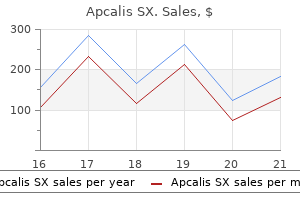
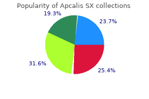
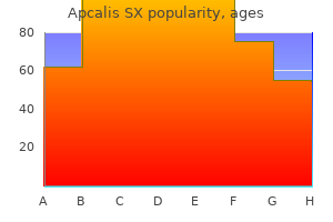
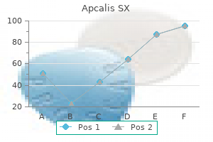
Apcalis SX dosages: 20 mg
Apcalis SX packs: 10 pills, 20 pills, 30 pills, 60 pills, 90 pills

Apcalis sx 20 mg generic free shipping
However erectile dysfunction patanjali medicine buy generic apcalis sx 20 mg on line, if the medical presentation suggests an infectious etiology erectile dysfunction herbs a natural treatment for ed 20 mg apcalis sx for sale, neurocysticercosis, with its scolex as the mural nodule, is the first diagnostic consideration. It is noteworthy that posterior fossa lesions are extra typically cystic (70%) and the uncommon supratentorial lesions are more not often cystic (20%). Approximately 76% appear within the posterior fossa; 9% are supratentorial; 7% seem within the spinal wire; and 5% appear within the brainstem. Pleomorphic xanthoastrocytoma: Fewer than 48% are a cyst with an enhancing mural nodule; 52% are stable; lower than 2% seem within the posterior fossa; and 98% are supratentorial. Only two case reports of spinal cord pleomorphic xanthoastrocytoma exist in the literature. The most typical location is the cerebellum, but when the lesion is supratentorial, it most commonly occurs in the optic nerve or diencephalon (chiasm/hypothalamus, flooring of the third ventricle), thalamus, and infrequently happens within the spinal cord. Ganglioglioma: Approximately 40% are a cyst with an enhancing mural nodule; 60% are solid, and the most typical location is supratentorial, with the temporal lobe as the most common website. The affiliation of optic pathway pilocytic astrocytomas with neurofibromatosis sort 1 is nicely documented. Hemangioblastoma: this extremely vascular lesion with a subpial nodule demonstrates related flow voids. Approximately 75% are sporadic, and 25% are related to von Hippel�Lindau illness. Ganglioglioma: this slow-growth lesion often is associated with a historical past of persistent seizures and most frequently is situated in the temporal lobe, although it may happen all through the cerebrum. Pleomorphic xanthoastrocytoma: this lesion is a rare astrocytoma variant affecting the superficial cerebral cortex and meninges. It typically demonstrates a superficial cortical location of a cystic part with an intensely enhancing nodule abutting the leptomeninges. Glioblastoma multiforme: this lesion is extra generally a heterogeneous, hemorrhagic, and necrotic mass with thick and irregular avidly enhancing components. Neurocysticercosis: In the initial vesicular stage of central nervous system an infection, lesions manifest as cystic parenchymal lesions with an inside nodule (the scolex), with little to no perilesional edema and minimal to no enhancement. Metastasis: Metastasis may be cystic or fairly heterogeneous on account of necrosis, hemorrhage, and liquefaction. More frequent patterns of enhancement include stable, nodular, and ringlike enhancement. Safavi-Abbasi S, Di Rocco F, Chantra K, et al: Posterior cranial fossa gangliogliomas, Skull Base 17(4):253�264, 2007. Heterogeneous T1 sign and diffuse enhancement is noted on sagittal T1 and T1 postcontrast images. Bone destruction, excessive T2 signal, and enhancement recommend the analysis of chordoma. Axial and sagittal thinsection T2 photographs reveal an intradural, prepontine, cystic retroclival lesion hooked up to the dorsal clivus by the osseous stalk/pedicle without proof of clival bony destruction. The lack of diffusion-weighted imaging hyperintensity rules out an epidermoid cyst as a diagnostic consideration. An axial thin-section T2 image demonstrates an intradural, prepontine, solid-appearing retroclival lesion attached to the dorsal clivus by the osseous stalk/pedicle with out evidence of clival bony destruction. The lack of bone destruction suggests the analysis of intradural/ benign chordoma. Clival chordomas usually are symptomatic, T2-hyperintense, enhancing, extradural, regionally invasive lesions demonstrating bone destruction and foci of calcification. Intradural chordomas seem to have a extra favorable prognosis than do extradural clival chordomas. This downside is particularly vexing contemplating the shortage of a extensively accepted gold standard for pathologic differentiation. This state of affairs has led some researchers to suggest the terms "intradural/benign chordoma" or "giant/symptomatic ecchordosis physaliphora" to embody all symptomatic intradural extraosseous physaliphorous lesions. A sagittal T1 postcontrast picture demonstrates a dural-based, avidly enhancing retroclival lesion with a dural tail consistent with a meningioma. Calcification and related hyperostosis was evident on computed tomography pictures (not shown). Ciarpaglini R, Pasquini E, Mazzatenta D, et al: Intradural clival chordoma and ecchordosis physaliphora: a challenging differential diagnosis: case report, Neurosurgery 64(2):E387�E388, 2009. Atlantoaxial separation may result from disruption of the transverse ligament, alar ligament, or tectorial membrane or from fracturesofC1andC2. Atlantoaxialsubluxation is outlined as an anterior shift of the atlaswithrespecttotheaxis. Inthecaseof basilarinvagination/impression,theC1arch maintains a comparatively regular relationship with C2. Trauma is responsible for the vast majority of circumstances of atlantoccipital and atlantoaxial separation. Craniometric Parameters and Measures Description Distance between the inferior tip of the basion to the superior edge of the dens; normally measures <12. Secondary basilar invagination, or basilar impression, is acquired and infrequently is related to situations that result in softening of the skull base. Although platybasia can occur in isolation, it usually coexists with basilar invagination. Several craniometric parameters have been devised to help characterize craniovertebral junction anatomy. Some of the more generally used measurements are listed and depicted in Table 30-1. Nevertheless, a qualitative evaluation is often adequate for characterizing the abnormality. Because basilar invagination and platybasia are findings, not diagnoses, it is important to seek for associated abnormalities to establish a prognosis (Table 30-2). Welcher basal angle Formed by the intersection of lines drawn from the nasion to the tuberculum sella and from the tuberculum sella to the basion; usually <140 degrees. For example, the basiocciput, which is separated from the basisphenoid by the sphenooccipital synchondrosis, is derived from fusion of the primary 4 sclerotomes. Failure of the final sclerotome to fuse leads to condylus tertius, whereas underdevelopment of the sclerotomes leads to condylar or basiocciput hypoplasia. Selected examples of how secondary findings may be helpful are described and depicted in Table 30-3. Imaging performs an essential function in evaluating patients who current with these complications. However, additional findings on the imaging study itself typically are current and might counsel a prognosis or can at least help narrow the differential prognosis. Lateral radiograph of the spine reveals a hypoplastic L1 vertebra with inferior beaking (arrow) and related gibbus deformity (focal kyphosis). Treatment ranges from utility of traction devices to surgical occipitocervical decompression (odontoidectomy and laminectomy) and fusion. Additional cerebral intraparenchymal lesions are also famous in a patient with a known metastatic cervical carcinoma. Note that the upper cervical lesion spans more than three vertebral physique segments and that wire expansion is gentle.
20 mg apcalis sx buy overnight delivery
Differential prognosis A benign cystic mass of the parotid gland erectile dysfunction drugs sales cheap apcalis sx 20 mg with mastercard, corresponding to obstructive or traumatic retention cyst rogaine causes erectile dysfunction discount apcalis sx 20 mg fast delivery, displays low T1 and high T2 signal without post-contrast enhancement. It lies in a plane parallel to the external auditory canal and lateral to the anticipated course of the facial nerve. No discernible tissue characteristics had been seen on postsurgical histopathologic examination. Serpiginous multilocular high T2 signal channels are seen bilaterally throughout the suprahyoid neck areas, together with parapharyngeal, masticator, and parotid space. There is patchy enhancement and enlargement of the best parotid gland with pericapsular and subcutaneous stranding (short arrows). They are often asymptomatic; although early reports of related occipital headache exist, this has not been substantiated [2]. These are normally smaller in size, in the order of some millimeters, and situated throughout the lymphoid tissue. Differential diagnosis Nasopharyngeal mass lesions must be thought-about in the differential analysis. They are well-defined lesions with fluid signal/attenuation and must be separated from strong nasopharyngeal masses. Thyroid most cancers nodal metastases can additionally be cystic and should be thought of within the differential diagnosis. Acutely infected lymph nodes (lymphadenitis or suppurated lymph nodes) can have cystic look, however these are tender to contact and related to erythema, fever, and leukocytosis. Note induration of the subcutaneous tissue (arrows) adjacent to the mass, suitable with suppurative adenitis. Hemangioma is a vascular tumor characterized by fast endothelial proliferation shortly after birth. The lesion is typically absent at start, demonstrates development in early infancy, adopted by a spontaneous resolution in childhood. On the other hand, vascular malformations are structural anomalies which have a traditional progress price and endothelial turnover. These are congenital, have an equal gender incidence, and almost never involute spontaneously. Venous and lymphatic malformations of the pinnacle and neck might present with a mass or facial deformity and should coexist. Both venous and lymphatic malformations sometimes demonstrate a brilliant signal on T2 imaging. For this cause, T2weighted sequences, ideally with fats suppression, are quite helpful in delineating the complete extent of the lesion. Lesions occurring in deep compartment might lead to difficulty in swallowing or chewing, or bleeding. T2-weighted axial photographs show a big, infiltrative, trans-spatial mass with tubular and rounded channels in addition to phleboliths (arrows). Note a heterogeneous appearance on T2-weighted imaging, mild circumferential enhancement and a fluid-fluid stage. Presence of in depth, tubular move voids is suggestive of high-flow vascularity related to these lesions (arrows). A neurogenic tumor, more than likely schwannoma, appears to be a well-defined oval delicate tissue mass with intermediate to hyperintense T2 signal and homogeneous post-contrast enhancement. If larger, it may present presence of curvilinear and serpentine flow-void areas inside it that seem like "pepper. The T1 signal is variable, with some of the hemorrhagic channels exhibiting hyperintense T1 sign, while predominantly lymphatic channels seem hyperintense. It extends as an inverted pyramid from the base of the cranium all the method down to the junction of the posterior stomach of the digastric muscle and the hyoid bone. Apart from fats, it accommodates solely vessels, and no different content material such as mucosa, muscle, lymph nodes, or bones, although ectopic minor salivary glands can occur in this space [1]. Depending on the path in which the parapharyngeal fat is displaced by a mass, the area of origin for that mass could be decided. Parapharyngeal space tumors: another consideration for otalgia and temporomandibular issues. Other symptoms embrace change in voice, abnormal pharyngeal sensation, dysphagia, dyspnea, otalgia, or facial ache [3]. Most lots are slow-growing and present in the grownup age group, peaking within the fifth decade. When large, however, it seems contiguous with the deep lobe of the parotid gland. The pharyngeal mucosal area is displaced medially, whereas the pterygoid muscular tissues within the masticator area are displaced laterally. There is presence of regular fat separating the lateral margin of the mass from the left parotid gland (arrow). Clinical indicators and signs include ache, tenderness, redness, fever, and leukocytosis. Imaging description Acute infections and abscesses of the neck are most commonly odontogenic in origin and happen within the neighborhood of the oral cavity, and they could unfold in some cases from there into the deep neck areas. Acute infections of the salivary glands and lymph nodes (suppurative adenitis) account for the majority of the remaining instances. An abscess occurring inside or within the neighborhood of the thyroid is rare and will elevate the suspicion of an underlying third branchial pouch anomaly, significantly in the pediatric inhabitants [1]. A sinus tract connecting the piriform sinus to the thyroid lobe has been recognized as the trigger of childhood thyroid/perithyroid abscesses, and it may lead to recurrent infections if left untreated [1,2]. However, exams performed through the acute inflammatory part may be false negative [2]. Differential analysis In the right clinical setting a loculated rim enhancing assortment in the neck ought to be recognized as abscess and requires no differential diagnosis. Teaching factors Left-sided neck abscess within the vicinity of the thyroid and acute suppurative thyroiditis in children ought to immediate an investigation for a sinus tract arising from the piriform sinus. A new function for computed tomography in the diagnosis and therapy of pyriform sinus fistula. Revisiting imaging features and the embryologic foundation of third and fourth branchial anomalies. Branchial sinus of the piriform fossa: reappraisal of third and fourth branchial anomalies. Third branchial pouch anomalies account for only 3� 10% of all branchial anomalies. The strong affiliation between a left-sided intra/perithyroid abscess and third branchial pouch sinus is essential to acknowledge, and may lead to a seek for the underlying sinus tract to stop recurrent abscesses.
20 mg apcalis sx with visa
The nurse should understand the distinctive actions of drugs of their pediatric patients to deliver protected and efficient pharmacotherapy effexor xr impotence cheap apcalis sx 20 mg with amex. Absorption: Two main factors have the potential to affect the oral absorption of medicine in pediatric sufferers: increased gastric pH and delayed gastric emptying erectile dysfunction treatment vacuum constriction devices apcalis sx 20 mg buy generic line. Low gastric acid manufacturing may improve the absorption of acid-labile medicine similar to ampicillin and penicillin and sluggish the absorption of weak acids like phenobarbital. Slowed gastric motility in very young kids will keep the drug within the abdomen longer. This will increase the absorption of medicine that are absorbed across the abdomen mucosa, but slow the rate of drug absorption for medicine that depend on the gut for absorption. The rate of bile salt secretion is diminished in untimely infants and neonates, which is able to delay the absorption of lipid-soluble medicine and vitamins. Distribution: Three major elements affecting drug distribution in youngsters are the proportion of water to fat, immature liver operate, and the underdeveloped blood�brain barrier. The higher proportion of water dilutes watersoluble medication such as furosemide (Lasix). The total effect is lower serum drug levels; greater doses could additionally be needed to maintain enough serum levels of the drug. Lipid-soluble medicine that would normally be distributed to fats tend to keep within the blood, raising serum drug ranges. Drugs that normally bind to plasma proteins will now be current as "free" medication in the serum. Drugs and endogenous substances similar to bilirubin compete for the few available protein-binding sites. In some circumstances, previously bound substances or drugs may be displaced into the serum, resulting in potential toxicity. Drugs with high levels of protein binding embody ampicillin, morphine, phenytoin (Dilantin), and propranolol (Inderal). Metabolism is significantly slower in youngsters, resulting in lowered clearance charges and prolonged half-lives for medication extensively metabolized by the liver. To keep away from toxicity, drug doses have to be precisely determined and administered at adequately spaced intervals to enable enough time for metabolic handling of the drug. The enzyme alcohol dehydrogenase is markedly decreased at start, steadily rising till age 5. This enzyme is answerable for detoxifying benzyl alcohol, a preservative present in parenteral drug formulations. Newborns are especially sensitive to the effects of benzyl alcohol and may develop "gasping syndrome," which may lead to respiratory and cardiovascular failure. For medicine to be excreted efficiently, the kidneys will need to have an enough glomerular filtration rate and energetic tubular secretion and reabsorption functions. Young children have immature renal methods with slower renal clearance, leading to accumulation of medication excreted by the kidneys. Although normal pediatric doses could be administered at approximately three to 5 months of age when the toddler is in a position to concentrate urine, serum ranges of nephrotoxic medication similar to gentamicin have to be monitored closely to forestall adverse reactions. The following particular nursing interventions and parental teaching factors are necessary for this age group: the toddler must be held and cuddled while administering drugs and offered a pacifier if the toddler is on fluid restrictions attributable to vomiting or diarrhea. Medications are sometimes administered to infants through droppers into the eyes, ears, nostril, or mouth. Oral medications must be directed to the inside cheek and the child given time to swallow the drug to avoid aspiration. If rectal suppositories are administered, the buttocks ought to be held collectively for five to 10 minutes to forestall expulsion of the drug before absorption has occurred. Some consider that is controversial because the infant may affiliate the nipple with medication and refuse feedings. Unlike adults, infants lack well-developed muscle plenty, so the smallest needle appropriate for the drug ought to be used. The gluteal website is often contraindicated due to potential injury to the sciatic nerve, damage to which can end in permanent incapacity. Because of the lack of decisions for injection sites, the nurse must rotate injection websites from one leg to the subsequent to avoid overuse and to prevent irritation and extreme pain. The nurse responsible for administering pediatric medications should think about the age, weight, and developmental level of the child. For the needs of medicine administration, the pediatric patient is defined as being any age from start to 16 years and weighing less than 50 kg (110 lb). Additionally, kids are categorized as neonates, infants, toddlers, preschoolers, school-age kids, and adolescents. Growth is a term that characterizes the progressive enhance in physical (body) measurement. Development refers to the useful evolution of the bodily, psychomotor, and cognitive capabilities of a residing being. Stages of progress and bodily development often go hand in hand in a predictable sequence, whereas psychomotor and cognitive improvement tend to be extra variable in nature. From delivery by way of adolescence, the growth and development of the pediatric patient mandate changes in pharmacotherapeutics above and past the calculation of doses. Pharmacotherapy of the toddler: Infancy is the interval from birth to 12 months of age. During this time, nursing care and pharmacotherapy are directed towards security of the infant, correct dosing of prescribed drugs, and teaching parents how Pharmacotherapy of the toddler: Toddlerhood ranges from 1 to three years of age. The 2-year-old is ready to metabolize medication at a very speedy rate; therefore, the health care supplier must regulate drug doses accordingly to preserve therapeutic levels. The child begins to explore, wants to attempt new things, and tends to place everything in the mouth. When prescription drugs are provided as flavored elixirs, it is important to train parents that the child not be given entry to the medications. Some of the most typical toxic substances concerned within the publicity of children less than 6 years are analgesics, cough and chilly preparations, topical ointments, and vitamins. Giving long, detailed explanations to the toddler will prolong the procedure and create further nervousness. Short, concrete explanations adopted by instant drug administration are greatest for this age group. Physical comfort within the type of touching, hugging, or verbal reward following drug administration is necessary. Use of the dorsogluteal web site has declined because of the danger of affecting the sciatic nerve. Like the toddler, the preschooler often physically resists medication administration. A brief clarification adopted instantly by medication administration is usually the best method. Uncooperative kids might have to be restrained, and patients over four years of age may require two adults to administer the treatment.
Order apcalis sx 20 mg fast delivery
To take this additional what do erectile dysfunction pills look like 20 mg apcalis sx order visa, I wish to erectile dysfunction drugs on nhs order apcalis sx 20 mg with visa see contrast-enhanced T1 and diffusion-weighted images to help further characterisation. However, the presence of restricted diffusion is suggestive of lymphoma, but not a glioma or metastasis. Pearls Key features to point out with all mind lots are their location, T2 signal depth, enhancement pattern and related oedema. There is an in depth abnormality throughout the distal spinal twine and conus that extends over a size of no less than five vertebral physique heights. It contains focal areas of high and low T2 signal, which can characterize cystic change and haemorrhage, but correlation with T1 pictures is needed. Discussion Intramedullary spinal lesions lie inside the substance of the spinal cord. Ependymomas are extra frequent in adults, whereas astrocytomas are the commonest spinal tumour in children. Bony remodelling is a useful signal that can be utilized to help distinguish between an ependymoma and an astrocytoma. Ependymomas are slow-growing tumours with an insidious onset of signs together with back ache, sensory loss, and bowel and bladder dysfunction. This gradual price of progress is associated with bony remodelling, similar to pedicle erosion, laminar thinning and posterior vertebral physique scalloping, all of which result in widening of the spinal canal. These situations have classical multisystem imaging features, so are seen more typically in the examination than in real life. They are associated with a disproportionately large space of cord oedema with respect to the scale of the lesion. Most intramedullary plenty are both ependymomas (more widespread in adults than children) or astrocytomas (more frequent in youngsters than in adults). There is a focal mass lesion inside the spinal canal on the degree of the conus medullaris. The lesion is separate from the twine, which it displaces anterolaterally to the proper. When compared to the spinal cord, the lesion demonstrates reasonably high T1 signal, and on T2 sequences is isointense. As that is an adult affected person, the more than likely analysis is a spinal metastasis, maybe secondary to melanoma. The differential diagnoses for an intradural extramedullary lesion also embody nerve sheath tumours and meningioma. Discussion Intradural extramedullary lots are positioned exterior of the spinal twine however throughout the dural sac. Neurofibromatosis is a vital multisystem disorder within the context of the exams. If you think you studied this condition, knowledge of the outlined diagnostic criteria and other associated manifestations ought to be used to formulate your administration plan for additional imaging. It will not be potential to distinguish reliably between a neurofibroma and schwannoma as they share related imaging traits. This look occurs when the tumour has both an extradural and an intradural component and is narrowed in the middle as it passes by way of the neural foramen. These features will allow you to distinguish a nerve sheath tumour from metastases and meningiomas. This case tests your capability to mix two diagnostic lists � one for the intradural extramedullary mass and the opposite for a lesion with excessive T1 sign. This state of affairs is comparatively frequent within the viva setting and tests your capacity to process info sensibly when confronted with a number of imaging options. Haemorrhage and fat (lipoma/dermoid) can have a excessive T1 signal however neither improve. Melanin produces a excessive T1 signal and melanoma metastases can improve, due to this fact a melanoma metastasis is a wise proposition on this case. Substances with a excessive signal on T1-weighted imaging embody subacute haemorrhage (methaemoglobin), fats, protein, melanin and distinction. There is extensive, intermediate T1 and T2 signal materials inside the spinal canal. This surrounds the cauda equina at the stage of L3 and L4, and extends superiorly in the posterior facet of the spinal canal to a minimum of the T10 degree and past the higher limit of the provided photographs. On the axial image, the lesion is positioned outdoors the thecal sac, in maintaining with an extradural location. Discussion Intraspinal lots could additionally be extradural (as on this case), intradural extramedullary (Case 43) or intramedullary (Case 42). It is significant to localise precisely a spinal mass to certainly one of these three compartments early in your description of a case, as this can lead you to the correct record of differential diagnoses. In daily practice, there are a quantity of circumstances that require pressing action when diagnosed. Good examples of those include a rigidity pneumothorax, leaking stomach aortic aneurysm, suspected non-accidental harm and spinal cord compression. In the viva, the examiner will wish to see that not only are you capable to diagnose these situations shortly and confidently but additionally that you just recognise the urgency of the scenario. A spinal epidural abscess is a surgical emergency because of the risk of spinal twine compression and subsequent paraplegia. The appearances are attribute and can be differentiated from the opposite diagnoses described in the desk. Failure to clearly reveal this understanding may raise doubts about your ability to practice safely. An extradural abscess normally occurs because of local extension from discitis, vertebral osteomyelitis or via haematogenous unfold from different sources. Plain films could show a loss of intervertebral disc house height, vacuum phenomenon and bony sclerosis of the adjoining vertebral finish plates. You should understand and use the outlined descriptions of disc illness corresponding to bulge, protrusion, extrusion and sequestration. In the presence of spinal trauma or anticoagulation, an extradural haematoma is a crucial additional differential diagnosis to consider. Pearls A diffuse multi-level epidural abnormality with peripheral enhancement is suggestive of epidural abscess. Lung, breast and prostate are the most common tumours to metastasise to the backbone. There is a thick asymmetrical periosteal response involving each tibial diaphyses, the right radial diaphysis and the entire mandible. The differential diagnoses embody infection, malignancy and prostaglandin remedy. To take this additional, I would review the scientific historical past and any related previous imaging. A periosteal reaction is defined as new subperiosteal bone shaped in response to gentle tissue or osseous illness, detectable by radiographs. There is bilateral symmetrical cortical thickening of various density starting from refined periosteal reaction to solid cortical new bone.
Diseases
- Hyperkeratosis palmoplantar localized epidermolytic
- Pancreatic lipomatosis duodenal stenosis
- Fitzsimmons Guilbert syndrome
- Ceroid lipofuscinois, neuronal 1, infantile
- Pleural effusion
- Congenital gastrointestinal disorder
- Antihypertensive drugs antenatal infection
- Mixed M?llerian tumor
Order 20 mg apcalis sx amex
The most well-liked therapy is surgical resection with a transcallosal transforaminal approach [9] impotence lotion 20 mg apcalis sx cheap with visa. However impotence drugs over counter discount apcalis sx 20 mg free shipping, the newer neuroendoscopic technique is related to decreased morbidity and problems [10]. One should bear in mind the possible uncommon locations and issues such as hemorrhage, calcification, rupture, and meningitis. Progressive enlargement or spontaneous regression of a colloid cyst is possible [12]. Given the potential of sudden deterioration and unpredictable outcomes, you will need to promptly talk the discovering of colloid cyst, even when incidental, to the referring physician. Colloid cyst of the third ventricle, hypothalamus, and heart: a dangerous link for sudden dying. Acute hemorrhage in a colloid cyst of the third ventricle: a rare cause of sudden deterioration. A colloid cyst within the fourth ventricle complicated with aseptic meningitis: a case report. Management outcome of the transcallosal, transforaminal strategy to colloid cysts of the anterior third ventricle: an evaluation of 78 circumstances. Long-term outcomes of the neuroendoscopic administration of colloid cysts of the third ventricle: a sequence of 90 circumstances. Note the incidental lipoma of corpus callosum (short black arrows) that ends just posterior to it. There is a subependymal nodule at posterior proper lateral ventricle (short black arrow). The cortical tubers (short white arrows) and subependymal nodule (short black arrow) are seen. En plaque and globular meningiomas are frequently related to hyperostosis of the overlying calvarium. Primary intraosseous cavernous hemangiomas are slowgrowing benign tumors arising from intrinsic vasculature of the bone. Metastatic deposits from prostate most cancers could be osteoblastic and may be related to reactive enhancement and thickening of underlying dura. Metastatic deposits from lymphoma or other cancer could also be multifocal, whereas blastic deposits from prostate could be focal or diffuse. Osteosarcoma of calvarium is a rare primary malignant bone tumor and is typically seen following long-term radiation remedy. Imaging description Meningiomas are frequent benign masses that are easily acknowledged on the premise of their typical dural-based location. About 1�2% of menigiomas arise from extradural areas and pose a diagnostic challenge [1]. They probably come up from multipotent mesenchymal cell precursors, likely as a response to an unidentified stimulus [4]. Soft tissue enhancement may be intra- and/or extracranial, and it could range from delicate dural thickening to sizable masses. Importance Even although biologically benign and slow-growing, the incidence of aggressive options is larger in intraosseous meningiomas (11%) than in intradural meningiomas (2%) [4,5]. Adjuvant radiotherapy may be carried out in patients who present progression of residual mass or development in symptoms [4]. Primary intraosseous meningioma of orbit and anterior cranial fossa: a case report and literature evaluate. Typical scientific state of affairs Sphenoid wings and frontoparietal convexity are the two most common locations for intraosseous meningiomas. Primary extradural meningiomas: a report on 9 circumstances and evaluate of the literature from the era of computerized tomography scanning. Note the intact cortical margins and abrupt transition between the conventional and irregular bone. Areas of increased fibrous content seem to be lytic and hypodense (short black arrows). Rounded high T2 sign within the calvarium probably represents intradiploic vessel (short black arrow). Ninety p.c of craniopharyngiomas exhibit cystic component, areas of calcification, and variable post-contrast enhancement. It expands the hypothalamus and optic chiasm and will prolong into the optic nerves and tracts. Germinoma is morphologically homologous to neoplasm arising in gonads and extragonadal sites. Proliferating Langerhans cell histiocytes kind granulomas inside the skull or infundibulum/hypothalamus area. Imaging description Tumors involving the sella and suprasellar cistern have diverse origin although their medical presentation is very comparable. Suprasellar meningiomas generally arise from diaphragma sellae or tuberculum sellae. The suprasellar meningiomas account for 10% of all the chiasmal tumors [2], and the place of the chiasm related to the tumor determines the pattern of visual loss [3]. Histologically, they include elongated bipolar cells with eosinophilic cytoplasm, arranged in syncytial configuration with whirls. When present, psammoma our bodies (concentrically laminated calcifications) are a distinguishing characteristic. They are generally isointense to cortical gray matter on each T1-weighted and T2-weighted images, however atypical options corresponding to cystic areas or hemorrhage are incessantly seen. On post-contrast examine, homogeneous and intense enhancement is seen, with frequent presence of a dural tail. Macroadenomas are treated by way of an endoscopic transsphenoidal approach, and non-pituitary sellar/suprasellar tumors are handled with transcranial or modified endoscopic approaches that shield the pituitary gland. When coping with a sellar and suprasellar mass, if even a small a part of the traditional pituitary gland is visualized it signifies a nonpituitary tumor, as large macroadenomas almost invariably substitute the whole gland, rendering it invisible on imaging. The significance of early diagnosis and treatment of the meningiomas of the planum sphenoidale and tuberculum sellae: a retrospective research of 105 instances. Importance Suprasellar lots without involvement of sella turcica have traditionally been surgically excised by a transcranial method, whereas pituitary adenomas that stretch to the suprasellar area are treated with endoscopic trans-sphenoidal approaches. For the non-pituitary suprasellar lots, just lately, supraorbital craniotomy [4], a supraorbital endoscopic approach, and endoscopic endonasal prolonged transsphenoidal approaches that protect the normal pituitary gland [5] have been advised. A giant suprasellar meningioma may additionally be embolized earlier than the surgical procedure to minimize blood loss [6]. With the recognition of minimally invasive surgical procedure that may shorten hospital stay, radiologists will be increasingly liable for figuring out the connection of adjacent crucial vascular structures and optic apparatus. Typical clinical scenario Suprasellar meningiomas commonly present with visual disturbance within the type of reduced visual acuity, loss of colour imaginative and prescient, and visual area defects, mostly bitemporal hemianopsia as a outcome of compression of the inferior chiasmatic fibers [2].
20 mg apcalis sx proven
Third branchial equipment lesions may be centered at the superior pole of the thyroid impotence vs impotence apcalis sx 20 mg cheap amex, whereas fourth branchial equipment lesions might lie extra anterior to the thyroid gland erectile dysfunction injections apcalis sx 20 mg buy with amex. Third branchial cleft cysts classically lie within the posterior cervical compartment, posterior to the sternocleidomastoid muscle and common or internal carotid arteries, but additionally they might lie alongside the lower, anterior border sternocleidomastoid muscle. Fourth branchial equipment lesions more typically lie throughout the tracheoesophageal groove. Rare reports have described mediastinal abscesses from fourth branchial equipment origin. A uncommon unilocular lymphatic malformation might have an look just like that of a branchial cleft cyst. Dermoid/epidermoid cyst: Although dermoid and epidermoid cysts might appear to be equivalent to branchial cleft cysts, dermoid cysts might contain fat. Thyroglossal duct cyst: In distinction to third and fourth branchial cleft cysts, thyroglossal duct cysts mostly lie in the midline, from the tongue base to the thyroid gland, and arise from remnants of the thyroid diverticulum. They are associated with the hyoid bone or strap muscle tissue and clinically elevate with swallowing or tongue protrusion. Thymic cyst: Rare remnants of the third branchial pouch, thymic cysts are remnants of the thymopharyngeal duct from the third branchial pouch that courses from the angle of the mandible to the upper mediastinum and usually is associated with the carotid sheath. Parathyroid cysts: the inferior and superior parathyroid glands derive from the third and fourth branchial pouches, respectively, and parathyroid cysts could additionally be located anyplace in affiliation with the thyroid gland. Bronchogenic cysts: Bronchogenic cysts are a developmental anomaly from abnormal budding of the ventral foregut. Midline cervical cleft: A uncommon lesion current at start, a midline cervical cleft is situated within the anterior lower midline neck with a cutaneous ulceration and a sinus tract to the sternum, mandible, or a blind pouch. Suppurative lymphadenitis: Suppurative lymph nodes within the parotid gland or along the cervical lymph node chain may be included in the differential for infected first and second via fourth branchial cleft cysts, respectively. In an identical trend to infected branchial equipment lesions, suppurative lymphadenopathy could develop after another head and neck an infection, beginning as reactive nodes and growing into an intranodal abscess. Cold abscess: A tuberculous or nontuberculous mycobacterial infection could present as cystic-appearing necrotic lymph nodes with out vital surrounding inflammatory change. Necrotic lymph node metastases: Most necrotic lymph node metastases are from head and neck squamous cell carcinomas or papillary thyroid carcinomas and ought to be the first consideration in a cystic neck mass in sufferers older than 40 years. Necrotic squamous cell carcinoma lymph node metastases also could also be considered in younger adults within the setting of human papillomavirus. Infrahyoid Neck Cystic Lesions 283 Postsurgical problems may relate to superior laryngeal, glossopharyngeal, spinal accent, hypoglossal, or facial nerve harm, depending on the lesion sort and site. Nicoucar K, et al: Management of congenital third branchial arch anomalies: a systematic evaluation, Otolaryngol Head Neck Surg 142:21�28, 2010. Lymph Nodes Enlarged lymph nodes of the retropharyngeal area can bulge anteriorly and compress the prestyloid parapharyngeal fats. They could also be solid or cystic appearing, depending on the etiology, measurement, and extent of inside necrosis. In addition to the aforementioned differential prognosis, rhabdomyosarcoma can happen on this location in pediatric patients. Lymphatic and venolymphatic malformations and branchial cleft cysts also can occur right here. It can be divided into two portions, the prestyloid and poststyloid spaces, by the tensor-vascular-styloid fascia running obliquely from the styloid course of and musculature to the tensor veli palatini. Delineation of the space of origin determines the differential diagnosis, which is very important for preoperative surgical planning. These lesions usually have an intermediate signal on T1-weighted images and a excessive sign on T2-weighted images with average homogenous or patchy enhancement. The lesion may be characteristically "bosselated" with slight undulation of the margin. The facial nerve passes just exterior the tunnel, making the tunnel an necessary landmark. Benign pleomorphic adenoma is the commonest solid neoplastic lesion on this space. Most of these lesions come up from the parapharyngeal means of the deep lobe of the parotid gland and protrude into the prestyloid area. Malignant salivary gland tumors such as mucoepidermoid carcinoma or adenoid cystic carcinoma are less widespread. Malignant degeneration of a pleomorphic adenoma can happen and is clinically manifested by a speedy enhance within the measurement of a preexisting adenoma. It may be divided into two parts, the prestyloid house and poststyloid area (also referred to as the carotid space is a few classifications). Differentiating lesions in these two areas is necessary for preoperative surgical planning because the 2 spaces have very distinct differentials. The two areas are divided by a fascia extending from the styloid process and styloid muscles to the tensor veli palatine (the tensor-vascular-styloid fascia). Neurogenic tumors are the most common type of tumors arising throughout the poststyloid compartment, with the majority representing schwannomas, though neurofibromas could also be thought of in patients with neurofibromatosis. Schwannomas on this location show an increased sign on T2-weighted photographs with a variable enhancement pattern and displace the interior carotid artery anteromedially. Neurogenic tumors also could arise from the sympathetic chain, which runs alongside the medial border of the carotid artery. A sympathetic schwannoma often displaces the interior carotid artery anterolaterally. Paragangliomas are benign tumors arising from chromaffin cells in glomus cells derived from the embryonic neural crest. The enlarged nodes at this degree actually reside in the retropharyngeal space and displace the carotid artery laterally. These retropharyngeal nodal lots might mimic lesions within the poststyloid compartment. The presence of distant metastasis is an indication for malignancy, no matter the histologic grade of a paraganglioma. Poststyloid Parapharyngeal Space 291 Neurogenic tumors that arise in the poststyloid house displace the carotid sheath constructions anteriorly. Schwannomas are easily marginated, have low to intermediate intensity on T1-weighted images, and have high signal intensity on T2-weighted photographs with either homogeneous or heterogeneous enhancement. Schwannomas arising from the sympathetic chain tend to push the internal carotid artery laterally. Nodes could additionally be cystic or strong and are medial to the carotid artery at this degree (nodes of Rouvi�re). These lesions nearly all the time can be differentiated based mostly on their attribute appearance and medical data. A ranula is a benign cystic mass that varieties, presumably, as the result of trauma or sublingual or minor salivary gland ductal obstruction. A simple ranula is a thin-walled cyst that occupies the ground of mouth above and medial to the paired mylohyoid muscular tissues and presents clinically as an intraoral mass lesion. A plunging ranula has herniated either via or around the posterior margin of the mylohyoid muscle into the submandibular space. Dermoid cysts are on a spectrum of true dermoid cyst, epidermoid cyst, and teratoid cyst, depending on the forms of tissues found histologically and the embryologic layers represented.
Order apcalis sx 20 mg with amex
If the mix potency lies below the line of additivity erectile dysfunction medication shots apcalis sx 20 mg cheap mastercard, this is ready to counsel a synergistic or supra-additive interplay impotence grounds for divorce in tn cheap 20 mg apcalis sx with visa. If the mixture potency lies above the road of additivity, then this would counsel a subadditive or antagonistic interplay. Other simpler analytical options to the isobolographic strategy have been used in the clinical setting to determine synergy [20,21]. Therefore, in a clinical setting, synergy is fascinating but 291 Section 6: the Management of Neuropathic Pain additivity, or in some instances even only some additive effects, could be adequate to justify a combination strategy so lengthy as there are completely different toxic or antagonistic results. The examine enrolled fifty six patients and confirmed that the combination therapy provided superior pain reduction to the monotherapy with fewer adverse results. Results are in line with the expectations and expertise of many clinicians who successfully mix tricyclic antidepressants in very low doses with alpha-2-delta ligands similar to gabapentin. Another randomized managed trial in contrast patients who acquired gabapentin plus venlafaxine with patients treated with gabapentin plus placebo and confirmed improvement in pain, temper disturbance, and high quality of life with the mixture therapy [26]. Thereby, this study supports both the notion of superiority of a combination therapy of gabapentin with venlafaxine and the clinically relevant knowledge that venlafaxine is often a useful add-on to gabapentin if the latter is of restricted efficacy. The newer alpha-2-delta ligand pregabalin, which shows superior pharmacokinetic properties with regard to bioavailability and period of action, has only been assessed as one arm of a non-inferiority comparability trial with duloxetine [27]. The primary aim of the trial was a head-to-head comparison between duloxetine and pregabalin on this indication with the study design based mostly on a non-inferiority assumption to scale back the number of subjects wanted. However, beside the two monotherapy arms there was additionally a combination arm with duloxetine and gabapentin. The main results of the trial was that duloxetine was non-inferior to pregabalin for the treatment of diabetic peripheral Human mixture studies Combinations between first-line treatments Most of the international therapy guidelines for neuropathic pain agree on a set of first-line therapies. It is, therefore, worthwhile to look first at these trials which combine two of such first-line therapies. In an excellent randomized controlled trial, which intently simulated medical apply, the group around Baron compared topical lidocaine patch in opposition to pregabalin in these two conditions [23]. Responders at week 4 were outlined as those that had a discount of their pain scores by more than 2 points on the numerical rating scale or had reached a mean ache of lower than four on the dimensions. Both medicine were equally efficient in these circumstances with responder charges within the range of 62�65%, however, not surprisingly, there were barely any patients who discontinued in the lidocaine patch arm, while 24% of sufferers discontinued pregabalin because of opposed events. In a second part of the study, non-responders to the one treatment were provided the addition of the opposite treatment, i. This mixture resulted in a larger reduction in ache scores and elevated affected person satisfaction compared to the monotherapy. These results of a wonderful trial recommend very strongly that, if monotherapy with a topical patch fails, the addition of pregabalin may be efficient and vice versa. A secondary consequence was the noninferiority between duloxetine monotherapy and duloxetine plus gabapentin. Adverse results have been comparable with the exception of considerably more insomnia in the duloxetine monotherapy group. The authors conclude that adding duloxetine to gabapentin was efficacious with sufferers experiencing a discount in pain just like that achieved with duloxetine or pregabalin monotherapy. Thereby, the research must be considered inconclusive with regard to the assist for combination remedy. Combination therapies with opioids Most of the current international guidelines regard opioids as second- or even third-line choices for the treatment of neuropathic ache, not because of their efficacy, which is normally good, but because of considerations about long-term safety and overarching issues with the growing use of opioids in our neighborhood. However, a selection of controlled trials have compared monotherapy with a first-line agent to a combination of this first-line agent with an opioid. One of the first and finest trials right here was a randomized controlled trial evaluating the efficacy of gabapentin with the efficacy of sustained-release morphine and their combination [29]. The study design was quite refined, comparing the combination remedy with monotherapy with either medicine, and monotherapy with an active placebo mimicking the side-effect profile of the 2 monotherapies. This enabled all patients to take part in all 4 remedy arms, further decreasing the influence of confounding factors. The total results of the trial was that the best therapy results were achieved by the mixture of gabapentin with sustained-release morphine. Another study has been carried out with pregabalin together with slow-release oxycodone [30]. This was an open-label prospective study carried out by enrolling 409 patients with a wide selection of neuropathic ache states and treated with pregabalin alone, oxycodone slow-release alone, and a combination of each. The combination and oxycodone alone had been superior to pregabalin monotherapy, however the combination resulted in higher enhancements of high quality of life and permitted dose reductions of each brokers with a median reduction of the oxycodone dose by 22% and the pregabalin dose by 51% compared with the monotherapies. The similar group has also reported the long-term statement (12 months) of patients on this mixture with fascinating results [31]; over the yr dose reductions of pregabalin and even more so oxycodone had been noticed and almost 20% of patients dropped out of the research because of full decision of their pain. It is of notice, nevertheless, that an identical quantity withdrew, mainly as a outcome of antagonistic events. The study design is rather unusual as sufferers had been initially started on oxycodone 10 mg per day or placebo. After a week, open-label pregabalin was uptitrated, whereas either oxycodone or placebo was continued. There is, nonetheless, one other adverse trial combining the first-line tricyclic antidepressant nortriptyline with morphine in lumbar radicular ache [33]. Patients had been randomized in 9-week blocks to morphine (15�90 mg), nortriptyline (25�100 mg), the mixture of both and an active placebo group receiving benztropine. This was a randomized doubleblind, placebo-controlled trial in 20 patients over forty nine days. Not solely was the combination the only effective therapy of ache at rest and through motion, however it additionally reduced disability and altered cerebral processing. Overall, most of the well-designed studies with adequate amounts of opioids in the mixture arm suggest that combining first-line remedies for neuropathic ache with an opioid results in superior analgesia with discount of adverse results. Add-on therapies A variety of randomized trials have added an additional proven therapy of neuropathic ache compared with placebo to sufferers on present therapy. Again ache management was higher within the pregabalin mixture remedy teams; nonetheless, in each trials this additionally brought on extra opposed results. Eight percent capsaicin patch has been registered for treatment of localized neuropathic pain. In one other add-on trial, lamotrigine, thought to be relieving neuropathic ache inconsistently, was added to pre-existing remedy (gabapentin, tricyclic antidepressant, or non-opioid analgesic) in sufferers with a broad range of neuropathic pain syndromes [43]. Lamotrigine in doses up to four hundred mg was not superior to placebo on this add-on trial. Other combos A variety of trials have looked at quite uncommon combination therapies, usually combining somewhat unconventional therapies. All three energetic therapies supplied related pain reduction; the onset of analgesia was higher with the mixture remedy. Another potential randomized doubleblind, placebo-controlled trial evaluated the mix of sodium valproate plus glyceryl trinitrate spray vs.
Discount apcalis sx 20 mg otc
More just lately erectile dysfunction medications cost apcalis sx 20 mg buy generic online, P2X4 erectile dysfunction treatment in unani discount apcalis sx 20 mg mastercard, P2X7, and P2Y12 receptors expressed on microglia have emerged as new essential gamers within the etiology of neuropathic ache. Indeed, spinal microglial activation is a crucial pathological process occurring as little as 4 hours following peripheral nerve injury and highly correlated with the release of pro-inflammatory cytokines and ache hypersensitivity [2]. Recent pharmacological, genetic, and behavioral research have indicated that P2X4 receptors are each necessary and sufficient for neuropathic ache. The mechanism by which microglia P2X4 receptor stimulation produces neuropathic ache entails changes in electrophysiological phenotype of lamina I neurons in the dorsal horn of the spinal wire. Hence, P2X4 is considered a core mediator of microglianeuron signaling, and its blockade reverses ache hypersensitivity. Recent evidence signifies that variation within the coding sequence of the gene encoding the P2X7 receptor pore formation affects persistent ache sensitivity in each mice and humans, indicating a brand new technique for individualizing the remedy of chronic pain [17]. P2Y12 receptors additionally activate microglia and contribute to neuropathic pain, with these receptors important to microglia engulfment of myelinated axons in the dorsal horn [18]. Furthermore, inhibition of those receptors utilizing P2Y12 antagonists, or genetic deletion of the P2ry12 gene, alleviates ache hypersensitivity in animal models of neuropathy [19]. The P2Y12 antagonist, clopidogrel, is used clinically for prevention of blood clots, which can expedite screening of therapeutic agents for clinical neuropathic ache. Therefore, the potential of concentrating on the purinergic system to dampen down nociceptive and inflammatory processes in neuropathic pain is extremely promising. Eicosanoids Arachidonic acid, the most important precursor of the eicosanoids, is a polyunsaturated fatty acid found in the cell membrane. These prostaglandins activate primary sensory afferents, and promote nociception within the spinal cord by depolarizing broad dynamic range neurons and blocking glycinergic neuronal inhibition [1]. Whilst agents developed to block leukotriene 79 Section 2: the Condition of Neuropathic Pain synthesis (Zileuton) and signaling (Montelukast and Zafirlukast) have been used clinically to deal with inflammatory ailments, such as bronchial asthma, their effectiveness to deal with neuropathic pain has but to be clinically evaluated. Histamine Histamine [2-(4-imidazolyl)-ethylamine] is an endogenous biogenic amine, which mediates its pleiotropic results via four distinct subtypes of G-proteincoupled receptors, designated H1 to H4, which are differentially expressed in varied cell varieties. Non-mast cell histamine is derived from numerous sources, corresponding to circulating leukocytes and neurons. It is believed that mast cell degranulation in the nerve is triggered by way of mechanical injury to tissue or elevated ranges of adenosine or bradykinin following damage. Histamine, as soon as launched, has the power to sensitize nociceptors, leading to elevated firing rates contributing to pain hypersensitivity [1]. Treatment with the mast cell stabilizer, cromoglycate, prevents the development of nerve injury-induced pain hypersensitivity, an effect in part as a result of decreased histamine launch [27]. Furthermore, native treatment of nerve-injured rats with H1 and H2 histamine receptor antagonists alleviates hyperalgesia. Mild analgesic results of antihistamines have been demonstrated clinically to deal with dysmenorrhea, trigeminal neuralgia, thalamic ache syndrome, and cancer ache, though usually together with opioids [2]. A newer generation of antihistamines, targeting H3 and H4 receptors, are actually available and have been examined in models of neuropathic pain producing conflicting results. The effect of blocking the H4 receptor in models of neuropathic pain can be unclear, with one study exhibiting local remedy with a H4 agonist abolished mechanical hypersensitivity, while others discovered Neurotrophic factors Neurotrophic elements are a family of proteins which may be central to many features of nervous system perform, from differentiation and neuronal survival to synaptogenesis and synaptic plasticity. In addition, neurotrophic elements regulate neuronal plasticity in response to injury or disease, and a few of them have been implicated in neuropathic pain. Nerve progress issue elicits peripheral sensitization by numerous mechanisms, together with modulating the expression of different algesic inflammatory mediators, receptors, and ion channels, and thus its administration to naive animals and wholesome topics induces speedy hyperalgesia [31]. However, whilst the outcomes confirmed promising analgesic effects (that have been simpler than non-steroidal anti-inflammatory drugs), several patients needed to withdraw as a end result of antagonistic events corresponding to abnormal peripheral sensations and progressive worsening of osteoarthritis due to bone necrosis [33]. In response to peripheral nerve injury, P2X4 receptors are upregulated in spinal microglia. Cytokines and chemokines Cytokines are small regulatory proteins which are produced by a wide variety of cells together with leukocytes, beneath both physiological and pathological circumstances. They modulate cell�cell interplay and regulate inflammation and immune responses of their native environment. Chemokines are small chemotactic cytokines, which direct the migration of leukocytes to the site of damage. Thus, the infiltration of immune cells to the spinal twine following nerve damage is considered extremely dependent upon cytokine launch. In basic, the steadiness between pro- and antiinflammatory cytokines following nervous system injury is important as to whether or not a person fully recovers, or develops continual neuropathic ache. Modulation of cytokine signaling by blocking pro-inflammatory cytokines and/or augmenting anti-inflammatory cytokines has shown considerable efficacy in fashions of neuropathy, and below is a discussion of a variety of the key cytokines implicated in neuropathic pain. These medication are also effective in reversing hyperalgesia in a rat joint pain mannequin [45] and nerve injury-induced neuropathic pain in mice [46]. Blocking this pathway can reduce the unfold of neuroinflammation within the spinal cord and attenuate ache hypersensitivity [52]. It is involved in the growth of neuropathic ache by selling infiltration of macrophages and T cells to the site of nerve harm, and the spinal wire [56]. These results have been alongside decreased T cell and macrophage infiltration to the sciatic nerve, and decreased glial activation in the spinal cord [58]. Based on these findings, modulation of those antiinflammatory cytokines warrants significant further investigation as potential treatments of chronic neuropathic pain. Interleukin-4 Interleukin-4, a prototypical anti-inflammatory cytokine, is launched by activated mast cells and some T-cell populations. It inhibits the main proinflammatory cytokines and might suppress macrophage/microglia activation. Chemokines It is well-known that chemokines and their receptors are essential in neuroinflammatory diseases. For example, chemokines have been shown to mediate the migration of leukocytes into sites of irritation within the nervous system. Indeed, proof has accrued that chemokines are concerned in neuronal migration and cell proliferation throughout mind improvement, modulation of synaptic transmission, neurodegenerative illnesses, and ache. This, in flip, contributes to regulation of synaptic plasticity and neuronal excitability in the spinal wire. Overall, these research suggest that launch of fractalkine from neurons, in response to injury or an inflammatory mediator, induces microglial activation and pain facilitation. Conclusion In abstract, preclinical research provide growing evidence that inflammatory mediators play pivotal roles within the pathogenesis of neuropathic pain. Targeting a few of these mediators, notably inhibiting proinflammatory and augmenting anti-inflammatory mediators, must be considered thrilling avenues for the therapy of neuropathic pain. This discrepancy seems to be due to the much larger reductions in proinflammatory cytokines that happen in rodent microglia and macrophages in response to propentofylline, than in the human equivalents [78]. Nevertheless, the continued growth of analgesic medication that modulate neuroinflammation could revolutionize the remedy of neuropathic ache in the close to future. The neuro-immune stability in neuropathic ache: involvement of inflammatory immune cells, immune-like glial cells and cytokines. Cellular localization of kinin B1 receptor in the spinal cord of streptozotocin-diabetic rats with a fluorescent [NalphaBodipy]-des-Arg9-bradykinin. Switching of bradykinin-mediated nociception following partial sciatic nerve injury in mice. Bradykinin produces ache hypersensitivity by potentiating spinal wire glutamatergic synaptic transmission.