

Aciclovir dosages: 800 mg, 400 mg, 200 mg
Aciclovir packs: 30 pills, 60 pills, 90 pills, 120 pills, 180 pills, 270 pills, 360 pills

400 mg aciclovir discount free shipping
The preliminary lesion is a red macule antiviral over the counter medicine 800 mg aciclovir mastercard, generally pruritic antivirus webroot buy aciclovir 400 mg cheap, evolving through papular and vesicular stages and eventually ulcerating, usually with a black eschar (the name "anthrax comes from the Greek word for"coal. The eschar is often surrounded by hanging edema, which normally is more extensive on the head or neck than on the trunk or extremities. Patients U$Ually are afebrile, with mild or no constitutional symptoms, however tender regional Iymphadenopathy, fatigue, fever, or chills could develop (ulceroglandular disease). The lesions heal spontaneously in 1 to 2 weeks in 8096 to 9096 of cutaneous cases, leaving very little scarring. Death from untreated cutaneous infection ranges as much as 20% however is near 0% with applicable early treatment. Inhalation anthrax is biphasic, starting with fever, malaise, myalgia, cough, and chest and stomach pain. Patients later quickly develop acute dyspnea, cyanosis, subcutaneous edema, and a characteristic mediastinal widening with adenopathy. Histopathologic Features Biopsies and cultures must be obtained from the perimeters of vesicles or eschars. Direct contact with the exudate must be averted, however the risk of well being care workers acquiring puhnonary anthrax from this materials is believed to be minimal. Biopsies generally present edema, necrosis, hemorrhage, and scattered diffuse dermallymphocytes more than neutrophils. They may be seen on smears from vesicular:8uid, eschars, or different body fluids. Immunohistochemistry could facilitate more definitive identification of the organisms. Differential Diagnosis Clinical lesions of anthrax may resemble staphylococcal infections, sporotrlchosis, orf, arthropod bites, or tularemia. Spider bites additionally usually tend to be painful Insect bites usually tend to comprise eosinophils. Brucellosls Several species of BruceUa may cause an infection in people (Brucella mellitensis, Brucella suis, Brucella abortus, and Brucella canis). The prognosis of brucellosis is usually made serologically as a outcome of cultures are hazardous and should take 6 weeks to grow. Brucellosis is usually an acute multisystemic influenza-like febrile situation occurring after the ingestion of food or inhalation of the organisms. About 1096 of patients develop nonspecific erythematous macular, morbillifonn, or eczematous eruptions or erythema multiforme, ulcers, bullae, o. The latter exposure typically results in a contact hypersensitivity response to Brucella antigens. Histopathologic Features the histology of skin lesions has seldom been described within the literature. Superficial and deep lymphohistiocytic perivascular dermatitis and septal or lobular panniculitis have been reported. Necrotic foci are frequent, and suppurative granulomatous inflammation with plasma cells may be present. Differential Diagnosis the differential prognosis of similar infections acquired from animals is discussed beneath �Ery5ipeloid. Wild rodents and their fleas are the traditional hosts for plague, and involvement in humans is incidental and has triggered epidemics. It still exists at present, with about a thousand instances and a hundred deaths per 12 months, including a couple of cases in the United States. Mortality decreases to 20% to 60% iftreabnent is began inside 24 hours of the onset of signs. Veterinarians could acquire an infection once they deal with plague abscesses of home cats. A Gram stain of lymph node aspirates or aspirates from different websites might reveal the organisms. Dermatologic manifestations of systemic an infection range from generalized macular erythema to petechiae and purpura. Histologie adjustments within the pores and skin are generally not diagnostic and range based on the sort oflesion biopsied. Differential Diagnosis Yersiniosis can mimic many infectious illnesses, and solely definitive serology or cultures allow a selected prognosis. Culture is feasible but is mostly not available as a routine test in most laboratories. Clinical Features Asymptomatic genital or perianal ulcers are inclined to develop ample friable granulation tissue, spreading in a serpiginous pattern (Table 19-12). Dissemination beyond the genital and inguinal regions to the liver and different organs ultimately may occur in neglected circumstances. Histopathologic Features the ulcerations present appreciable granulation tissue, with asuppurative granulomatous infiltrate of macrophages (histiocytes), plasma cells, and neutrophils. The borders of the ulcers might exhibit acanthosis or pseudoepitheliomatous hyperplasia. Very massive macrophages, up to 20 �m in diameter, could contain the 1- to 2-�m organisms (Donovan bodies), which have a safety-pin appearance when stained with Warthin-Starry or Giemsa stain (bipolar staining surrounded by a vacuole). Smears made from crushed biopsies stained with Wright or Giemsa stain are higher for demonstrating the organisms than tissue sections. Semithin 1-�m sections prepared for electron microscopy and stained with toluidine blue may be used to identify the organisms. Differential Diagnosis Chancroid, syphilis, herpes simplex, and lymphogranuloma venereum also generally produce genital ulcerations (see the discussion that follows for the differential prognosis of chancroid below). Parasitized macrophages are also noted with rhinoscleroma, histoplasmosis, and leishmaniasis, however the staining and tradition characteristics, clinical presentation, and site of the lesions normally easily permit a distinction. Clinical Features the standard presentation is solitary, painful, nonindurated ulcers (soft chancre) on the genitals 2 to 5 days after sexual contact (Table 19-13). Last, the lesions become fibrotic or sclerotic, generally resulting in life-threatening obstruction of the airway. The center zone consists of granulation tissue, swollen endothelial cells, and vessels that may comprise thrombi. On smears, they sometimes line up in a pattern known as a "college of fish" or "railroad tracks. Differential Diagnosis In the early phases, patients have nasal congestion, crusting, and discharge. Later, large deforming tissue proliferation occurs, adopted by indurated scarring. Ulcers of secondary syphilis are clinically less painful and have a tendency to be extra indurated. Syphilis, like several ulcer on mucous membranes or genital skin, also tends to exhibit plasma cells, but the three zones of irritation are inclined to be more distinct in chancroid. Macrophages containing gram-negative and Giemsa- and Warthin-Starry-positive bacilli clumped into collections of 10 to 20 organisms (Donovan bodies) are found. The histologic modifications of lymphogranuloma venereum are nonspecific, and Chlamydia species are troublesome to establish with Giemsa stain. Herpes simplex ulcers are inclined to present the characteristic herpetic cytopathic epithelial changes (nuclear molding, steel-gray nuclei with margination of chromatin), multinucleated epithelial big cell formation, and eosinophilic intranuclear inclusions.
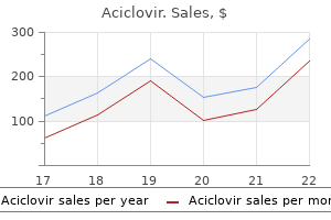
400 mg aciclovir cheap overnight delivery
Keratosis punctata palmaris et plantaris as an expression offocal acantholytic dyskeratosis hiv infection youth purchase aciclovir 800 mg on-line. Piepkorn � Armita Bahrami � Almut Boer-Auer � Artur Zembowicz � Thomas Wiesner the histopathological recognition of the exceptionally various benign antiviral for herpes purchase aciclovir 400 mg on line. The gravitas of the diagnostic drawback rests with the potential lethality ofcutaneous melanoma, the phenomenon of event. The causes for the inordinate issue of melanocytic lesions relate to the profound lack of expertise about their biological nature. The goals ofthis chapter are (1) to present histopathologic and other criteria for the interpretation of melanocytic lesions following a pragmatic strategy to interpretation in order that melanoma is recognif. The photo voltaic lentigo may transition to reticulated seborrheic keratosis, giant cell acanthoma, and lichenoid keratosis. The proliferation of melanocytes as clusters or nests within the epidermis defines the junctional nevus. These cells are closely related to melanocytes however perhaps functionally somewhat altered by the aggregative pattern of progress. Clinical Features the easy lentigo is an everyday, circumscribed macule of 1 to 5 mm diameter and is uniformly light brown, brown, or darkish brown. Differential Diagnosis Simple lentigo requires discrimination from a freckle and photo voltaic lentigo. Simple lentigo differs from freckles (ephelides) because of elongated epidermal rete ridges and an elevated frequency ofbasilar melanocytes. Occasional lesions on the genitalia specifically are notable for a mottled appearance and enormous dimension as much as 50 mm in diameter. Basilar melanoc:ytes (clear cells of Masson) are focally elevated in density on the base of the elongated rete. Accumulating investigative proof has uncovered limitations of the normal diagnostic processes that considerably affect reproducibility and accuracy and that have contributed to putative melanoma overdiagnosis and the notion of a melanoma epidemic. What is presently known in regards to the neoplastic course of because it pertains to the transformation of melanocytes provides the context for describing common and atypical melanocytic nevi and for contemplating the boundaries of histopathology in their prognosis. On the character of melanocytlc proltferatlons By widespread observations, melanocytic nevi and melanoma are related situations, overlapping both clinically and histologically in appearance. Further, the microscopic disa:imination ofmelanoma from nevi may be exceptionally diflicult due to overlapping standards, contributing to limitations of diagnostic reliability however illustrating a organic re1ationship. Acquired nevi and melanomas share a histogenesis in reaction to ultraviolet damage, and mutations in them share, at some oncogenic foci. Differential Dfagnosls the differential analysis consists of melanocytic nevi of mucosa! Histologically, atypical melanocytic lesions and melanoma on mucosae are characterized by progressively greater density of basilar melanoqtes and prominent eytologic atypia. In the case of melanoma, the basilar melanocytes usually reach confiuence along the basal layer in a lentiginous sample, and the melanocytes have steady marked cytologic atypia. Limits of hlstopathology In the diagnosis of melanocytlc proliferations Histological imagery is a highly artifi. The histopathological criteria for nevi and melanoma by frequent observation are steady variables; amongst these are dimension (diameter), architectural disorder, pagetoid scatter, and nuclear atypia. Interpretation requires a subjective setting of threshold along the factors scales. It is this subjective, observer-based threshold setting that accounts, in part, for discordance in melanocytic lesion prognosis,forty three for the putative overdiagnosis of melanoma, and for artifactual inflation of melanoma incidence data. Thereafter follows the crafting of observer-specific nomenclature to mirror and effectuate the two phases. One of the prior research quantifying diagnostic discordance reported a pair-wise settlement fee between experienced pathologists of 50% using a 5-point scale no dysplasia, slight dysplasia, average dysplasia, extreme dysplasia, melanoma), whereas a condensed 3-point scale not dysplastic, dysplastic, melanoma) modestly improved the agreement price to 63%. Epidemiological elements and natural historical past of common and atypical melanocytic nevi Definitions and Nomendature Nevus Cells and the Common Nevus. The nevus cell could be outlined biologically as a lesional cell type that originates from indigenous melanocytes following from environmental stimuli, genetic influences, and different poorly understood processes. The Natural History and Epidemiology of Melanocytic Nevi differs morphologically from native melanocytes and may be acknowledged as a unique cell form by lack of cytoplasmic dendrites, retention of pigment inside their cytoplasms, a finite capacity to proliferate, and tendency to combination as nests or theques inside the epidermis. A lesion comprised of such cells is termed a "nevus" (synonyms: nevocellular nevus, mole), which has a life history evolving through several clinicopathological developmental phases throughout the epidermis and later the dermis as the element cells migrate from epithelium to stroma, lose the capacity to proliferate, and senesce. A coherent definition of the dysplastic nevus and the dysplastic nevus syndrome is difficult due to disputed criteria for his or her recognition, their biological nature, and their medical significance. Any such efforts, nonetheless, are confounded by the reality that the atypical nevus phenotype is a continuous, not a dichotomous, trait, with no pure breakpoints and are thus points on a spectrum. Because few observers would dispute the relationship of enormous numbers of enlarged nevi as a biomarker for melanoma danger, the qualitative definition for the dysplastic nevus devolves to a bigger, extra disordered-irregular, and more variegated variant of a common nevus, however without exact quantitative boundaries. Regarding nomenclature, a Consensus Conference convened by the National Institutes of Health in 1992 deliberated the controversy and advocated that the term "dysplastic nevus" be deserted in favor of "nevus with architectural disorder and cytologic atypia. Current understanding posits that nevi observe a pathway of improvement that will arrest at any stage. Inferences as to their natural historical past derive largely from cross-sectional research, and confirmation by prospective information is incomplete. The population prevalence of nevi correlates with age, race, surroundings, and genetic factors. Few nevi are present early in childhood, a mean estimate being 2-3 in complete number within the first decade of life. Maximum numbers peak within the third decade; thereafter, nevi could disappear with aging. The development of nevi and the associated traits of complete nevi, appearance of nevi, and nevus density correlate with genetic and environmental factors. Numbers, size, and distribution of nevi correlate positively with melanoma threat along steady phenotypic scales, and melanoma is extra prone to occur at anatomical areas with increased nevus counts, particularly at websites such because the torso which might be subject to intermittent solar exposure. Clark and colleagues envisioned a stepwise course of involving "intermediate lesions" that constitute simulators, biomarkers of threat, and potential precursors of melanoma, though development at each stage was presumed uncommon and to require extra genetic mutations. The lack of standardized scientific and histological standards, or minimal criteria, limits the reliability of any such estimates. As nevi and melanoma one to one other is that they reflect pleiotropic (ie, alternative) sequelae of common antecedent stimuli. From the emerging genetic advances, common and atypical nevi could comprise a large, silent disease pool of genetically initiated lesions obtainable for, however not obligated to , additional transformation25�36. This article, as revised from prior editions, gives credence to the biological, in addition to the medical, existence of an intermediate class, regardless of the difficulties in defining it by histological criteria alone33 and regardless of sure inconsistencies with the classical linear development from common nevi to invasive melanoma (vide infra). One of probably the most potent markers for elevated relative danger of melanoma is complete variety of nevi on the individual, no matter clinical atypia. The relative danger is a continuous variable, associated to family history and private historical past of melanoma, whole nevi, total atypical nevi, and different components. A meta-analysis of 46 case-control research previous to 2002, for example, reported cited, the concordance between medical atypia and histological dysplasia is poor. Population-based estimates within the Utah inhabitants was 53% in the original analysis32 and 7% to 32% in a follow-up multiobserver study, the speed varying with observer opinion.
Diseases
- Lower mesodermal defects
- Swyer syndrome
- Multiple pterygium syndrome
- Neuropathy, hereditary sensory, type I
- Fernhoff Blackston Oakley syndrome
- Neuronal intestinal pseudoobstruction
800 mg aciclovir with visa
Attention to the constellation of histologic findings offered in live performance with procurement of clinic::al information (regarding duration of development and macroscopic appearance of the lesion usually allows for passable recognition of trichilemmal carcinoma antiviral otc 400 mg aciclovir discount mastercard. Tr1choblastic cardnoma Scattered stories may be discovered in the literature on trichoblastic carcinomas (also named malignant trichoepithelioma hiv infection via kissing aciclovir 400 mg visa, trichoepitheliocarcinoma, malignant transformation of trichoblastoma, malignant trichoblastoma). In any event, the scientific evolution of such tumors has been uneventful after easy excision. Clinical Features From the few instances reported in the literature, it seems that trichoblastic carcinoma is especially positioned on the face however can affect the trunk and the limbs. It appears in adults as an indurated plaque or ulcerated nodule with fast growth. In some circumstances, trichoblastic carcinoma developed in sufferers with a quantity of familial trichoepitheliomas. Lobular clear cell neoplasm in dermis with central cystic space and central stellate zones of necrosis. Histopathologic Featlres the lesion is often poorly restricted, infiltrating the subcutaneous tissue and presumably the fascia or skeletal muscle. Neoplastic aggregates are composed of germinative follicular cells with focal peripheral palisading. In cases where malignancy is related to the infiltrative sample, the cytological options are those of trichohlastoma. The stroma is plentiful and composed of plump fibroblasts in fibrillary collagen with no or few peritumoral clefting. Lobules and cords of extremely atypical basaloid cells, with focal keratinization, trichohyaline granules, and prominent stroma with numerous plump fibrocytes. Often, these manifest multilineage differentiation alongside multiple appendageal route and subsequently embody components exhibiting a combination of sudoriferous, sebaceous, and pilar traits. It resembles a multifocal superficial trichoepithelioma, changing each hair follicle in a confined phase of pores and skin and featuring an affiliation with hyperplastic sebaceous glands. It likewise may be linear or regionalized, pale or erythematous, plaquelike or papular, and solitary or generalized. Also, clinical information obviously is extremely useful in making the interpretative separation beneath dialogue. Hair follicle nevus Rare lesions in the literature have been described underneath this name. Hair follicle nevi are seen in sufferers of all ages, as nondescript, 1- to 5-mm,:tlesh-colored, smooth-surfaced papules in hair-bearing skin, mostly on the face. Constituent cell populations are additionally totally different in these lesions, with trichoepithelioma featuring far more basaloid elements. It could also be present in association with various other situations, together with vascular abnormalities, ocular changes, cerebral anomalies, and skeletal abnormalities, with none consistent sample of these associated circumstances. The follicular wall may be atrophic, or have bulbous projections, then resembling dilated pore of Winer. The intervening stroma contains a mixture of mesenchymal tissue sorts, including neural, vascular, and adipocytic components. It is unclear whether these parts are integral to the lesion or just symbolize entrapped or metaplastic tissues. Sebaceous gland hyperplasia and senile comedones: a prevalence research in aged hospitalized sufferers. Circumscribed sebaceous gland hyperplasia: autoradiographic and histoplanometric studies. Giant solitary sebaceous gland hyperplasia clinically simulating epidermoid cyst J Cutan PathoL 1988;15:396-398. A papular plaquelike eruption of the face as a end result of naevoid sebaceow gland hyperplasia. Sebaceous hyperplasias, keratoacanthomas, epitheliomas of the face, and cancer of the colon: a new entity Multiple sebac:eow neoplasms of the skin: an association with a number of visceral carcinomas, especially of the colon. Multiple sebaceous adenomas and inside malignant illness: a case report with chromosomal evaluation. Muir-Torre syndrome: reevaluation of the dermatological options and consideration of its relationship to the family most cancers syndrome. Cystic sebaceous tumors as marker lesions for the Muir-Torre syndrome: a histopathologic and molecular genetic study. Molec:ular pathologic: analysis enhances the analysis and administration ofMuir-Torre syndrome and offers insight into its underlying molecular pathogenesis. Adipophilin expression in sebaceous tumors and different cutaneous lesions with clear cell histology: an immunohistochemical research of 117 circumstances. Sebac:eow carcinoma: clinicopathologic features and diagnostic position of immunohistochemistry (including androgen receptor). Eyelid sebaceous carcinoma: clinic:opathologic and multiparametric immunohistochemical analysis that includes adipophilin. Sebaceoma: a distinctive benign neoplasm of adnexal epithelium differentiating toward sebaceous cells. Rippled-pattern sebaceoma: a report of a lesions on the back with a review ofthe literature. Carcinoid-like pattern in sebaceous neoplasms: one other distinctive, previously unrecognized sample in extraoc::ular sebaceous carcinoma and sebac:eoma. Sebocrine adenoma: an adnexal adenoma with sebaceous and apocrine poroma-like differentiation. Bowen disease with sebaceous differentiation: a case report and immunohistochemical evaluation of adipophilin and cytokeratin 1. Nevus sebaceus: a report of 140 instances with special regard to the development of secondary malignant tumors. Metastasizing eccrine porocarcinoma creating in a nevus sebaceus ofJadassohn: report of a case. Eruptive infundibulomas: a distinctive presentation of the tumor of follicular infundibulum. Sebaceous carcinoma of the ocular adnexa: a clinicopathologic research of 104 circumstances, with five-year followup data. Primary sebaceous carcinoma of lacrimal gland: a previously unreported main neoplasm. Sebaceous carcinoma arising in nevus sebaceus of Jadassohn: a clinicopathologic study of five instances. Sentinel lymph node biopsy for sebaceous cell carcinoma and melanoma of the ocular adnen. Comparative examination of androgen receptor reactivity for differential prognosis of sebaceous carcinoma from squamous cell and basal cell carcinoma. Sebaceous carcinoma of meibomian gland origin: the diagnostic importance of pagetoid unfold of neoplastic cells. Sebaceous carcinoma in situ masquerading clinically and histologically as Paget illness of the breast. Sebaceous carcinoma, with particular reference to the histopathologic differential analysis.
800 mg aciclovir purchase otc
The shaft is flattened so it has two surfaces-outer and inner; and two borders-upper and decrease quimioterapia antiviral 200 mg aciclovir cheap amex. This floor is marked by a ridge which is continuous behind with the decrease border of the neck hiv infection time period aciclovir 200 mg buy on line. The thoracolumbar fascia and the lateral fibres of the sacrospinalis muscle are hooked up to the angle. It articulates with the transverse strategy of first thoracic vertebra to type the costotransverse joint. The upper surface is marked by two shallow grooves, separated near the inner border by the scalene tubercle. The higher part of the first digitation of the serratus anterior, just behind the groove for the subclavian artery. For the shaft there are solely two secondary centres, one for the head and the other for the tubercle. These secondary centres seem at puberty and unite with remainder of bone after 20 years. Second Rib Features 1 Anteriorly, the neck is said from medial to lateral aspect to: a. The eighth cervical nerve 3 the anterior groove on the superior floor of the shaft lodges the subclavian vein, and the posterior groove lodges the subclavian artery and the lower trunk of the brachial plexus. The attachment of the costoclavicular ligament at the anterior end behind the subclavius. The insertion of the scalenus medius on the elongated tough space behind the groove for the subclavian artery. Attachments 1 the rough tubercle on the outer floor offers origin to 1� digitations of the serratus anterior muscle. The angle and costal groove are poorly marked within the eleventh rib and are absent within the twelfth rib. Attachments and Relations of the Twelfth Rib 3 the next are attached to the outer surface. Lumbocostal ligament close to the head, extending to the transverse process of first lumbar vertebra. The fascia masking the quadratus lumborum can be connected to this part of the rib. The costodiaphragmatic recess of the pleura is expounded to the medial three-fourths of the costal surface. The eleventh and twelfth ribs ossify from one primary centre for the shaft and one secondary centre for the pinnacle. The medial ends of the costal cartilages of the first seven ribs are hooked up directly to the sternum. The eighth, ninth and tenth cartilages articulate with each other and kind the costal margin. Each cartilage has two surfaces-anterior and posterior; two borders-superior and inferior; and two ends-lateral and medial. Attachments Anterior Surface Posterior Surface 1 the first cartilage provides origin to the sternothyroid muscle. Superior and Inferior Borders 1 Anterior floor of the first costal cartilage articulates with the clavicle and takes part in forming the sternoclavicular joint. The inner oblique muscle is hooked up to the, eighth, ninth and tenth cartilages; and the rectus abdominis to the fifth, sixth and seventh cartilages. Lateral End the lateral finish of every cartilage varieties a major cartilaginous joint with the rib involved. Medial End 1 the primary cartilage forms a main cartilaginous joint with the manubrium. The lowest tapering half forming the purpose of the sword is the xiphoid course of or xiphisternum. It has two surfaces-anterior and posterior; and 4 borders- superior, inferior, and two lateral. The posterior floor is concave and types the anterior boundary of the superior mediastinum. It is marked by the suprasternal notch or jugular notch or interclavicular notch in the median part, and by the clavicular notch on both sides. The inferior border forms a secondary cartilaginous joint with the physique of the sternum. The manubrium makes a slight angle with the physique, convex forwards, called the sternal angle of Louis. Each lateral border forms a main cartilaginous joint with the primary costal cartilage, and current a demifacet for synovial articulation with the upper part of the second costal cartilage. The higher half is said to the left brachiocephalic vein, the brachiocephalic artery, the left common carotid artery and the left subclavian artery. The lateral portions of the surface are related to the corresponding lung and pleura. It is at first cartilaginous, but in the adult it turns into ossified close to its upper end. It is widest close to its lower end reverse the articulation with the fifth costal cartilage. It has two surfaces-anterior and posterior; two lateral borders; and two ends-upper and decrease. It is marked by three ill-defined transverse ridges, indicating the traces of fusion of the 4 small segments referred to as sternebrae. Third and fourth sternebrae ossify from paired centres which seem in fifth and 6th months. The manubriosternal joint is a secondary cartilaginous joint and usually persists throughout life. It is finished in its higher half to prevent injury to arch of aorta which lies behind its decrease half. Partial fusion of the plates might lead to the formation of sternal foramina, bifid xiphoid process, etc. The secondary or compensatory curves are cervical and lumbar, Section 2 Thorax the vertebral column can also be known as the spine, the spinal column, or back bone. It supports the body weight and transmits it to the ground via the lower limbs. The vertebral column is made up of 33 vertebrae: Seven cervical, twelve thoracic, 5 lumbar, 5 sacral and 4 coccygeal. In the thoracic, lumbar and sacral regions, the variety of vertebrae corresponds to the variety of spinal nerves, every nerve mendacity beneath the corresponding vertebra. In the cervical area, there are eight nerves, the higher seven mendacity above the corresponding vertebrae and the eighth below the seventh vertebra.
Aciclovir 800 mg purchase free shipping
Atypical mycohacterial infeaion and sporotrichosis could resemble tularemia clinically and histologically typical timeline hiv infection aciclovir 400 mg purchase free shipping, except that the causative agents are different antiviral classification aciclovir 400 mg with amex, as demonstrated by cultures and particular stains. Prominent Iymphadenitis additionally develops in cat-scratch illness, however the localized pores and skin lesions are usually a lot much less impressive. Cat-Scratch illness the etiologic agent of this illness is now thought to be a gram-negative badllus similar to or similar to that of bacillary angiomatosis. It is dosely associated to Bartonella quintana, the etiologic agent of louse-home trench fever. Afipia felis, an unrelated organism, has also been implicated in cat-scratch disease, but current pondering favors B. Palisading granuloma within the dermis, surrounding a necrotic focus on the web site of the scratch. The necrosis turns into extra in depth in older lymph nodes, and this turns into surrounded by a more granulomatous infiltrate. Differential Diagnosis the primary lesion begins at the website of a cat scratch or bite, mostly in children. The lesions could also be solitary or multiple, often smaller than 5 mm, and so they mostly occur on the hand or forearm. Rarely, numerous morbilliform rashes, different skin eruptions, or inside organ involvement occurs. The diagnosis often turns into suspected when tender regional adenopathy develops a number of weeks later. Involvement of the conjunctiva and preauricular lymphadenopathy is identified as "Parinaud oculoglandular syndrome. Histopathologic Features the palisading granulomas in the pores and skin lesions of patients with cat-scratch illness could also be confused with granuloma annulare, necrobiosis lipoidica, rheumatoid nodules, rheumatic fever nodules, foreign physique granuloma. The suppurative granulomatous irritation in the nodes may be confused with all kinds of infectious illnesses, especially tularemia, brucellosis, mycobacterial infections, infectious mononucleosis. Mononucleosis and lymphomas tend to be bilateral Demonstration of the organisms with Warthin-Starry silver stains is crucial. Lymphocytes and eosinophils may surround the macrophages and multinucleated large cells. Sometimes ulceration or epithelial hyperplasia is current Other skin lesions show only nonspecifi. The cat flea (Ctenocephalides felis) has been implicated as a possible vector in bacillary angiomatosis. The term bacillary angiomatosis" refers to pyogenic granuloma-like vasoproliferative papules and nodules of the pores and skin, which comprise colonies of numerous organisms. Clinical Features Friable grouped nodules or papules resembling pyogenic granulomas or Kaposi sarcoma develop on the pores and skin, sometimes after a history of a cat scratch. Some patients have introduced with subcutaneous nodules, fungating lots, and hyperpigmented indurated plaques. Systemic dissemination might occur (especially within the gastrointestinal tract, or bacillary peliosis of the liver or spleen), which may result in demise. It is essential to distinguish bacillary angiomatosis from Kaposi sarcoma, which may happen in the same immunosuppressed population. A wluable due is that the H&E-stained sections of bacillary angiomatosis often include smudgy amphophilic areas that contain the organisms. Pyogenic granulomas, odd granulation tissue, and different vascular neoplasms could cause some confusion, but these are excluded easily so long as the index of suspicion is high sufficient to do the stain for organisms. The lesions of bacillary angiomatosis have some medical similarity to the nodules of verruga peruana, which are the chronic manifestation of infecti. Infection by this organism is sometimes called "bartonellosis� (even although Bartonella species additionally trigger cat-scratch disease, trench fever, and bacillary angiomatosis). The acute febrile, hemolytic phase of bartonellosis comes about 3 weeks after a chunk by the sandfly Phlebotomus verrucarum and is called Carrion disease or Oroya fever. The verrous nodules occur three to 6 months later and are related histologically to bacillary angiomatosis besides that the micro organism are usually fairly sparse. Some of the patients are on immunosuppressive remedy for lymphomas or renal transplantation. Ulcerated pyogenic granuloma-like nodule with amphophilic: smudgy stroma containing the bacilli. Patients current with furuncular, tender, purple nodules or papules, fluctuant abscesses or sinus tracts, or ulcers, mostly within the groin. Hlstopathologlc Featlres the histology of pores and skin biopsies has been poorly documented in the literature. Most probably, this varies relying tremendously on the type of lesion biopsied, as described underneath �clinical Features. Diffuse infiltrate of macrophages with outstanding calcified Michaelis-Gutmann bodies. Histopathologic Features A diffuse infiltrate of macrophages accommodates fantastic eosinophilic granules in the cytoplasm (von Hansemann cells) and small eccentric nuclei. Partially digested bacteria may be discovered in the phagolysosomes and are seen greatest with electron microscopy. The macrophages of malakoplakia could also be confused with macrophages that are digesting any nonspeciftc material. Parasitized macrophages are also seen with rhinoscleroma, granuloma inguinale, and histoplasmosis. Subsequent unfold via the lymphatics ends in very distinguished inguinal lymphade. Fibrosis of the lymphatics might cause obstruction, leading to genital elephantiasis, rectal strictures, or fibrotic vegetating ulcerations of the pudenda (esthioment, which is Greek for Psittacosis Psittacosis (omithosis) is an an infection of birds attributable to Chlamydia psittaci. Occasionally, it could be transmitted to people by the respiratory route, normally resulting in a fever, pneumonitis, and systemic disease. About 85% of infected humans have a historical past ofcontact with birds, but the birds themselves could also be asymptomatic. The diagnosis is troublesome to make and is confirmed most frequently serologically because the organism is tough to tradition. Clinical Features the severity of sickness varies greatly from mild to severe and will end in dying. Symptoms and signs embody fever, extreme headache, cough, chest pain, pleurisy, pericarditis, myocarditis, epistuis, myalgia, malaise, and hepatosplenomegaly. Cutaneous botryomycosis and staphylococcus aureus: prognosis, management, and a systemic: literature evaluation. Ecthyma gangrenosum: a rare manifestation of Pseudomonas aeruginosa sepsis in a c::ritically ill grownup patient. Staphylococcal scalded pores and skin syndrome in adults: a medical evaluate illustrated with a new case. Staphylococcal scalded skin syndrome in an adult: affect of immune and renal components. Staphylococcal scalded pores and skin syndrome in an especially premature neonate: a case report with a quick evaluation of the literature. Toxic shock syndrome in paediatric thermal injuries: a case collection and systematic literature evaluation.
Syndromes
- Coma
- Dried fruit, served with nuts or sunflower or pumpkin seeds
- Magnesium blood test
- Sleep problems
- Down syndrome (trisomy 21)
- Bronchitis
- A vacuum device can be used to pull blood into the penis. A special rubber band is then used to keep the erection during intercourse.
- The health care provider makes a tiny surgical cut in the groin, then inserts the catheters into a blood vessel and up into the heart.
- Calcium
800 mg aciclovir buy with visa
Warts are more papillomatous than arsenical keratoses antiviral research 400 mg aciclovir generic visa, typically have columns of parakeratosis hiv infection symptoms fever aciclovir 200 mg cheap mastercard, might possess massive keratohyaline lots, and often show dilated capillaries within the uppermost papillary dermis. Bowen disease has "full-thicknessn atypia involving all the layers of the dermis except the stratum corneum, whereas arsenical keratoses rarely demonstrate such features. Additionally, Bowen illness has dyskeratotic keratinocytes and a less cornpact stratum corneum. These lesions are associated with chronic photo voltaic injury, radiation therapy, and arsenic or paraquat exposure. Ss-lsa A case ofvulvar Bowen illness with abnormalities ofchromosome eleven and systemic erythematosus have been desc. Presentation as a cutaneous horn, inside a lesion of porokeratosis, porocarcinoma, sebaceous carcinoma, and atypical fibroxanthoma and related to a concomitant Merkel cell carcinoma have also been described. A pigmented variant has additionally been reported and may simulate melanocytic neoplasms. Terminal hair follicles may defend in opposition to this illness, but vellus follicles are more usually concerned. Immunoperoxidase staining for p27 and Ki-67 have proven usefulness in separating Bowen disease from actinic keratoses. However, the atypia is usually not of full thickness, epidermal modifications consists of"knuckling" somewhat than a psoriasiform sample, and koilocytic cells may be current. Keratinocytes may be multinucleated and are slightly eosinophilic is a few sections. However, these tumors are smaller and have often been completely eliminated by the biopsy. The presence ofcytokeratin 10 in Bowen illness however not bowenoid actinic keratoses has been reported. The dermis is irregularly acanthotic with occasional psoriasiform changes and reveals a loss of polarity. Frank koilocytosis is wicommon401; nonetheless, small basophilic our bodies with a surrounding halo could also be current within the stratum granulosum and stratum comewn. The presence of an intraepithelial unfold of atypical keratinocytes was initially described by Borst in 1904 and Jadassohn 22 years later. Montgomery first coined the time period "intraepithelial epithelioma of Borst-Jadassohn� in 1929. Some authors imagine that virtually all instances constitute infected seborrheic keratoses, squamous cell carcinomas, hidroacanthoma simplex, Bowen illness, epidermal nevi, or clear cell acanthomas. These nests proliferate in a microscopic sample often known as acervate (growing in clusters or heaps). They are commonest on the decrease extremity however can also be present on the top and neck. In the dermis are nests of keratinocytes with slight atypia proliferating in an �acervateH pattem. The papillary dermis might demonstrate mild thinning and a superficial perivascular lymphocytic infiltrate. Paget illness and melanoma in situ symbolize a proliferation of single cells or small nests somewhat than the bigger ones present in intraepithelial epithelioma. They were initially described in 1889 by Sir Jonathan Hutchinson as a crateriform ulcer of the face" and at present are believed to characterize a low-grade squamous cell carcinoma. This disorder have to be differentiated from the generalized eruptive keratoacanthomas of Grzybowski. Another situation, a number of persistent keratoacanthomas, occurs sporadically and consists of slow-healing tumors. Two syndromes, the Muir-Torre syndrome and xeroderma pigmentosum, show an elevated incidence of tumor formation. The subsequent stage, the mature kind, demonstrates a dome- or berry-shaped nodule with a central crater containing a plug of compact keratin. Over weeks to months, they turn out to be progressively extra depressed and eventuate into a scar with variable atrophy. Subungual keratoacanthomas are painful, persistent lesions that arise beneath the nails, often on the fingers. The thumb and fifth finger are most commonly involved, and bony destruction might take place. Mucosal keratoacanthomas could develop in the mouth and on different mucous membranes, including the bulbar conjunctiva, nasal mucosa, and genitalia (Table 26-24). Adjacent epithelium reveals acanthosis, hypergranulosis, and untimely cornification. The overlying dermis develops "lips" (collarettes) surrounding the crateriform plug that has expanded and consists of parakeratotic material. Mitotic activity and atypical mitotic figures are extra common, notably on the edges ofthe epithelial proliferation. Intraepidermal neuuophilic and eosinophilic microabscesses are current, as are horn pearls. The stroma may be granulomatous if epithelium has ruptured into the dermis and is being resorbed. Atypical eccrine sweat duct hyperplasia may be current Melanocyte proliferation with elevated dendricity has been described in proliferative epithelium. The epithelial lining can be benign without the standard eosinophilic changes seen in keratoacanthomas. The "lips" or buttressing commonly present in keratoacanthomas shall be absent from these lesions, as will the eosinophilic features ofthe proliferating epithelium, the neutrophilic microabscesses, and the infiltrating eosinophils. It seems doubtless that involvement of vessel walls by tumor cells is extra of a passive phenomenon dictated by the proximity oflarge vessels than one of malignant invasion. There has been appreciable discussion concerning the malignant potential of keratoacanthomas,442. This turnor can also be termed "basal cell epithelioma," "basalioma," and "rodent ulcer" and was initially described by Jacob in 1824. Persons with blue or green eyes, simple freckling, blond or purple hair, and with vital outdoor exposure are at elevated danger for these tumors. Atypical eccrine ductal hyperplasia, intraepidermal microabscesses, tissue eosinophilia, and intraepithelial elastic fibers are extra commonly found in keratoacanthomas than squamous cell carcinomas! These may be little more than coincidence, however in some cases the cutaneous malignancy could have arisen from extended irritation and inflammation. Warts, porokeratomas, varicella scars, neurofibromas, lesions oflupus vulgaris, tattoos, leishmania scars, nevi sebaceus, linear epidermal nevi, seborrheic keratoses, nevomelanocytic nevi, condyloma acuminata, main melanomas, metastatic melanoma deposits, port wine stains, infundibular cysts, hemangiomas, pilomatricomas, atypical fibroxanthomas, melanomas in situ, osteosarcomas, granular cell-type fibrous papules, trichoepitheliomas, blue nevi, indeterminate cell histiocytosis, and leukemia cutis have all been associated with basal cell carcinomas. These modifications might symbolize proliferating epidermal basal cells or follicle induction by underlying stroma.
Discount aciclovir 400 mg online
Students during lectures assist their head (for sleeping) with their hands with flexed elbows hiv infection no fever aciclovir 400 mg with mastercard. Because of this purpose even individuals who have lost terminal flexion or extension after a fracture/trauma are able to antiviral supplements for hpv cheap aciclovir 800 mg on-line accomplish these personal tasks with out much issues. The two bones are additionally related by the oblique cord which extends from the tuberosity of the radius to the tuberosity of the ulna. It is hooked up to the margins of the radial notch of the ulna, and is steady with the capsule of the elbow joint above � the quadrate ligament extends from the neck of the radius to the decrease margin of the radial notch of the ulna Anastomoses around the lateral aspect of the elbow joint Musculocutaneous, median, and radial nerves Supination and pronation Inferior radioulnar joint Pivot sort of synovial joint � Head of ulna � Ulnar notch of radius � the capsule surrounds the joint. It transmits forces (including weight) applied to the radius (through the hand) to the ulna. In a semiflexed elbow, the palm is turned upwards in supination, and downwards in pronation (kings pronate, beggars supinate). As radius with the hand comes medially throughout the decrease part of ulna, the interosseous membrane is spiralised. As a results of this motion, the axis (defined above) is displaced laterally in pronation, and medially in supination. Morphologically, pronation and supination had been advanced for selecting up meals and taking it to the mouth. Around 50� of supination and 50� of pronation are generally required to carry out most of the routine actions. It is aided by the pronator teres when the motion is speedy and against resistance. This will expose the superior and inferior radioulnar joints together with the interosseous membrane. Lastly reduce via the capsule of inferior radioulnar joint to locate the intra-articular fibrocartilaginous disc of the joint. It begins above from the base of the styloid process of the ulna and the anterior margin of the articular disc, runs downwards and laterally, and is attached to the lunate and triquetral bones. Relations 1 Scaphoid 2 Lunate three Triquetral bones Ligaments 1 the articular capsule surrounds the joint. It is attached above to the decrease ends of the radius and ulna, and under to the proximal row of carpal bones. A protrusion of synovial membrane, called the recessus sacciformis, lies in entrance of the styloid process of the ulna and in entrance of the articular disc. It is bounded inferiorly by a small meniscus projecting inwards from the ulnar collateral ligament between the styloid course of and the triquetral bone. It begins above from the anterior margin of the decrease end of the radius and its styloid process, runs downwards and medially, and is connected below to the anterior surfaces of the scaphoid, the lunate and triquetral bones. However, it nonetheless behaves as a practical unit in all actions of the wrist joint. In addition to the congruency and the shape of the articular surfaces of radius and carpal bones, the size of the ulna can also have an result on the amount of motion available at the wrist joint. In the ulnar negative variance, the distal finish of ulna is shorter than the radius and vice versa in ulnar optimistic variance. Abductor pollicis longus and extensor pollicis brevis 4 Adduction (ulnar deviation): It happens mainly at the wrist joint. Extensor carpi ulnaris 5 Circumduction: the vary of flexion is more than that of extension. Similarly, the vary of adduction is larger than abduction (due to the shorter styloid process of ulna). The midcarpal joint permits actions between the 2 rows of the carpus as already described with the wrist joint. Blood Supply Radial vessels provide blood to the synovial membrane and capsule of the joint. Nerve Supply First digital branch of median nerve supplies the capsule of the joint. Movements at this joint are, due to this fact, much more free than at some other corresponding joint. Type Saddle variety of synovial joint (because the articular surfaces are concavoconvex). The articulating floor of trapezium is concave in the sagittal aircraft and convex in the frontal plane. Flexion and extension of the thumb take place in the aircraft of the palm, and abduction and adduction at right angles to the plane of the palm. Flexion is related to medial rotation, and extension with lateral rotation on the joint. Opposition is unique to human beings and is certainly one of the most necessary actions of the hand considering that this movement is used in virtually all kinds of gripping actions. The adductor pollicis and the flexor pollicis longus exert stress on the opposed fingers. The numerous palmar ligaments of the metacarpophalangeal joints are joined to each other by the deep transverse metacarpal ligament. Each runs downwards and forwards from the pinnacle of the metacarpal bone to the base of the phalanx. This is the joint of the thumb and a wide variety of functionally useful actions take place right here. For their dissection, remove all of the muscular tissues and tendons from the anterior and posterior aspects of any two metacarpophalangeal joints. The proximal muscles of upper limb are provided by proximal nerve roots forming brachial plexus and distal muscles by the distal or lower nerve roots. In shoulder, abduction is finished by muscles provided by C5 spinal phase and adduction by muscular tissues innervated by C6, C7 spinal segments. Supination is caused by muscle innervated by C6 spinal segment even pronation is finished via C6 spinal segment. The interphalangeal joints also are flexed and prolonged by similar spinal segments, i. There was swelling and a bend simply proximal to wrist with lateral deviation of the hand. The backward bend just proximal to the wrist is as a outcome of of the pull of extensor muscular tissues on the distal segment of radius. A review of the surface and intra-articular anatomy of the wrist, the approach of building a secure portal and the particular uses of the radiocarpal, metacarpal and special-use portals. Movements at metacarpophalangeal joint of center finger with the muscles answerable for them. Which of the next muscle tissue is equipped by two nerves with completely different root values Which of the next muscle tissue is flexor, adductor and medial rotator of shoulder joint At its termination, the axillary artery, along with the accompanying nerves, varieties a prominence which lies behind another projection brought on by the biceps and coracobrachialis.
Purchase 400 mg aciclovir otc
The superior and inferior notches of adjoining vertebrae be a part of to type Thoracic Vertebrae Identification the thoracic vertebrae are identified by the presence of costal sides on the sides of the vertebral bodies antiviral herpes zoster trusted aciclovir 800 mg. There are 12 thoracic vertebrae kale anti viral buy cheap aciclovir 200 mg online, out of which the second to eighth are typical, and the remaining 5 (first, ninth, tenth, eleventh and twelfth) are atypical. Section 2 Thorax the intervertebral foramina which give passage to the dorsal and ventral rami of the spinal nerves rising from the spinal cord. The superior costal demifacet is larger and placed on the higher border of the physique close to the pedicle. The inferior costal demifacet is smaller and placed on the decrease border in front of the inferior vertebral notch. The superior vertebral notch is shallow, whereas the inferior vertebral notch is deep and conspicuous. The superior articular processes project upwards from the junction of the pedicles and laminae. The transverse processes are giant, and are directed laterally and backwards from the junction of the pedicles and laminae. The anterior surface of each process bears a aspect near its tip, for articulation with the tubercle of the corresponding rib. In the higher six vertebrae, the costal sides on the transverse processes are concave, and face forwards and laterally. In the final two vertebrae, the articular sides are absent (see costotransverse joints below). They also give attachment to several muscular tissues together with the trapezius, the rhomboids, the latissimus dorsi, the serratus posterior superior and the serratus posterior inferior, and tons of deep muscles of the again. First Thoracic Vertebra 1 the body of this vertebra resembles that of a cervical vertebra. Ninth Thoracic Vertebra the ninth thoracic vertebra resembles a typical thoracic vertebra besides that the body has solely the superior costal demifacets. However, the physique bears a single costal aspect on both sides, which lies more on the decrease part of the pedicle than on the physique. These are everted and are directed laterally, but the superior articular sides are thoracic in kind. The three major centres-one for the centrum and one for every half of the neural arch, appear during eighth to ninth weeks of fetal life. At start, the vertebra consists of three parts, the centrum and two halves of the neural arch. Sometimes the physique ossifies from two primary centres, and if one centre fails to develop, one half, proper or left of the body is lacking. This results in a hemivertebra and lateral bend in the vertebral column or scoliosis. However, after the second decade of life, degenerative adjustments set in leading to weakness of the annulus fibrosus. When such a disc is subjected to strain, the annulus fibrosus may rupture leading to prolapse of the nucleus pulposus. In addition to prolapse of the nucleus pulposus, inside derangements of the disc can also take place. The prolapsed nucleus pulposus presses upon adjoining nerve roots and gives rise to ache that radiates alongside the distribution of the nerve. It is also widespread within the decrease cervical region from fifth to seventh cervical vertebrae. The superior costotransverse ligament has two laminae which prolong from the crest on the neck of the rib to the transverse means of the vertebra above. The inferior costotransverse ligament passes from the posterior floor of the neck to the transverse means of its personal vertebra. The lateral costotransverse ligament connects the lateral non-articular a half of the tubercle to the tip of the transverse strategy of its personal vertebra. Rotation of rib-neck backwards causes elevation of second to sixth ribs with transferring forwards and upwards of the sternum. The articular surfaces of the seventh to tenth ribs are flat, allowing up and down gliding actions or bucket-handle actions of the decrease ribs. It permits slight actions of the physique of the sternum on the manubrium during respiration. This ligament is hooked up to the ridge on the pinnacle of the rib and to the intervertebral disc. Other ligaments of the joint embrace a capsular ligament and a triradiate ligament. Each joint has a single cavity besides within the second joint where the cavity is divided in two parts. Interchondral Joints the fifth to ninth costal cartilages articulate with each other by synovial joints. The interrupted strains indicate the place of the rib in inspiration Adjoining vertebrae (Th 5 and Th 6) are related to each other at three joints. There is a median joint between the vertebral our bodies, and two joints-one on the right side and one on the left side-between the articular processes. The joint between the vertebral our bodies is a symphysis (secondary cartilaginous joint). The surfaces of the vertebral our bodies are lined by skinny layers of hyaline cartilage. Section Chondrosternal Joints 2 Each rib is steady anteriorly with its cartilage, to kind a main cartilaginous joint. These are fibrocartilaginous discs which intervene between the bodies of adjoining vertebrae, and bind them collectively. The thickness of the disc varies in several regions of the vertebral column, and in several elements of the identical disc. The discs are thinnest in the higher thoracic area, and thickest in the lumbar region. The contribution is greater in the cervical and lumbar regions than in the thoracic region. It is made up of a narrower outer zone of collagenous fibres and a wider inside zone of fibrocartilage. Functions Movements between adjacent vertebrae occur concurrently at all the joints connecting them. However, when the actions between several vertebrae are added together the total vary of movement becomes considerable. The movements are these of flexion, extension, lateral flexion and a certain amount of rotation. This is influenced by the thickness and suppleness of the intervertebral discs and by the orientation of the articular aspects. Flexion and extension occur freely within the cervical and lumbar areas, but not within the thoracic area.
Aciclovir 400 mg order with amex
Chagas disease in Latin America: an epidemiological replace based mostly on 2010 estimates hiv new infection rates discount aciclovir 400 mg with amex. In Gutierrez Y hiv infection rates nigeria 400 mg aciclovir order with amex, ed Diag- nostic Pathology of Parasitic Infections with Clinical Correlations. Enzyme-linked immunosorbent assay for serological prognosis of Chagas illness employing a Trypanosoma cruzi recombinant antigen that consists of four completely different peptides. Correlation oflatent toxoplasmosis with particular illness burden in a set of 88 countries. Cutaneous acquired toxoplasmosis in a toddler: a case report and review of the literature. Disseminated cutaneous acanthamebiasis: a case report and review of the literature. Cutaneous protothecosis and different extraordinary �aquatic-borne" cutaneous infections. Disseminated cutaneous protothecosis in an immunocompromised host: a case report and literature evaluation. Papular protothecosis of the chest: immunologic evaluation and remedy with a mix of oral tetracycline and topical amphotericin B. Pathology of microsporidiosis: rising parasitic infections in sufferers with acquired immunodeficiency syndrome. Most helminths related to the pores and skin that make the most of people as a definitive host (ie, the host during which maturation and sexual copy take place) are filarial nematodes. There are several inherent challenges with figuring out helminths in tissue specimens, especially as regards to (I) situation of the specimen (if the worm is dead and degrading, sure morphologic options possibly be tough to discern), (2) the angle of the minimize (oblique or tangential cuts could alter parasite morphology), and (3) helminths within the pores and skin are sometimes rare or uncommon zoonotic species that might not be acquainted to the microscopist. When suspect helminths are observed in biopsy specimens, such specimens may be despatched to a parasitologist for consultation. The following morphological standards and terminology may be helpful in figuring out helminths in tissue specimens: 1 1. With most nematodes, the infectious stage is the L3 (sometimes referred to as a filariform larva). Externally, most nematodes have an analogous morphology, being long and slender and lacking true segmentation. However, nematodes vary considerably in the form of their inside organs and organ systems, and the shape and association of inside constructions is taxonomically and diagnostically important. Occasionally, nematodes may have external projections that might be diagnostically helpful, corresponding to bosses (Loa loa), cuticular ridges (Onchocerca and Dirofilaria), and alae (Toxocara and Lagochilascaris). Between the cuticle and the musculature is a skinny layer of cells referred to as the hypodermis. The muscles mendacity underneath the hypodermis are somatic and include a basal contractile portion and a cytoplasmic noncontractile portion. Muscle cells may also be described when it comes to their number, being numerous per quadrant (polymyarian) or fewer per quadrant (meromyarian). The digestive system of nematodes is a relatively straight tube that runs from the mouth (stoma) to the anus. Often, the morphology of the esophagus and intestine (especially almost about the quantity and arrangement of intestinal cells and their nuclear arrangement) could be diagnostically essential. The reproductive tract of nematodes can additionally be highly variable, but with most parasitic species the male has a single testis and the feminine has paired uterine tubes. Often, eggs or microfilariae may be noticed in utero and may be diagnostically helpful. The cestodes (tapeworms) are segmented worms that as adults reside in the intestinal tract of the definitive host. As such, cestodes observed in extraintestinal tissue specimens (including all these in skin) are larval. Cestodes have advanced life cycles involving a quantity of hosts, and people manifesting in humans as cutaneous larvae are often zoonotic in origin. Calcareous corpuscles are a hallmark of cestode morphology, and their presence in helminth tissue is pathognomonic for a cestode an infection. Larval cyclophyllidean cestodes (eg, T~nia) have 1 or more buildings generally recognized as a protoscolex, which serves because the precursor of the scolex (head) of the grownup worm and consists of 4 suckers, 1 or more rings of refractile booklets, and an internal spiral canal. The number of protoscole<:es and the quantity and arrangement of booklets could be diagnostically helpful 3. The trematodes (flukes) are a bunch ofparasitic flatworms which have complex life cycles involving a number of hosts. All species parasitic to humans require a freshwater or terrestrial snail as an intermediate host Skin involvement with trematodes normally includes 1 of3 scenarios: (1) a papular erythematous rash brought on by the penetrating cercariae of avian sc. Schlstosoma species have particular person female and male worms but function as a single entity with the feminine residing in a gynecophoral canal along the physique of the male. Beneath the tegument is a layer ofcircular muscle followed by a layer of longitudinal muscle. Between the musculature and the organ techniques is the parenchyma, containing vitelline glands and interstitial fibers. Although the worms are hermaphroditic, sexual copy between 2 worms is optimal. In sexually mature adults, eggs may be noticed in utero, which may help with the analysis, even in histologic sections. CiniC<11I Features Onchocercal nodules (onchocercomata) are discrete, soft, and palpable, as a lot as 5 cm throughout in measurement, and are sometimes positioned close to bony prominences, though they may be found anywhere on the body. Endemic onchocercal dermatitis is intensely itchy; predominantly papular and lichenoid; and if extreme and witreated, could lead to cachexia and demise from intercurrent infection. Lesions are most frequently generalized; less regularly, particularly in Yemen and the savannah areas of Africa. S Depending on their length, the lesions can be spottily depigmented (leopard skin�), scaly and atrophic ("lizard skin"), or thickened and hyperkeratotic ("elephant pores and skin"). In superior illness, sufferers can also have lymphedema of the groin ("hanging groin"). Onchocerciasis acquired by vacationers to endemic areas is seen as a lot as 2 years later as an itchy rash with marked peripheral eosinophilia, sometimes affecting 1 extremity only, as in sowda. In cross-section, the females measure as a lot as four hundred �m in diameter, malting them one of the largest human parasitic filariae nematodes; males are markedly smaller at 200 iim. Gravid feminine worms present prominent paired uteri containing maturing microfilariae; the reproductive tubes of the males might present spermatowa. Musculature is decreased, with just a few cells per quadrant Lateral chords are short however conspicuous. The cuticle contains transverse cuticular ridges, that are finest visualized in lateral cuts somewhat than cross-sections. Onchocerciasis is a persistent dermatitis that may trigger extreme itching and disfiguring skin lesions; ocular involvement can lead to blindness. Most human infections are associated with Onchocerca volvulus, which is endemic to tropical Africa.
Purchase aciclovir 200 mg on-line
The anterior border of the left lung shows a wide cardiac notch under the extent of the fourth costal cartilage hiv infection by needle stick cheap 200 mg aciclovir free shipping. It extends from the extent of the seventh cervical backbone to the tenth thoracic spine hiv infection condom buy generic aciclovir 800 mg online. The medial floor is split right into a posterior or vertebral part, and an anterior or mediastinal part. The mediastinal part is expounded to the mediastinal septum, and exhibits a cardiac impression, the hilum and numerous other impressions which differ on the two sides. Various relations of the mediastinal surfaces of the 2 lungs are listed in Table 16. The right lung is split into three lobes (upper, middle and lower) by two fissures (oblique and horizontal). The indirect fissure cuts into the whole thickness of the lung, besides at the hilum. It passes obliquely downwards and forwards, crossing the posterior border about 6 cm under the apex and the inferior border about 5 cm from the median airplane. Due to the oblique airplane of the fissure, the lower lobe is more posterior and the higher and center lobes more anterior. In the right lung, the horizontal fissure passes from the anterior border up to the indirect fissure and separates a wedge-shaped middle lobe from the higher lobe. The fissure runs horizontally at the degree of the fourth costal cartilage and meets the indirect fissure in the midaxillary line. The tongue-shaped projection of the left lung below the cardiac notch is called the lingula. The lungs expand maximally within the inferior direction as a outcome of movements of the thoracic wall and diaphragm are maximal in path of the bottom of the lung. The presence of the indirect fissure of every lung permits a extra uniform growth of the whole lung. Surface Marking of the Lung Surface marking of lung is same as that of visceral pleura described in Chapter 15. Left ventricle, left auricle, infundibulum and adjoining part of the proper ventricle 2. Anterior pulmonary plexus, lymph nodes and lymph vessels in the anterior and inferior components. Single bronchus with bronchial vessels and posterior pulmonary plexus alongside its posterior wall. A point on the anterior border of the best lung at the stage of the fourth costal cartilage. Posterior pulmonary plexus 2 On left aspect: Descending thoracic aorta Superior 1 On proper facet: Terminal a part of azygos vein 2 On left facet: Arch of the aorta. Differences between the Right and Left Lungs Root of the Lung Root of the lung is a short, broad pedicle which connects the medial surface of the lung to the mediastinum. It is shaped by buildings which either enter or come out of the lung at the hilum (Latin depression). The roots of the lungs lie reverse the bodies of the fifth, sixth and seventh thoracic vertebrae. Deoxygenated blood is dropped at the lungs by the 2 pulmonary arteries and oxygenated blood is returned to the guts by the four pulmonary veins. The greater part of the venous blood from the lungs is drained by the pulmonary veins. The vessels move around the borders of the lung and margins of the fissures to attain the hilum. The superficial vessels have numerous valves and the deep vessels have just a few valves or no valves at all. Nerve Supply 2 Sympathetic nerves are derived from second to fifth sympathetic ganglia. That is how sympathomimetic medication, like adrenaline, cause bronchodilatation and relieve signs of bronchial bronchial asthma. Sensory fibres are answerable for the stretch reflex of the lungs, and for the cough reflex. The trachea divides on the level of the lower border of the fourth thoracic vertebra into two primary principal bronchi, one for every lung. Inhaled particles or overseas bodies, subsequently, are probably to cross extra regularly to the best lung, with the result that infections are more widespread on the right aspect than on the left. Right bronchus makes an angle of 25� with tracheal bifurcation, while left bronchus makes an angle of 45� with the trachea. Each principal bronchus enters the lung through the hilum, and divides into secondary lobar bronchi, one for every lobe of the lungs. Thus there are three lobar bronchi on the right aspect, and solely two on the left side. Each lobar bronchus divides into tertiary or segmental bronchi, one for every bronchopulmonary segment; that are 10 on the proper facet and 10 on the left facet. The segmental bronchi divide repeatedly to kind very small branches known as terminal bronchioles. Each respiratory bronchiole aerates a small part of the lung known as a pulmonary unit. Apical Posterior Anterior Lateral Medial Superior Medial basal Anterior basal Lateral basal Posterior basal B. Lower Bronchopulmonary segments are well-defined anatomical segments aerated by tertiary/segmental bronchus. These are pyramidal in form with apex directed in path of hilum and base directed in the path of periphery having their very own arterial provide; but venous drainage is shared by adjoining bronchopulmonary segment. Superior lingular Inferior lingular Superior Medial basal Anterior basal Lateral basal Posterior basal 2 Section 1 these are well-defined anatomic, functional and surgical sectors of the lung. Relation to Pulmonary Artery the branches of the pulmonary artery accompany the bronchi. Thus each section has a couple of vein and each vein drains a couple of section. It forms the lining of the larynx, the trachea, the bronchi and the pulmonary alveoli. The connective tissue, cartilage and clean muscles of those constructions develop from splanchnic mesenchyme surrounding the foregut. As growth progresses, the diverticulum separates from the foregut by the tracheo-oesophageal septum (except on the entrance to the larynx). The respiratory diverticulum beneath the larynx grows caudally and forms the trachea within the midline. In the fifth week of intrauterine life, the proximal components of every lung bud varieties the principal bronchi. Each of those grows laterally and invaginates the pericardioperitoneal canals (primitive pleural cavities). Following this, the first bronchi divide into secondary bronchi (3 on the right facet and a pair of on the left side). Each tertiary bronchus with its surrounding mesenchyme types a bronchopulmonary phase.